October 2018 — Environmental Engineering Research published a paper presenting a new method for cell counting Microcystis colonies using the FlowCam. Researchers from Korea Water Resources Corporation, University of Central Florida, and Kyungbook National University developed a three-dimensional image processing method using an algorithm to count colonial Microcystis cells.
Current methods for counting Microcystis include optical microscopy, which is time-consuming, subjective, and labor-intensive. The FlowCam analyzes surface areas in two dimensions. Therefore, if algae are not distributed as single cells, cell enumeration of a three-dimensional colony could be miscalculated. Mechanical methods of cell separation have been employed, such as sonication or alkaline hydrolysis. However, up until Park et al. (2018), an image processing technique had not yet been developed for colonial cell enumeration.
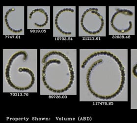
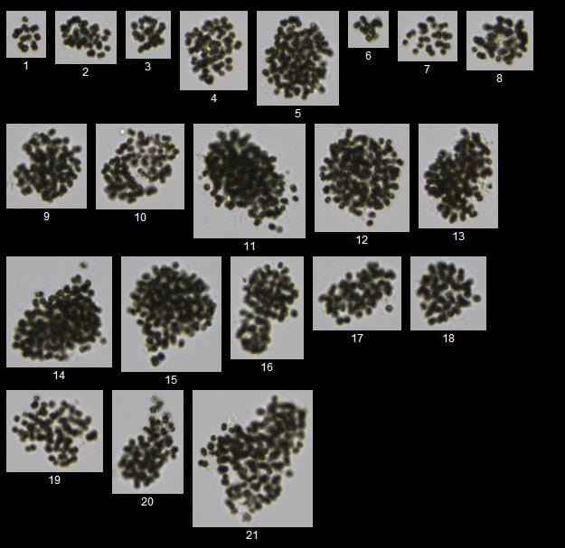
First, Microcystis colonies within a water sample were identified with a FlowCam using a local FlowCam image library of Microcystis colonies collected from rivers and reservoirs in South Korea. Second, the total sum of the areas of the Microcystis colonies within the water sample was obtained using the FlowCam. Then, a model algorithm (i.e., the relationship between the number of cells vs. the 2D surface area of the algal colonies) developed in this study was applied to estimate the total number of individual cells of Microcystis sp. within the water sample. The accuracy of the model algorithm in counting the total number of individual cells in Microcystis colonies within the water sample was then evaluated by comparing the result from the FlowCam with the result from a conventional microscopic method for algal cell counting (Park et al., 2019).
"The developed model algorithm showed satisfactory performance for Microcystis sp. cell counting in water samples collected from two rivers, and can be used for algal management in fresh water systems," (Park et al., 2019).
Access the paper by Park et al. here.
Citation:
Park, J., Kim, Y., Kim, M., and W. H Lee, 2018, A novel method for cell counting of Microcystis colonies in water resources using a digital imaging flow cytometer and microscope. Environmental Engineering Research, doi: 10.4491/eer.2018.266.