Can Zooplankton Be Entangled by Microfibers in the Marine Environment?: Laboratory Studies


1
Risk Assessment Research Center, South Sea Research Institute, Korea Institute of Ocean Science & Technology (KIOST), Geoje 53201, Korea
2
Department of Marine Environmental Sciences, University of Science and Technology, Daejeon 34113, Korea
3
Oil and POPs Research Group, Korea Institute of Ocean Science & Technology (KIOST), Geoje 53201, Korea
*
Author to whom correspondence should be addressed.
Water 2020, 12(12), 3302; https://doi.org/10.3390/w12123302
Submission received: 5 October 2020
/
Revised: 13 November 2020
/
Accepted: 20 November 2020
/
Published: 24 November 2020
(This article belongs to the Special Issue Terrestrial Microplastics in Soil and Water)
Abstract
:We investigated the probability of copepod entanglement in microfibers in a laboratory experiment. This experiment was inspired by an accidental observation of entangled copepods with microfibers during isolating copepods for grazing experiments. The exposure of microfibers to copepods was designed by applying conditions similar to environmentally relevant concentrations of microfibers to zooplankton in the Yellow Sea as well as highly elevated concentrations of microfibers compared to the previously reported maximum natural concentration. Copepod entanglement in microfibers was reproduced in laboratory. The entanglement was not observed in the condition which simulated the environmental scenario of copepods outnumbering microfibers observed in the Yellow Sea, while it occurred inconsistently in the conditions of the maximum and 10-fold maximum natural concentrations of microfibers. However, consistent entanglement of copepods by microfibers was found in the 100-fold maximum environmental concentration of microfibers. These results suggest that copepod entanglement by microfibers can happen accidentally under the maximum natural concentration of microfibers occurring in the marine environment; however, copepods are likely to be entangled in microfibers consistently if copepods encounter highly enhanced concentration of microfibers.
1. Introduction
A large amount of waste debris, of which the most abundant material is plastic, enters marine environments from sea- and land-based sources due to human activities [1,2]. Increased plastic pollution is a potential threat to marine biota because of the potential for entanglement and transfer of ingested contaminants to higher organisms [3,4]. The negative physical impacts of interactions with macroplastics (e.g., net, rope, monofilament line, packing straps, six-pack rings, and plastic sheeting) include debilitation, starvation, mistaken sense of fullness, injury, choking, and death of marine birds, mammals, fish, and reptiles due to plastic entanglement and ingestion [3,4,5,6,7].
On the other hand, numerous previous studies of the effects of microplastic exposure have focused primarily on the deleterious effects of ingested microplastic on aquatic invertebrates in terms of four types of response: consumption, growth, reproduction, and survival [8]. The particles (<5 mm in size) resulting from the fragmentation of macroplastics and primary manufactured microplastics, defined collectively as microplastics, exhibit worldwide distribution patterns from background levels to localized hotspots in sediments, beaches, and coastal and open oceans [9]. Among the microplastics, fibers are among the most prevalent types of microplastic debris observed in the marine environment [10]. Fibers are found in the range of 0–100 particles/L, accounting for 8–100% of total microplastics in the water column in marine environments [9,10,11,12,13,14,15,16,17,18]. Although these fibers occur in oceans worldwide, studies about fibers and microfibers are very hard because those are easily contaminated from diverse routes while sampling and treatment in the in situ condition [18]. Thus, studies of the effects of microfibers on aquatic invertebrates remain scarce, except a few studies considering ingested microfibers by some crustaceans. Toxicity from microplastic fibers was reported to be greater compared to other microplastic particles, stemming from their longer residence times of fibers in the gut in the freshwater amphipod Hyalella Azteca [19]. Moreover, tightly tangled balls of plastic strands were found in the gut of the decapod crustacean Nephrops norvegicus, and this configuration likely influences the gut retention time of fibers, causing a false feeling of satiation and leading to gut blockage [19,20]. In addition, plastic microfibers have been found inside deposit-feeding organisms inhabiting the deep-sea floor [21].
However, observations of zooplankton entangled in microfibers in marine environments have not been reported; thus, understanding about their occurrence can serve as a starting point for crucial research on issues such as impeded zooplankton behavior and consequent effects on survival. The widespread distribution and relatively high proportion of fibers compared to other types may increase the chance of interaction with marine biota. In particular, rates of encounter between microfibers and zooplankton can be high in localized hotspots, such as industrial drainage systems, runoff from rivers, and storm drains where microplastic abundances were known to be especially high at coastal waters [9,16,22]. However, far from our expectation, we accidentally observed live copepods entangled in microfibers during isolating live copepod Calanus sinicus for grazing experiments via on-board bottle incubations in the offshore waters of the Yellow Sea of Korea. There could be opportunities for entanglement by copepods in microfibers during the sampling process rather than in situ entanglement. There is a high probability of entanglement stemming from “net entanglement” due to vigorous mixing in the cod-end bucket while the net is being towed gently. Therefore, we aimed to assess the probability of copepod entanglement in microfibers under the laboratory experiments with no possibility of “net entanglement”. We tested the probability of copepod entanglement in microfibers under conditions close to environmentally relevant concentration of microfibers to zooplankton as well as highly elevated numbers of microfibers compared to maximum natural concentration of microfibers found so far in the marine environment.
2. Materials and Methods
2.1. Accidental Observation of Copepods Entangled in Microfibers
Live zooplankton samples were originally collected for grazing experiments on board R/V EARDO at stations (A03, D03) located at 35.0–36.0° N and 124.30° E in the Yellow Sea. The live copepod Calanus sinicus were isolated for an analysis of grazing rates per day via the on-board bottle incubation method. The rate can be calculated by the difference of prey concentration as a result of grazing for a day. Samples were caught by gentle, oblique hauling of a net (diameter, 45 cm; mesh size, 200 µm) within the surface mixed layer at night in April (spring) and August (summer) of 2013 (Figure 1). After recovery of the net, samples were washed carefully in a cod-end bucket and transferred to a 1-L beaker containing pre-filtered seawater at the same temperature as the seawater. While the catch was sorted as soon as possible under a stereomicroscope, C. sinicus copepods entangled in microfiber were randomly separated, transferred to a 10-mL vial, and preserved immediately in 5% (final concentration) borax-neutralized formalin.
2.2. Analysis for Exposure Condition Considering Environmentally Relevant Concentration
We analyzed microfibers and zooplankton from archived samples collected from stations where zooplankton entangled by microfibers was found in order to get environmentally relevant condition of microfibers to zooplankton for laboratory exposure. Archived samples, which had been originally collected to understand distribution of mesozooplankton community, were acquired by vertical net towing (diameter, 60 cm; mesh size, 200 μm) through the surface mixed layer (0–10 m) from 12 stations located at 34.3–36.0° N and 124.30–125.40° E in the Yellow Sea in April (spring) and August (summer) of 2013 (Figure 1). Half of each preserved sample (final concentration (5%) of borax-neutralized formalin) was totally used for microplastic analysis, and a sub-sample of the other half was used for analysis of zooplankton community and existence of zooplankton entangled by microfibers for determination of exposure condition considering environmentally relevant concentration (Table 1). After treatment for analysis of microplastics, the samples were processed and observed based on the previous studies [16,23,24,25]. The specific polymer types of entangling fibers on the zooplankton were analyzed using Fourier-transform infrared spectroscopy (FT-IR) microscopy (Thermo Nicolet 6700 and Continuum; Thermo Scientific, Waltham, MA, USA). All spectra of samples were recorded as the average of 64 scans in the spectral range of 550–4000 cm−1 at a resolution of 8 cm−1 (Figure 2; Table 2). We employed procedural blanks during the laboratory analyses to check for background contamination. Blanks were also subjected to the entire experimental procedure and filtered through GF/F filters (diameter, 47 mm; Whatman, Buckinghamshire, UK) after the experiment was terminated. The GF/F filters were also processed and observed under a stereomicroscope (Discovery SV8; Zeiss, Zeiss, Göttingen, Germany) as above described for the experimental treatments.
2.3. Microfiber Exposure Experiment
2.3.1. Preparation of Seawater for Exposure
For the microfiber exposure experiment, natural seawater was collected from the surface of Jangmok Bay, located in the South Sea of Korea, and then screened through 100-µm mesh into a polycarbonate carboy for transport to the laboratory. The seawater was further filtered through a 0.45-μm filter (GTTP 04700; Merck Millipore, Burlington, MA, USA). The pH, DO, and salinity of the filtered seawater were 7.8–8.1 (average, 7.9), 7.5–8.5 mg/L (average, 8 mg/L), and 30–35 psu (average, 32.5 psu), respectively. The filtered seawater was stored until the exposure experiment.
2.3.2. Preparation of Copepod for Exposure
It is practically difficult to get the copepod Calanus sinicus consistently for laboratory exposure experiments because the copepod lives in the offshore waters. So, we selected and utilized a promising marine model organism, Tigriopus japonicus, which also belongs to copepods and is easy to rear and maintain for continuous exposure experiments in the laboratory [26].
Harpacticoid copepod T. japonicus cultures were acquired from the Department of Biology, College of Life Sciences, Hanyang University, Seoul, Republic of Korea. The copepods was maintained at the Korea Institute of Ocean Science and Technology since 2011. They were cultured to an average abundance of 400 individuals/L in a 10-L volume of filtered seawater (pore size, 0.22 μm; GTBP04700; Millipore, Darmstadt, Germany) in an incubator under a 12:12-h light:dark cycle (150–160 µmol/m2 s) at 20 ± 1 °C. The pH, dissolved oxygen (DO), and salinity of the filtered seawater were 7.8–8.1, 7.5–8.5 mg/L, and 30–35 psu, respectively. Tetraselmis suecica was supplied as a food source once weekly. T. suecica was cultured at 20 ± 1 °C under a 12:12-h light:dark cycle (150–160 µmol/m2 s) in Walne’s medium.
2.3.3. Preparation of Microfiber for Exposure
The polymer types of microfiber that are dominant in the surface waters of the Yellow Sea include polyester, cotton, and polypropylene. We selected 100% polyester fibers from a blanket used for approximately 2 years for the exposure experiment to assess the possibility of copepod entanglement by microfibers under laboratory conditions. The blanket microfibers were washed carefully with distilled water (Milli-Q Gradient A10; Merck Millipore, Burlington, MA, USA) and manually cut into the smallest lengths possible (0.86 ± 0.26 mm, n = 50) using sharp scissors. The lengths of the microfibers used for exposure of microfibers to copepods in the laboratory were set considering the approximate ratio between body lengths of entangled zooplankton and lengths of entangling fibers on zooplankton found in the Yellow Sea (Table 2).
2.3.4. Exposure Design for Entanglement Experiment
- Experimental consideration for exposure of microfibers to copepods
Probability of copepod entanglement in microfibers was first tested under the condition explaining environmental scenario of higher zooplankton with quite low of microfibers generally observed in the natural field [12,16,17,23,27]. Further, if entanglement can happen in that condition, a possible condition affecting the consistent occurrence of entanglement was explored by enhancing the possibility of encounter between microfibers and copepods such as bubbling and reduction of incubation volume under enhanced microfibers to copepods.
The concentrations of microfibers relative to zooplankton reported in previous studies were too low to recreate the same conditions in the laboratory. So, experimental condition simulating natural concentration of microfibers (0.05 and 0.02) was set almost close to the ratio of microfibers to zooplankton observed in the Yellow Sea (Table 3). In addition, elevated microfibers to copepods are experimentally practical in terms of an easier way to differentiate entangled copepods from the incubating containment and enhance effectively the encounter rate between microfibers and copepods, although that condition is the opposite way to the environmental scenario of low microfibers with higher zooplankton observed in the marine environment [16,17,28].
Therefore, in order to understand the possibility of copepod entanglement in microfibers in the laboratory, first, we adopted exposure condition close to the in situ ratio of microfibers to zooplankton found in the offshore waters of the Yellow Sea (Table 3) and the maximum concentration of microfibers observed in nature so far (100 particles/L) [11]. Then, if entangled copepods by microfibers occurs in the conditions abovementioned, we conducted the experiment to understand which exposure condition can be significant to consistent entanglement of copepod with microfibers by treating approximately 10-fold and 100-fold the maximum natural microfiber concentration.
- Exposure of microfibers to copepods under environmentally relevant concentration (50 mL)
This experiment was designed to determine whether copepod entanglement can occur under the laboratory conditions nearly similar to the natural ratio of microfiber to zooplankton in the Yellow Sea (Table 3). Twenty and fifty adults of Tigriopus japonicus were transferred to glass beakers (Duran, Wertheim, Germany) containing 50 mL filtered seawater with 1 microfibers from the blanket (polyester) to achieve ratios of microfiber to copepod (0.05 and 0.02), respectively (Table 3). All beakers were washed and rinsed three times with distilled water before use. Tetraselmis suecica was provided three times (0, 72, and 144 h) as food sources at concentrations of 12 × 105 cells/mL during the entire period. Each treatment has three replicates. The glass beakers were incubated at 20 ± 1 °C under a 12:12-h light:dark cycle (155–168 μmol/m2s) during 168 h and covered with aluminum foil to prevent evaporation and contamination by other microfibers. All other experimental conditions and processes such as procedural blanks were done as described above.
- Effects of bubbling under elevated microfibers (500 mL)
This experiment was designed to determine whether copepod entanglement can occur or be enhanced under laboratory conditions when applying a bubbling treatment to enhance the probability of encounter between microfibers and copepods under elevated microfibers to copepod abundances. Thirty T. japonicus adults were transferred to glass beakers (PYREX Griffin beaker, Duran, Wertheim, Germany) containing 500 mL filtered seawater with 50 and 5000 microfibers prepared using stock solutions (an average of 5.5–6.0 and 100 polyesters/mL) to achieve the maximum natural concentration and 100-fold the maximum environmental concentration, respectively (Table 4).
All beakers were washed and rinsed three times with distilled water before use. T. suecica and natural phytoplankton were provided three times at 0, 72, and 144 h as food sources at concentrations of 9 × 10⁴ cells/mL and 3–5 × 10⁴ cells/mL, respectively, during the entire experimental period (168 h).
To enhance the rate of encounter between microfibers and copepods, the water in the beakers was bubbled and no-bubbled using an air pump (DK-9000; Daegwang, Seoul, Korea) in the control (no microfiber) and treatment (polyester microfibers) groups for comparison purposes. Each treatment had nine replicates. We compared the abundance of copepods entangled in microfibers between treatments. All other experimental conditions and processes such as procedural blanks were prepared as described above.
- Reduced volume of incubation under elevated microfibers (10 mL)
This experiment was conducted to determine the probability of copepod entanglement under an enhanced encounter between microfibers and copepods by reducing the incubation volume under elevated microfibers to copepod abundances compared to the maximum natural concentration during short-term (96 h) exposure (Table 4).
To achieve the maximum natural microfiber concentration (100 particles/L), 10- and 100-fold maximum environmental microfiber concentration, 10 Tigriopus japonicus adults were added to each well of six-well cell culture plates (SPL Life Sciences), of which each contained 10 mL filtered seawater with 0 (control), 1, 10, 25, 50, 250, or 500 polyester microfibers. Additionally, the condition of 500 microfibers + 20 T. japonicus was additionally conducted, simultaneously. Each treatment had nine replicates. T. japonicus were observed in the plates under a stereomicroscope at 6, 12, 18, 24, 48, 72, and 96 h. Shortly before the start of the experiment, Tetraselmis suecica and natural phytoplankton at concentrations of 9 × 10⁴ and 3–5 × 10⁴ cells/mL, respectively, were provided to the copepods for 1 day as a food source and were subsequently maintained at these abundances for observation under a stereomicroscope. All other experimental conditions and processes were treated as described above.
2.3.5. Observation and Confirmation of Entangled Copepods
Live T. japonicus entangled in microfibers were observed under a stereomicroscope (Discovery SV8; Zeiss) at ×10, ×20, and ×80 magnification every 24 h after initiation of the experiments. In the experiment of reduced volume of incubation (10 mL), observations were made every 6 h to determine whether zooplankton became entangled within 24 h of exposure and every 24 h after 24 h exposure. Magnified images of T. japonicus entangled in microfibers were taken using an image analysis system with a light microscope under magnifications of ×100, ×200, and ×400 (AxioCam ICc 3, ZEN 2010 program; Axio Imager A2; Zeiss, Dresden, Germany).
For observations of copepod entanglement in microfibers under a stereomicroscope, the supernatant in the incubated beakers (500 or 50 mL) was carefully transferred to a clean beaker, taking care to avoid loss of microfibers as much as possible. Subsequently, T. japonicus and the microfibers in the beakers were observed directly under a stereomicroscope (Discovery SV8; Zeiss, Göttingen, Germany). After observation, the supernatant was carefully transferred back into the beakers for consistent incubation. In the experiment using six-well plates, each covered plate was observed directly under a stereomicroscope. To ensure true entanglement, rather than the positioning of a copepod above or below a fiber, we defined entanglement as follows. First, to differentiate between entanglement and non-entanglement, we gently touched all copepods with a dissection needle and observed their response. When a microfiber had not completely entangled a copepod, it separated from the copepod when the latter moved. When a copepod moved together with an entangled microfiber for more than at least 1 min, we determined that it was entangled in the microfiber. In the case of archived samples, we confirmed it as the entangled zooplankton by microfibers if microfibers entangled on zooplankton are still tightly stuck to the body after rinsing out with a squeeze bottle containing filtered seawater.
2.3.6. Statistical Analysis
To assess the effect of bubbling on copepod entanglement in microfibers, Student’s t-test was used to compare mean abundances of copepods entangled in microfibers between bubbling and no-bubbling treatments. Differences in the mean numbers of individual copepods entangled in microfibers among treatments were examined using one-way ANOVA. All statistical analyses were performed using SPSS ver. 12.0 (SPSS, Chicago, IL, USA), with a significance level of p < 0.05.
3. Results
3.1. Entangled Copepods in Microfibers: On-Board Observation
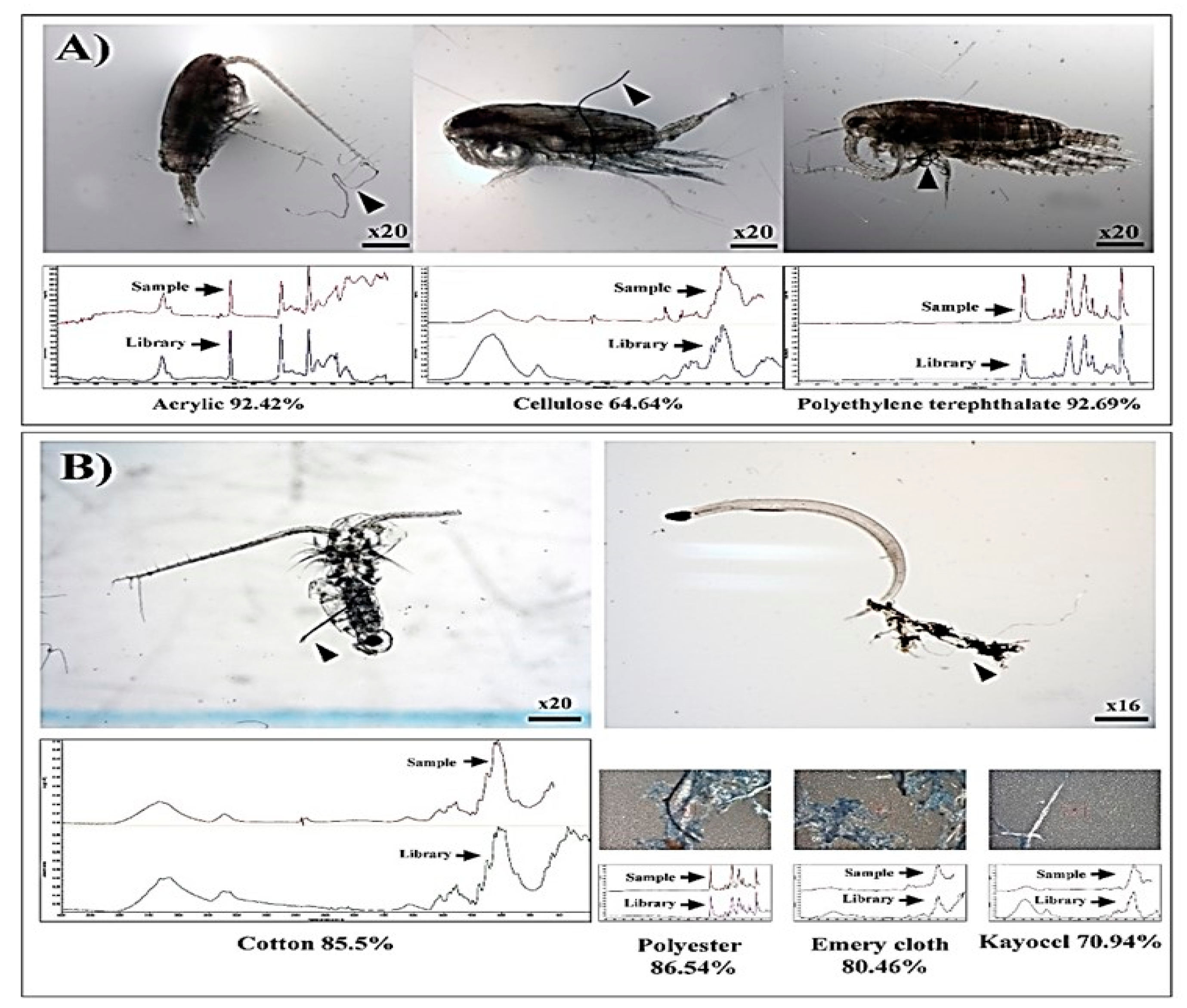
We accidentally observed several live Calanus sinicus entangled in microfibers during isolating live copepods under a stereomicroscope for on-board bottle incubations at stations A03 and D03 in the Yellow Sea in April and August 2013 (Figure 1). The polymer types of the microfibers entangling C. sinicus were identified as acrylic, cellulose, and polyethylene terephthalate (Figure 2A; Table 2). The body parts of the copepod entangled by microfibers included appendages, antennae, and the side of the body (Figure 2A).
3.2. Determination for Exposure Considering Environmentally Relevant Concentration
For designing the exposure scheme to simulate the natural condition of microfibers and copepods, we analyzed microfibers and zooplankton from all archived samples, and then extracted the ratio of microfibers to zooplankton at stations where zooplankton entangled by microfibers were found (Table 2 and Table 3). Zooplankton entangled in microfibers was found at only two stations (A03 and D06) from a total of 12 stations collected in August in the Yellow Sea. In addition, proportion of the entangled zooplankton at those stations was less than 2% of the total zooplankton abundances and the ratios of microfibers to zooplankton were 0.002 and 0.005, with remarkably low microfibers compared to zooplankton (Table 3). The ratio is too low to recreate the same conditions in the laboratory. So, the exposure condition simulating the environmental relevant concentration of microfibers to zooplankton was set as 0.05 and 0.02, close to the ratio of microfibers to zooplankton observed in the Yellow Sea (Table 3). Fiber clumps entangling a chaetognath (Sagitta sp.) were identified as polyester, emery cloth, and Kayocel, while a copepod was entangled in cotton (85.5%) (Figure 2B; Table 2). The body parts of zooplankton entangled by microfibers included the waist of the copepod and the tail of the chaetognath (Figure 2B).
3.3. Laboratory Experiments
3.3.1. Probability of Copepod Entanglement in Microfibers: Environmentally Relevant Concentration of Microfibers (50 mL)
Copepod mortality in the control treatments was less than 10% and no microfiber was detected in the blank treatments. Entangled copepods by microfibers were not found during the entire exposure time (168 h) under the condition close to the natural ratio of microfiber to zooplankton found in the Yellow Sea (Table 3).
3.3.2. Probability of Copepod Entanglement in Microfibers: Effects of Bubbling Under Elevated Microfibers (500 mL)
No microfiber was detected in the blank samples and copepod mortality in the control treatments was less than 10%. Entanglement by microfibers was observed for only a small number of copepods (average, 0.3 individuals/replicate) on day 7 under the conditions of 50 microfibers with 30 copepods with no bubbling in 500-mL beakers (Table 4), corresponding to the maximum natural microfiber concentration (100 particles/L). The proportion of entangled copepods was 1.0% or 0% at no-bubbling or bubbling treatments, respectively, with an encounter rate of 1.6 (Table 4).
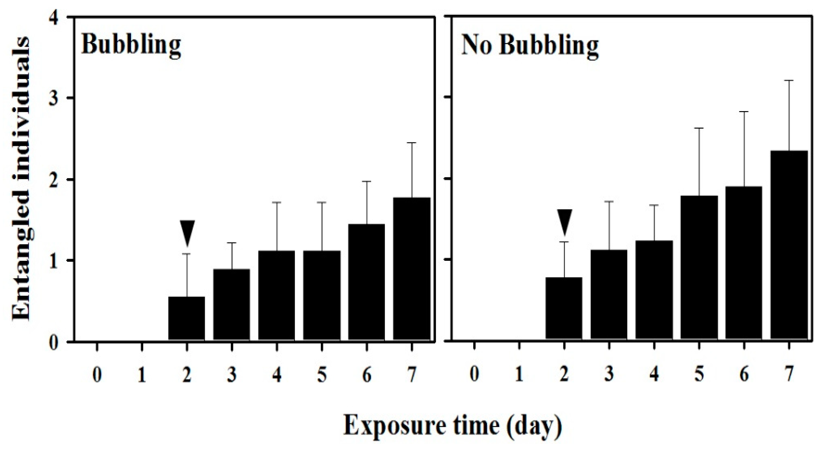
However, for the combination of 5000 microfibers and 30 copepods corresponding to 100-fold maximum natural concentration of microfibers, entanglement of T. japonicus in microfibers was consistently observed under all experimental conditions. Additionally, the mean abundances of entangled copepods increased gradually from 0.6–1.8 individuals with bubbling and from 0.8–2.3 individuals with no-bubbling during the course of the experiment (Figure 3). The first entanglement was observed 48 h after exposure to the microfibers under conditions with and without bubbling (Figure 3). The maximum average abundances and proportion of entangled copepods were 1.8 individuals (6.0%) with bubbling and 2.3 individuals (7.7%) with no bubbling at 7 days (168 h) after exposure (Figure 3; Table 4) with an encounter rate of 166.6. The mean abundances of copepods entangled in microfibers did not differ significantly between the bubbling and no-bubbling treatments (p > 0.05). Student’s t-test indicated that bubbling did not significantly affect the abundance of copepods entangled in microfibers.
3.3.3. Probability of Zooplankton Entanglement in Microfibers: Reduced Volume of Incubation under High Microfiber Concentrations (10 mL)
Copepod mortality in the control treatment was less than 10% and no microfiber was contaminated in the procedural blank samples. When exposed to the maximum natural concentration of microfibers reported previously in the marine environment and 10-fold the maximum natural concentration, no copepod entanglement in microfibers was observed under conditions of 1, 10, and 25 microfibers with 10 Tigriopus japonicus adults (Table 4). However, mean entangled copepods with microfibers and proportion were observed as 0.3 individuals (3.0%) under condition of 50 microfibers with 10 T. japonicus adults at 96 h after exposure (Table 4).
Under conditions with 100-fold the maximum microfiber concentration, no entangled copepod was observed prior to 24 h after exposure in any treatment (Figure 4). However, mean entangled copepods with microfibers and proportion ranged from 1.0 individuals (10.0%) to 2.3 individuals (11.5%) under conditions of 250, 500, and 500 microfibers with 10, 10, and 20 T. japonicus adults at 96 h after exposure, respectively (Table 4). Entangled copepods in microfibers occurred consistently in the conditions with 100-fold the maximum natural concentration. In addition, mean entangled copepods with microfibers in the 100-fold the maximum natural concentration were higher than those found in the conditions of maximum and 10-fold the maximum natural concentrations of microfibers at 96 h after exposure. Especially, the results for 500 microfibers with 20 T. japonicus differed significantly from those for the other treatments at 72 and 96 h after exposure (p < 0.05), indicating that increased encounter between microfibers and copepods can lead to increment of entangled copepods in microfibers (Figure 4).
3.4. Body Parts of Copepods Entangled in Microfibers
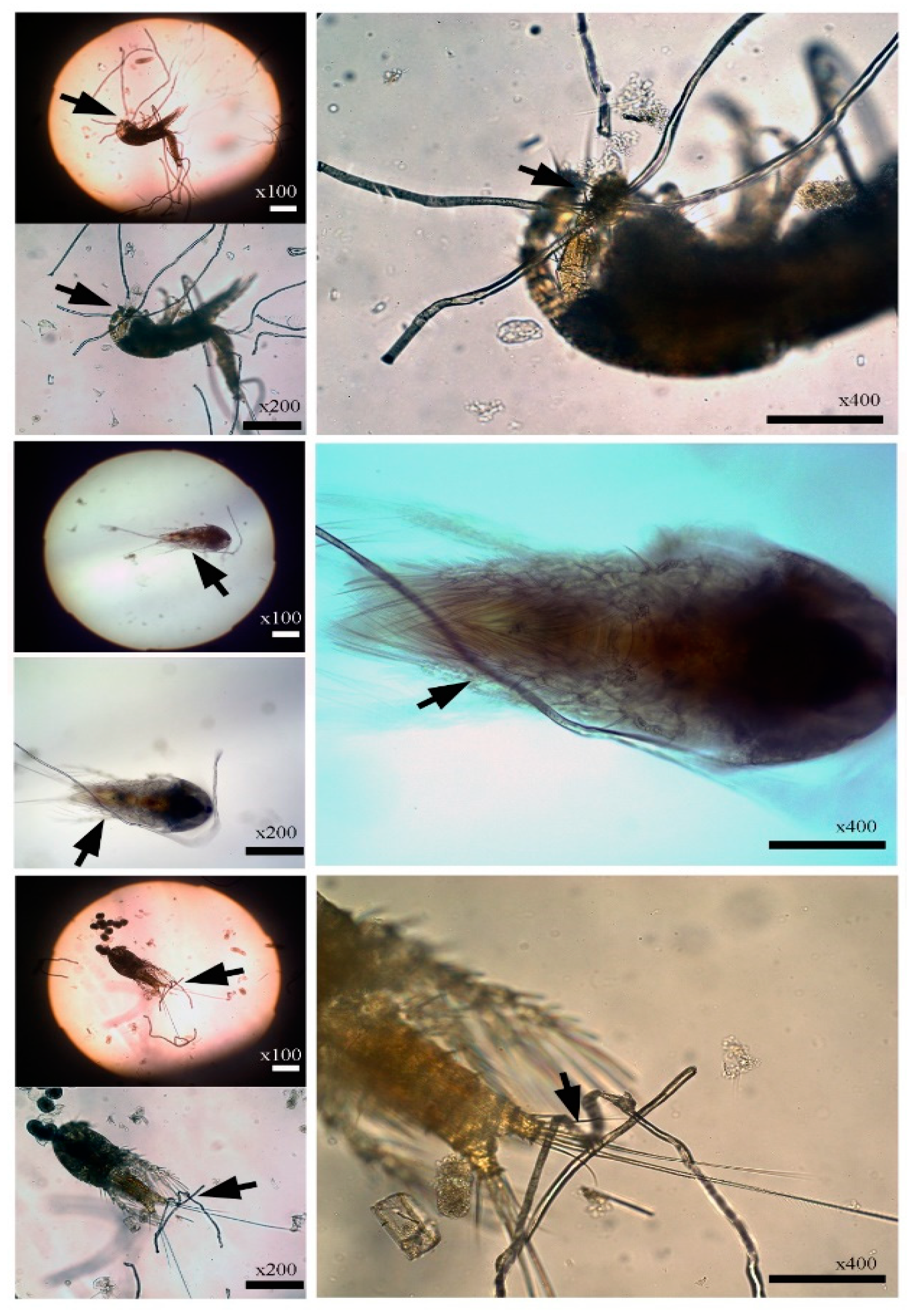
In images of T. japonicus entangled in microfibers in the laboratory studies, the entangled microfibers were observed primarily on the antennae, setae, and bristles of appendages, as well as on the caudal rami (Figure 5).
4. Discussion
The impact of microfibers on marine invertebrates had been studied mainly in terms of the presence of microfibers in the guts of freshwater amphipods, marine decapod crustaceans, and deposit-feeding organisms inhabiting the deep-sea floor [8,19,20,21,29], whereas the external effects of microplastics on marine invertebrate have not been reported in terms of entanglement. Marine copepods entangled in microfibers were by chance observed first while isolating live copepods for an on-board grazing experiment in the Yellow Sea in 2013 (Figure 2; Table 2). There could be several opportunities for entanglement by zooplankton in microfibers during the sampling process and storage of zooplankton samples rather than in situ entanglement in the marine environment. There is a high probability of entanglement stemming from “net entanglement” due to vigorous mixing in the cod-end bucket while the net is being towed, as well as entanglement during fixation and settling out of the sample in sample bottles. However, prior to mentioning it as the in situ entanglement, we need to explore the probability of copepod entanglement by microfibers under the laboratory condition with no “net entanglement” or any other causes [30]. We analyzed microfibers and zooplankton from all archived samples in order to design the exposure scheme to simulate natural condition of microfibers and copepods. Even though the presence of entangled zooplankton in the archived samples does not mean entangled zooplankton in the field, it can be a clue for determination of exposure condition considering environmentally relevant concentration of microfibers and zooplankton because we need to select appropriate exposure condition of microfibers to zooplankton in the laboratory. In addition, highly enhanced concentrations of microfibers to copepods for the laboratory experiment were set based on the maximum concentration of microfibers reported in the marine environment (100 particles/L). Thus, the probability of copepod entanglement in microfibers was experimented under exposure conditions close to natural concentration of microfibers and zooplankton as well as highly enhanced concentrations of microfibers to copepods in a laboratory. Laboratory experiments showed that copepods can be entangled in microfibers with no “net entanglement” (Figure 3 and Figure 4; Table 4).
However, the entanglement was not observed in the environmentally relevant condition which simulated the ratio of microfibers to zooplankton found in the Yellow Sea (Table 3), while it occurred inconsistently in the condition of the maximum natural microfibers reported so far (Table 4). Even though the extremely high concentration of microfibers has not been reported so far in the marine environment, entanglement of copepods by microfibers was consistently found in 100-fold the maximum environmental concentration of microfibers in the laboratory.
Our results suggested that copepod entanglement in microfibers cannot occur under the current environmental scenario based on the ratio of microfiber to zooplankton (relatively high abundances of zooplankton compared to microplastics) found in the Yellow Sea (Table 3). Therefore, entangled copepods by microfibers found in the archived and live samples collected from the Yellow Sea was likely to be associated with the “net entanglement” during the sampling process or storage-related entanglement. However, copepod entanglement in microfibers can occur at maximum natural concentration of microfibers (100 particles/L) [11] observed in the marine environment (Table 4). Therefore, the observation of entanglement was not necessarily consistent or generalizable under the current concentration of microfibers observed in the Yellow Sea as well as the condition of maximum natural concentration of microfibers so far (Table 3 and Table 4). On the other hand, our findings indicated that copepods became entangled by microfibers consistently under elevated abundances of microfibers (approximately 100-fold the maximum natural concentration) compared to copepods with encounter rates ranging from 0.1 to 5.0 (Table 4).
The “encounter rate” used in the present study means that the opportunity that zooplankton encounter microplastics in the water column, comparing the ratio of microplastics to zooplankton based on abundance [22,31,32,33]. The concept meant that the higher the encounter rate is, the worse risk of microplastic to zooplankton is. Even though the rate is the relative value calculated from abundances between microplastics and zooplankton, it has helped us to understand the potential impact of microplastic to zooplankton at sites of interest.
Until recently, the ratios reported in marine environments, which indicate the ‘rate of encounter’ between microplastics and zooplankton, ranged from 0.004 (Jinhae Bay, Korea) to 0.6 (southern California coast, USA) [17,28,32], corresponding to the relatively low microfiber to high zooplankton ratios in the experiment simulating the environmentally relevant concentration of microfibers, as well as those observed at stations where zooplankton entangled in microfibers were found in archived samples from the Yellow Sea (Table 3 and Table 4). These results suggest that abundances of copepods entangled with microfibers can occur at negligible values in current marine environments. However, until recently, high ratios of microfibers to zooplankton (i.e., 5.0 to 166.6) have not been reported in marine environments, indicating that the consistent occurrence in the abundances of copepods entangled with microfibers are not likely to occur under the current levels of microfibers and zooplankton in marine environments.
The ratios observed in nature generally result from relatively high abundances of zooplankton and low abundances of microplastic; in contrast, marine environments with high ratios caused by relatively high abundances of microplastic and low abundances of zooplankton (i.e., conditions similar to our experimental treatments) are likely to be quite scarce, probably except in environmental discontinuity. Our results showed that copepods can be entangled significantly in the condition of 100-fold maximum natural concentration of microfibers, suggesting that rates of the entanglement will be probably high if conditions of relatively high abundances of microfibers and low abundances of copepods occur at a certain ratio in marine environments, such as in environmental discontinuities (Figure 3 and Figure 4; Table 4). Environmental discontinuities, such as river run-off after heavy precipitation from coastal storms, as well as in convergence zones, inside of gyres, fronts, eddies, and meanders in offshore locations, all of which can potentially enhance rates of encounter between microplastics and zooplankton through the retention and accumulation of microplastics [22,27,34,35,36].
Microfiber shapes vary from entangled to straight, curved, and twisted [37]. Fibers are likely to be the most frequently encountered form of microplastics globally, accounting for 8–100% of total microplastics in the water column of marine environments [9,11,12,14,15,18]. Given that microfibers of various shapes drift in currents and accumulate in enclosed areas, such as harbors and ports, as well as in retention zone such as environmental discontinuities, these microfibers constantly could interact with zooplankton and other marine organisms. The complex shapes of microfibers can readily entangle moving marine zooplankton, which have morphological characteristics such as setae on their antennae and appendages. Some shapes of the entangled microfibers were similar to those observed in our laboratory studies (Figure 5), although such shapes varied somewhat between the laboratory and in situ environment. When entanglement involves the feeding appendages of zooplankton and the zooplankton are unable to escape, they could face inefficient foraging and reduced predator avoidance capacities. In zooplankton, feeding appendages include antennules, antennae, mandibles, maxillules, and maxillae, which are important for the detection and selection of motile prey organisms in association with mechano- and chemosensory abilities [38] Even though Tigriopus japonicus, which have been used in laboratory experiments, are an intertidal copepod unlike the planktonic copepod Calanus sinicus observed in the Yellow Sea, we are able to anticipate similar results to the C. sinicus due to their morphological characteristics in terms of the same function of foraging and avoidance capacities. However, there could be one thing to consider when we understand present results in terms of swimming patterns of the intertidal T. japonicus. Unlike calanoid copepods swimming always only in the water column, marine harpacticoid T. japonicus not only remain usually near the bottom of the incubation containers but also can swim up and down through the water column [39]. That was the same to the exposure experiment of microfiber to copepods in the beakers conducted in the present study, which means that T. japonicus can have more possibility to be entangled with microfibers distributing in the bottom and water column of the beakers compared to the case of calanoid copepods. So, we need to consider characteristics between T. japonicus and calanoid copepods having different swimming traits when we understand the meaning of entangled copepods in microfibers observed in the present study. The ultimate effects of entanglement on marine zooplankton have not yet been documented at the individual or population level, and the consequences of entanglement must be explored in terms of reduced survival due to the disruption of key behaviors. For instance, diel vertical migration, a zooplankton survival strategy, could be hampered by the negative impacts of entanglement on swimming and feeding abilities.
5. Conclusions
Copepod entanglement in microfibers was reproducible in the laboratory. However, there was no entanglement or negligible under the conditions mimicking the environmentally relevant concentrations of microfibers found in the Yellow Sea as well as the maximum natural microfibers reported so far in the marine environment (100 particles/L) [11]. It indicates that copepod entanglement with microfibers is not common in the current conditions of microfibers in the marine environment. However, if microfibers outnumber copepods under the condition of 100-fold the maximum natural microfibers, consistent entanglement of copepod in microfibers can happen in the laboratory. These results suggest that copepod entanglement in microfibers is currently uncommon under natural microfiber concentrations with relatively high copepods; however, copepods will become entangled in microfibers consistently at highly elevated microfiber abundances relative to the abundance of copepods.
Author Contributions
W.J.S. and S.H.H. conceived and acquired the project and contributed to the manuscript preparation. O.-Y.K. performed the experiments. J.-H.K. wrote and edited the manuscript. All authors have read and agreed to the published version of the manuscript.
Funding
This study was funded by research project titled ‘‘Environmental Risk Assessment of Microplastics in the Marine Environment’’ from the Ministry of Ocean and Fisheries, Korea.
Acknowledgments
We would like to thank Minju Kim (Korea Institute of Ocean Science & Technology) for the help in the revise of figures and tables in this manuscript.
Conflicts of Interest
The authors declare no conflict of interest.
References
- GESAMP. GESAMP (2010, IMO/FAO/UNESCO-IOC/UNIDO/WMO/IAEA/UN/UNEP Joint group of experts on the scientific aspects of marine environmental protection). In Micro-Plastics in the Marine Environment, Proceedings of the GESAMP International Workshop on Plastic Particles as a Vector in Transporting Persistent, Bioaccumulating and Toxic Substances in the Oceans. GESAMP Reports & Studies; Paris, France, 28–30 June 2010; Bowmer, T., Kershaw, P., Eds.; UNESCO-IOC: Paris, France, 2010; Number 82; pp. 18–27. Available online: http://www.gesamp.org/publications/proceedings-of-the-gesamp-workshop-on-microplastic-particles (accessed on 28 June 2018).
- Andrady, A.L. Microplastics in the marine environment. Mar. Pollut. Bull. 2011, 62, 1596–1605. [Google Scholar] [CrossRef] [PubMed]
- Derraik, J.G.B. The pollution of the marine environment by plastic debris: A review. Mar. Pollut. Bull. 2002, 44, 842–852. [Google Scholar] [CrossRef]
- Gregory, M.R. Environmental implications of plastic debris in marine settings-entanglement, ingestion, smothering, hangers-on, hitch-hiking and alien invasions. Philos. Trans. R. Soc. B Biol. Sci. 2009, 364, 2013–2025. [Google Scholar] [CrossRef] [PubMed]
- Jiménez, S.; Domingo, A.; Brazeiro, A.; Defeo, O.; Phillips, R.A. Marine debris ingestion by albatrosses in the southwest Atlantic Ocean. Mar. Pollut. Bull. 2015, 96, 149–154. [Google Scholar] [CrossRef] [Green Version]
- Nicolau, L.; Marçalo, A.; Ferreira, M.; Sá, S.; Vingada, J.; Eira, C. Ingestion of marine litter by loggerhead sea turtles, Caretta caretta, in Portuguese continental waters. Mar. Pollut. Bull. 2016, 103, 179–185. [Google Scholar] [CrossRef]
- Ryan, P.G.; Nico de Bruyn, P.J.; Bester, M.N. Regional differences in plastic ingestion among Southern Ocean fur seals and albatrosses. Mar. Pollut. Bull. 2016, 104, 207–210. [Google Scholar] [CrossRef] [Green Version]
- Foley, C.J.; Feiner, Z.S.; Malinich, T.D.; Höök, T.O. A meta-analysis of the effects of exposure to microplastics on fish and aquatic invertebrates. Sci. Total Environ. 2018, 631–632, 550–559. [Google Scholar] [CrossRef] [Green Version]
- Wright, S.L.; Thompson, R.C.; Galloway, T.S. The physical impacts of microplastics on marine organisms: A review. Environ. Pollut. 2013, 178, 483–492. [Google Scholar] [CrossRef]
- Browne, M.A.; Crump, P.; Niven, S.J.; Teuten, E.; Tonkin, A.; Galloway, T.; Thompson, R. Accumulation of microplastic on shorelines worldwide: Sources and sinks. Environ. Sci. Technol. 2011, 45, 9175–9179. [Google Scholar] [CrossRef]
- Buchanan, J. Pollution by synthetic fibres. Mar. Pollut. Bull. 1971, 2, 23. [Google Scholar] [CrossRef]
- Thompson, R.C.; Olsen, Y.; Mitchell, R.P.; Davis, A.; Rowland, S.J.; John, A.W.G.; McGonigle, D.; Russell, A.E. Lost at sea: Where is all the plastic? Science 2004, 304, 838. [Google Scholar] [CrossRef] [PubMed]
- Claessens, M.; Meester, S.D.; Landuyt, L.V.; Clerck, K.D.; Janssen, C.R. Occurrence and distribution of microplastics in marine sediments along the Belgian coast. Mar. Pollut. Bull. 2011, 62, 2199–2204. [Google Scholar] [CrossRef] [PubMed]
- Desforges, J.P.W.; Galbraith, M.; Dangerfield, N.; Ross, P.S. Widespread distribution of microplastics in subsurface seawater in the NE Pacific Ocean. Mar. Pollut. Bull. 2014, 79, 94–99. [Google Scholar] [CrossRef] [PubMed]
- Ivar do Sul, J.A.; Costa, M.F. The present and future of microplastic pollution in the marine environment. Environ. Pollut. 2014, 185, 352–364. [Google Scholar] [CrossRef]
- Kang, J.-H.; Kwon, O.-Y.; Lee, K.-W.; Song, Y.K.; Shim, W.J. Marine neustonic microplastics around the southeastern coast of Korea. Mar. Pollut. Bull. 2015, 96, 304–312. [Google Scholar] [CrossRef]
- Aytan, U.; Valente, A.; Senturk, Y.; Usta, R.; Sahin, F.B.E.; Mazlum, R.E.; Agirbas, E. First evaluation of neustonic microplastics in Black Sea waters. Mar. Environ. Res. 2016, 119, 22–30. [Google Scholar] [CrossRef]
- Gago, J.; Carretero, O.; Filgueiras, A.V.; Viñas, L. Synthetic microfibers in the marine environment: A review on their occurrence in seawater and sediments. Mar. Pollut. Bull. 2018, 127, 365–376. [Google Scholar] [CrossRef]
- Au, S.Y.; Bruce, T.F.; Bridges, W.C.; Klaine, S.J. Responses of Hyalella Azteca to acute and chronic microplastic exposures. Environ. Toxicol. Chem. 2015, 34, 2564–2572. [Google Scholar] [CrossRef]
- Murray, F.; Cowie, P.R. Plastic contamination in the decapod crustacean Nephrops norvegicus (Linnaeus, 1758). Mar. Pollut. Bull. 2011, 62, 1207–1217. [Google Scholar] [CrossRef]
- Taylor, M.L.; Gwinnett, C.; Robinson, L.F.; Woodall, L.C. Plastic microfiber ingestion by deep-sea organisms. Sci. Rep. 2016, 6, 33997. [Google Scholar] [CrossRef] [Green Version]
- Moore, C.J.; Moore, S.L.; Leecaster, M.K.; Weisberg, S.B. A comparison of plastic and plankton in the North Pacific central gyre. Mar. Pollut. Bull. 2001, 42, 1297–1300. [Google Scholar] [CrossRef]
- Song, Y.K.; Hong, S.H.; Jang, M.; Kang, J.-H.; Kwon, O.Y.; Han, G.M.; Shim, W.J. Large accumulation of micro-sized synthetic polymer particles in the sea surface microlayer. Environ. Sci. Technol. 2014, 48, 9014–9021. [Google Scholar] [CrossRef] [PubMed]
- Lusher, A.L.; Hernandez-Milian, G.; O’Brien, J.; Berrow, S.; O’Connor, I.; Officer, R. Microplastic and macroplastic ingestion by a deep diving, oceanic cetacean: The True’s beaked whale Mesoplodon mirus. Environ. Pollut. 2015, 199, 185–191. [Google Scholar] [CrossRef] [PubMed]
- Tagg, A.S.; Sapp, M.; Harrison, J.P.; Ojeda, J.J. Identification and quantification of microplastics in wastewater using focal plane array-based reflectance micro-FT-IR Imaging. Anal. Chem. 2015, 87, 6032–6040. [Google Scholar] [CrossRef] [PubMed] [Green Version]
- Raisuddin, S.; Kwok, K.W.H.; Leung, M.Y.; Schlenk, D.; Lee, J.S. The copepod Tigriopus: A promising marine model organism for ecotoxicology and environmental genomics. Aqut. Toxicol. 2007, 83, 161–173. [Google Scholar] [CrossRef] [PubMed]
- Doyle, M.J.; Watson, W.; Bowlin, N.M.; Sheavly, S.B. Plastic particles in coastal pelagic ecosystems of the Northeast Pacific ocean. Mar. Environ. Res. 2011, 71, 41–52. [Google Scholar] [CrossRef]
- Frias, J.P.G.L.; Otero, V.; Sobral, P. Evidence of microplastics in samples of zooplankton from Portuguese coastal waters. Mar. Environ. Res. 2014, 95, 89–95. [Google Scholar] [CrossRef]
- Lee, K.-W.; Shim, W.J.; Kwon, O.Y.; Kang, J.-H. Size-dependent effects of micro polystyrene particles in the marine copepod Tigriopus japonicus. Environ. Sci. Technol. 2013, 47, 11278–11283. [Google Scholar] [CrossRef]
- Davison, P.; Asch, R.G. Plastic ingestion by mesopelagic fishes in the North Pacific Subtropical Gyre. Mar. Ecol. Prog. Ser. 2011, 432, 173–180. [Google Scholar] [CrossRef] [Green Version]
- Collignon, A.; Hecq, J.H.; Galgani, F.; Collard, F.; Goffart, A. Annual variation in neustonic micro- and meso-plastic particles and zooplankton in the Bay of Calvi (Mediterranean-Corsica). Mar. Pollut. Bull. 2014, 79, 293–298. [Google Scholar] [CrossRef] [Green Version]
- Kang, J.-H.; Kwon, O.-Y.; Shin, W.J. Potential threat of microplastics to zooplanktivores in the surface waters of the southern sea of Korea. Arch. Environ. Contam. Toxicol. 2015, 69, 340–351. [Google Scholar] [CrossRef] [PubMed]
- Botterell, Z.L.R.; Beaumont, N.; Dorrington, T.; Steinke, M.; Thompson, R.C.; Lindeque, P.K. Bioavailability and effects of microplastics on marine zooplankton: A review. Environ. Pollut. 2019, 245, 98–110. [Google Scholar] [CrossRef] [PubMed]
- Moore, C.J. Synthetic polymers in the marine environment: A rapidly increasing, long-term threat. Environ. Res. 2008, 108, 131–139. [Google Scholar] [CrossRef] [PubMed]
- Law, K.L.; Moret-Ferguson, S.; Maximenko, N.A.; Proskurowski, G.; Peacock, E.E.; Hafner, J.; Reddy, C.M. Plastic accumulation in the North Atlantic Subtropical Gyre. Science 2010, 329, 1185–1188. [Google Scholar] [CrossRef] [Green Version]
- Carson, H.S.; Nerheim, M.S.; Carroll, K.A.; Eriksen, M. The plastic-associated microorganisms of the North Pacific Gyre. Mar. Pollut. Bull. 2013, 75, 126–132. [Google Scholar] [CrossRef]
- Nel, H.A.; Froneman, P.W. A quantitative analysis of microplastic pollution along the south-eastern coastline of South Africa. Mar. Pollut. Bull. 2015, 101, 274–279. [Google Scholar] [CrossRef]
- Mauchline, J.; Blaxter, J.H.S.; Southward, A.J.; Tyler, P.A. Advances in Marine Biology: The Biology of Calanoid Copepods, 1st ed.; Academic Press: San Diego, CA, USA, 1998; pp. 140–174. Available online: https://www.sciencedirect.com/bookseries/advances-in-marine-biology/vol/33 (accessed on 28 June 2018).
- Kim, H.-J.; Yamade, T.; Iwasaki, K.; Marcial, H.S.; Hagiwara, A. Phototactic behavior of the marine harpacticoid copepod Tigriopus japonicus related to developmental stages under various light conditions. J. Exp. Mar. Biol. Ecol. 2019, 518, 151183. [Google Scholar] [CrossRef]
Figure 1.
Map showing the 12 sampling stations (black dot) for analysis of zooplankton and microfibers in the Yellow Sea in 2013.
Figure 1.
Map showing the 12 sampling stations (black dot) for analysis of zooplankton and microfibers in the Yellow Sea in 2013.

Figure 2.
Isolated zooplankton entangled in microfibers (▲) and polymer types of the entangling microfibers and clumps. (A) Calanus sinicus during on-board grazing experiments in the Yellow Sea in April and August 2013. (B) Unknown copepod and Sagitta sp. observed in archived samples collected in the Yellow Sea in August 2013. Scale bar: 500 μm.

Figure 3.
Tigriopus japonicus entangled in microfibers in 500-mL beakers under the conditions of 5000 microfibers (polyester) and 30 T. japonicus with and without bubbling. ▼: Times when entangled copepods in microfibers were first observed. Error bars indicate standard deviation of nine replicates with and without bubbling treatment.

Figure 4.
Tigriopus japonicus individuals entangled in microfibers in reduced volume of incubation (10 mL) containing enhanced concentrations of microfibers. ▼: Times when copepods entangled in microfibers were first observed, *: Significant differences among treatments (p < 0.05). Each condition has nine replicates.

Figure 5.
Images of copepods entangled in microfibers in the laboratory experiments (scale bar: 100 μm).
Figure 5.
Images of copepods entangled in microfibers in the laboratory experiments (scale bar: 100 μm).


Table 1.
Information for analysis of microplastic, zooplankton, and entangled zooplankton with microfibers in the archived samples collected from the Yellow Sea in 2013. Subsampling indicates the proportion of sample examined to the total samples. Spring: April, Summer: August.
Table 1.
Information for analysis of microplastic, zooplankton, and entangled zooplankton with microfibers in the archived samples collected from the Yellow Sea in 2013. Subsampling indicates the proportion of sample examined to the total samples. Spring: April, Summer: August.
| Year | Station | Sampling Strata | Filtered Volume (m3/sample) | Subsampling for Analysis of Entanglement (%) | Subsampling for Analysis of MP (%) | |||
|---|---|---|---|---|---|---|---|---|
| Spring | Summer | Spring | Summer | Spring | Summer | |||
| 2013 | A03 | 10 m to surface (surface mixed layer) | 5.0 | 3.1 | 3.5 | 1.8 | 50 | 50 |
| A05 | 3.9 | 6.6 | 1.9 | 1.8 | 50 | 50 | ||
| A06 | 4.1 | 4.4 | 3.8 | 1.6 | 50 | 50 | ||
| A07 | 4.0 | 4.7 | 4.1 | 1.7 | 50 | 50 | ||
| D03 | 3.9 | 4.2 | 1.8 | 1.7 | 50 | 50 | ||
| D05 | 3.4 | 5.5 | 100.0 | 2.5 | 50 | 50 | ||
| D06 | 3.8 | 3.9 | 100.0 | 2.0 | 50 | 50 | ||
| D07 | 4.5 | 4.7 | 5.8 | 1.7 | 50 | 50 | ||
| E01 | 4.9 | 3.5 | 100.0 | 3.8 | 50 | 50 | ||
| E03 | 5.5 | 5.8 | 7.3 | 0.9 | 50 | 50 | ||
| E05 | 4.6 | 8.3 | 100.0 | 1.9 | 50 | 50 | ||
| E07 | 4.6 | 5.7 | 3.7 | 0.9 | 50 | 50 | ||
Table 2.
Information of entangled microfibers (length and thickness) and zooplankton (body length) in the archived samples and laboratory studies. n: number of samples analyzed.
Table 2.
Information of entangled microfibers (length and thickness) and zooplankton (body length) in the archived samples and laboratory studies. n: number of samples analyzed.
| Condition | Zooplankton | Body Length of Entangled Zooplankton (mm) | Length of Entangled Microfibers (mm) | Thickness of Entangled Microfibers (µm) | Polymer Types of Entangled Microfibers |
|---|---|---|---|---|---|
| Archived samples | Calanus sinicus, Unknown copepod, Sagitta sp. | 2.94 ± 1.81 (n = 5) | 2.14 ± 0.76 (n = 5) | 41.5 ± 10.7 (n = 4) | Acrylic, Cellulose, Polyethylene terephthalate, Cotton, Polyester, Emery cloth, Kayocel |
| Laboratory Studies | Tigriopus japonicus | 0.85 ± 0.02 (n = 20) | 0.83 ± 0.13 (n = 20) | 27.1 ± 0.3 (n = 20) | Polyester |
Table 3.
Information of abundance of entangled zooplankton with microfibers and exposure condition in the archived samples and laboratory studies.
Table 3.
Information of abundance of entangled zooplankton with microfibers and exposure condition in the archived samples and laboratory studies.
| Information of Study | Month/Year | Station Which Entangled Zooplankton Is Observed | Ambient Condition | Abundance (Individuals/m3) and Proportion of Entangled Zooplankton | Microfiber/Zooplankton Ratio | |
| Microfiber (number/m3) | Zooplankton (individuals/m3) | |||||
| In-situ study (Yellow Sea) | 08/2013 | A03 | 8.00 | 3258 | 37/1.1% | 0.002 |
| 08/2013 | D06 | 6.94 | 1359 | 26/1.9% | 0.005 | |
| Laboratory Study (Beaker) | Exposure Time (h) | Treatment | Experimental condition | Mean abundance (n = 3) (individuals) and Proportion of Entangled T. japonicus | Microfiber/Copepod Ratio | |
| Microfiber (number) | T. japonicus(individuals) | |||||
| 50-mL beaker | 168 | No bubbling | 1 | 20 | 0/0.0% | 0.05 |
| 1 | 50 | 0/0.0% | 0.02 | |||
Table 4.
Abundances of entangled zooplankton with microfibers and exposure condition under elevated microfibers in the laboratory studies.
Table 4.
Abundances of entangled zooplankton with microfibers and exposure condition under elevated microfibers in the laboratory studies.
| Instruments for Exposure | Exposure Time (h) | Treatment | Number of Microfibers Treated | Inserted T. japonicus | Mean Abundance (n = 9) and Proportion of Entangled T. japonicus | Microfiber/Copepod Ratio |
|---|---|---|---|---|---|---|
| Beaker (500 mL) | 168 | Bubbling | 50 * | 30 | 0.0/0.0% | 1.6 |
| No bubbling | 50 * | 30 | 0.3/1.0% | 1.6 | ||
| Bubbling | 5000 *** | 30 | 1.8/6.0% | 166.6 | ||
| No bubbling | 5000 *** | 30 | 2.3/7.7% | 166.6 | ||
| 6-well plate (10 mL) | 96 | No bubbling | 1 * | 10 | 0.0/0.0% | 0.1 |
| 10 ** | 10 | 0.0/0.0% | 1.0 | |||
| 25 ** | 10 | 0.0/0.0% | 2.5 | |||
| 50 ** | 10 | 0.3/3.0% | 5.0 | |||
| 250 *** | 10 | 1.0/10.0% | 25.0 | |||
| 500 *** | 10 | 1.0/10.0% | 50.0 | |||
| 500 *** | 20 | 2.3/11.5% | 25.0 |
*: maximum natural concentration of microfibers, **: 10-fold maximum natural concentration of microfibers, ***: 100-fold maximum natural concentration of microfibers.
Publisher’s Note: MDPI stays neutral with regard to jurisdictional claims in published maps and institutional affiliations. |
© 2020 by the authors. Licensee MDPI, Basel, Switzerland. This article is an open access article distributed under the terms and conditions of the Creative Commons Attribution (CC BY) license (http://creativecommons.org/licenses/by/4.0/).
Share and Cite
MDPI and ACS Style
Kang, J.-H.; Kwon, O.-Y.; Hong, S.H.; Shim, W.J. Can Zooplankton Be Entangled by Microfibers in the Marine Environment?: Laboratory Studies. Water 2020, 12, 3302. https://doi.org/10.3390/w12123302
AMA Style
Kang J-H, Kwon O-Y, Hong SH, Shim WJ. Can Zooplankton Be Entangled by Microfibers in the Marine Environment?: Laboratory Studies. Water. 2020; 12(12):3302. https://doi.org/10.3390/w12123302
Chicago/Turabian StyleKang, Jung-Hoon, Oh-Youn Kwon, Sang Hee Hong, and Won Joon Shim. 2020. "Can Zooplankton Be Entangled by Microfibers in the Marine Environment?: Laboratory Studies" Water 12, no. 12: 3302. https://doi.org/10.3390/w12123302
Note that from the first issue of 2016, this journal uses article numbers instead of page numbers. See further details here.