Article

Cultivation and Molecular Studies to Reveal the Microbial Communities of Groundwaters Discharge Located in Hungary

1
Department of Microbiology, Eötvös Loránd University, 1117 Budapest, Hungary
2
Faculty of Sciences, Hassan II University of Casablanca, Casablanca 20000, Morocco
3
Centre for Ecological Research, Danube Research Institute, 1113 Budapest, Hungary
4
Faculty of Water Sciences, University of Public Service, 1083 Budapest, Hungary
*
Author to whom correspondence should be addressed.
Water 2021, 13(11), 1533; https://doi.org/10.3390/w13111533
Submission received: 10 April 2021 / Revised: 10 May 2021 / Accepted: 27 May 2021 / Published: 29 May 2021
(This article belongs to the Special Issue Algae: Indices of Water and Ecological Quality)

Abstract

:
In the present study 12 water samples of five sampling sites (Tatabánya, Dandár, Szentendre, Szent Flórián and Ciprián groundwaters) known as nutrient-depleted aquatic environments were studied using amplicon sequencing (NGS) and cultivation techniques. Diversity indices and cell counts were determined to assess the species richness in relation to the cell counts within the samples, and the oligocarbophile growth capability of the isolated bacteria was tested in microtiter plates. Altogether, 55 bacterial phyla were identified from the samples by amplicon sequencing. The microbial communities of the different sampling times of the same sites did not differ significantly. Patescibacteria and Proteobacteria were present in all samples. Ciprián sample was dominated by Bacteroidetes, while in Dandár sample a high ratio of Chloroflexi was detected. Rokubacteria and WOR-1 dominated Szent Flórián sample and Tatabánya had a high number of Epsilonbacteraeota. Nine archaeal phyla were also detected; the samples were characterized by the presence of unclassified archaea and Nanoarchaeota, among them Woesearchaeia, as the most dominant. Crenarchaeota and Altiarchaeota were detected in high ratios in Dandár water samples. Among Thaumarchaeota the family Nitrosopumilaceae, and orders of Nitrosotaleales and Nitrososphaerales appeared in Szent Flórián and Tatabánya samples. Key organisms of the different biogeochemical cycles were discovered in these nutrient-depleted environments: methanogenic archaea, methanotrophic bacteria, ammonia oxidizer, nitrate reducers, diazotrophs, sulfate reducers, and sulfur oxidizer. Diversity indices and cell counts of the samples show negative correlation in case of bacteria and positive in case of archaea in Ciprián sample. The high diversity indices in Szentendre samples are connected to low cell counts, most probably due to the vulnerability of the groundwaters to the external environment factors which lead to the infiltration of soil microbes and contaminants to the water. The isolated bacteria were affiliated into four phyla, most of them belonging to Proteobacteria (59%) followed by Actinobacteria (21%), Firmicutes (17%) and Verrucomicrobia (1%). The members of the facultative chemolithotrophic genera of Sphingobium, Sphingomonas, Sphingopyxis were characterizing only Szentendre, Szent Flórián and Tatabánya samples. Only 10% of the isolated species showed an obligate oligocarbophile character. From the samples, a high number of novel bacterial taxa were cultivated. As a conclusion, our results confirmed the predominance of unclassified and unknown taxa in subsurface water, pointing to the importance and necessity of further studies to characterize these microbial populations.

1. Introduction

Oligotrophic environments are characterized by low nutrient flux and low concentrations of organic material [1]. According to the review of Kuznetsov [2], the total contents of dissolved and suspended organic substances are the most important ecological factors for the development of oligotrophic microorganisms in fresh waters. The value of suspended organic substance should be around 1.36 mg/L and the amount of dissolved organic material 15.24 mg/L, respectively. In these environments the members of the microbial communities are strongly dependent on each other, most of them belonging to the uncultivable diversity of microorganisms: they are often in a VBNC (Viable but Non Cultivable) state e.g., due to starvation [3], and they can grow only on multiple substrates [4]. They grow in the presence of a mutualistic partner, e.g., iron reducers, sulfate reducers, methanogens, or methanotrophs [5], or require special co-aggregations to multiply [6]. They can also compete with each other for substrates [7] etc. In order to survive the stress of low-nutrient content, oligotrophic microorganisms possess different strategies to cope with these conditions. Higher substrate affinity makes possible the uptake of inorganic and organic nutrients presented in “nano” and “pico” molar concentrations. The efficient transport systems are characterized by unique metabolic regulation [8]. Cells in nutrient-depleted conditions often increase their surface-to-volume ratio to optimize the uptake of the sparse nutrients [9]. A host-dependent lifestyle [10] also helps them to survive in these environments. The cultivation of microorganisms inhabiting low-nutrient-content environments under laboratory conditions is hard and many bacteria are classified as “yet to be cultured bacteria” [11]. The challenge of cultivation still exists [12] though many attempts have recently been made to cultivate previously uncultured microorganisms, e.g., cultivation with a diffusion growth chamber [13], modified plating methods [14] or high-throughput culturing (HTC) using dilution-to-extinction approaches [15]. In the last decade high-throughput sequencing methods have been used to study microbial diversity in groundwater, however, studies investigating the entire bacterial community in groundwater and wells have rarely been conducted [16].
The aim of the present study was to 1: check the prokaryotic diversity of nutrient-depleted aquatic environments located in Hungary: well waters of Tatabánya, Dandár, Szentendre and Szent Flórián, and Ciprián groundwater (all are characterized as nutrient depleted environments but differ in their chemical composition) based on cultivation and cultivation-independent techniques (e.g., by NGS: New Generation Sequencing), 2: test cultivated bacteria for oligocarbophile characters, and 3: estimate the ecological roles of the detected prokaryotes based on literature data.
Knowing that these environments are used or considered as potential sources of drinking water or bathing, the obtained results from this study can be useful for environmental policymakers to formulate water management strategies (e.g. biomonitoring, water distribution systems).

2. Materials and Methods

2.1. Description of the Hydro-Geological Properties of the Sampling Sites

The Dandár well is located in the southern discharge zone of the thermal karst region of Budapest. It is characterized as a confined region: all the springs in this zone are hot springs, with a temperature range between 33 and 47 °C [17]. The seasonal discharge variations are negligible [17]. Due to the long travel time of the water through the pores of the host rock, the high temperature and the geological nature, the water is characterized by high SO42− content and high conductivity. The Ciprián groundwater is located on the northern shore of Lake Balaton on Tihany Peninsula. It is an unconfined aquifer: sands and silts form the rocks above the groundwater with remarkable porosity and permeability [18]; therefore, the retention time of the water is short. Moreover, intensive agricultural activity is observed in this region. The Szentendre spring is located within the Dunazug mountains where the water comes from a confined aquifer formed by agglomerates of andesite. Szent Flórián is an artesian well (semi-confined aquifer with siliciclastic cover) near the center of Nagytétény; its host rock is a Miocene carbonate [19,20]. Tatabánya well belongs to the Komárom–Štúrovo reservoir, and this area is situated in the north-eastern part of the Transdanubian Range in Hungary. Limestone and dolomite karst aquifer form the area of outcrops of the Upper Triassic rocks [21]. At the time of sampling, gas bubbles were observed in the water of the aquifer.

2.2. Collection of Water Sample

Water samples of Dandár (47.476453 N 19.070999 E) were collected from the underground pipe alimenting the thermal bath, and Szentendre (47.698721 N 19.047149 E), Szent Flórián (47.396307 N 18.985843 E) and Ciprián (46.921588 N 17.886032 E) water samples were collected from the spring’s water outflow. Concerning the Tatabánya site (47.5692 N 18.4048 E), the water sample was collected from a former mine aquifer. Sampling was repeated four times from the Dandár bath, three times from the Szentendre and Szent Flórián springs, and once from the Ciprián spring and Tatabánya well during the period from 2017 to 2019. The water samples (2-2 L) were aseptically collected into clean, sterile, glass bottles according to ISO 19458:2006 standard, transferred at 4 °C (maximum 2 h) and filtered for cell count determination and molecular studies immediately upon arrival at the laboratory.

2.3. Determination of the Physical and Chemical Parameters

The pH and temperature were measured on site using a Hach HQ40D portable multimeter (Hach, Loveland, CO, USA). All other parameters were determined in the laboratory according to standard methods [22]. Nitrate ion (ASTM 4500-NO3--B) was measured by applying the UV-spectrophotometric screening method using a Perkin Elmer Lambda 35 UV/VIS spectrophotometer (Waltham, MA, USA). Sulfate ion was precipitated in an acidic medium with barium chloride and the absorbance of the resulting barium sulfate suspension (ASTM 4500-SO42--E) was measured with a Hach DR2000 spectrophotometer (Loveland, CO, USA). Iron (3500-Fe-B) was brought into the ferrous state by boiling with acid and hydroxylamine, then 1,10-phenantroline was added. The absorbance of the resulting red complex was determined with the Hach DR2000 spectrophotometer (Loveland, CO, USA). The amount of total organic carbon (TOC) was measured after the removal of inorganic carbon by acidification and sparging applying the combustion-infrared method (ASTM 5310-B). The samples were injected into a heated reaction chamber packed with platinum group metals, where their organic carbon content was oxidized to carbon dioxide and water. The amount of the carbon dioxide was measured by an infrared detector. The type of TOC analyzer was a Multi N/C 2100S (Analytik Jena, Jena, Germany). Hardness was measured using the EDTA titrimetric method applying a Eriochrome Blact T indicator (ASTM 2340-C Hardness).

2.4. Determination of Total Cell Count

In order to determine the total cell counts of the samples, 200 mL from each water sample were filtered on a polycarbonate membrane filter (0.2 μm GTTP, Millipore, Burlington, MA, USA). Then, the filters were fixed in a solution of 2% paraformaldehyde (Sigma-Aldrich, Darmstadt, Germany) dissolved in 0.1 M phosphate buffer (NaH2PO4 3.2 g, Na2HPO4 10.9 g in 1000 mL distilled water, pH 7.2) overnight. The obtained filters were stored at −20 °C until further analysis. Microscopic cell counts were determined using Nikon80i epifluorescent microscopy and NisElements program package according to Kéki et al. [23].

2.5. Isolation of Bacterial Strains

To isolate bacterial strains, a new medium (named M5) was developed using 0.05 g/L yeast extract, 0.05 g/L proteose peptone, 0.05 g/L casamino acids, 0.05 g/L glucose, 0.05 g/L soluble starch, 0.03 g/L sodium pyruvate, 0.03 g/L K2HPO4, and 0.005 g/L MgSO4▪7H2O, adding 15 mL/L of growth factor solution (composition: sodium acetate: 0.5 g, sodium formiate: 0.5 g, sodium succinate: 0.5 g, L-D glucosamine: 0.5 g and glycerin: 0.5 mL dissolved in 100 mL of distillated water) and 15 mL/L of trace element solution (FeSO4▪7H2O: 2 g, H3BO3: 0.03 g, MnCl2▪4H2O: 0.1 g, CoCl2▪6H2O: 0.19 g, NiCl2▪6H2O: 0.024 g, CaCl2▪2H2O: 0.002 g, ZnSO4▪7H2O: 0.144 g, Na2MoO4▪2H2O: 0.036 g and EDTA: 5.2 g dissolved in 1 L of distilled water). pH was adjusted to 7.0–7.2 and tap water was added to the medium until the final volume 1 L was reached, and then finally autoclaved at 121 °C for 20 min. The media were solidified with either agar or gellan gum, respectively. Isolation happened in a random manner after direct spreading of 100 μL water samples and also after enrichment of 50 mL of water sample in 250 mL of R2A and M5 media for 2 weeks using polyurethane foam-based traps [24]. Plates were incubated (9 days at 25 °C). Isolates from the different samples were purified and grouped based on their MALDI-TOF profile [25], and the group representatives and ungrouped bacterial strains were subjected to 16S rRNA gene sequencing.

2.6. Molecular Analysis

2.6.1. DNA Isolation and Identification of the Isolated Bacterial Strains

DNA was extracted from the isolated bacterial strains as described by Szuróczki et al. [24]. PCR amplification was done on the 16S rRNA gene using the primers 27F (5′-AGA GTT TGA TCM TGG CTC AG-3′) and 1492R (5′-TAC GGY TAC CTT GTT ACG ACT T-3′) following the protocol of Kalwasińska et al. [26]. The 16S rRNA gene sequencing was carried out at LGC (Berlin, Germany). The identification of the sequenced strains was performed using EzBioCloud’s online identification system as described by Yoon et al. [27]. The sequences of the bacterial strains were deposited in the NCBI GenBank database and are available under the accession numbers from MN684211 to MN684320.

2.6.2. DNA Extraction from the Water Samples and Amplicon Sequencing

The total DNA was extracted from 250 mL of water sample after filtration using a 0.22 μm pore size sterile mixed cellulose filter (MF-Millipore GSWP04700, Billerica, MA, USA) using a DNeasy® PowerSoil® DNA Isolation Kit (QIAGEN, Hilden, Germany) according to the manufacturer’s instructions. The mechanical cell disruption was performed by shaking at 30 Hz for 2 min using a Retsch Mixer Mill MM400 (Retsch, Haan, Germany). For PCR reactions, a 3 μL quantity of the template DNA was used. The PCR reaction to amplify the 16S V4 region was done based on the following protocol: 98 °C for 3 min; 25 cycles: 95 °C for 30 s, 55 °C for 30 s and 72 °C for 30 s; and 72 °C for 5 min for bacteria and 98 °C for 3 min; 25 cycles: 95 °C for 30 s, 60 °C for 30 s and 72 °C for 30 s; and 72 °C for 10 min for archaea using the following primers: CS1-TS-B341F and CS2-TS-805NR [28] for bacteria and CS2-TS-Arch-855R and CS1-F-A519F [29] for archaea. Before sequencing, DNA concentration of the PCR products was determined using a Qubit meter (Invitrogen Life Technologies, CA, USA) and a minimal concentration of 4 ng/μL and 50 μL of PCR product was respected. Sequencing was performed on an Illumina MiSeq platform using MiSeq standard v2 chemistry by the Genomics Core Facility RTSF, Michigan State University. The forward and reverse fastq files obtained from the Illumina sequencer were processed and analysed using the Mothur v1.40.5 software [30]. The contigs were obtained using the make.contigs with a deltaq value of 10, in order to keep the sequences with high quality scores. To keep only the sequences fulfilling the expected length and number of polymers and ambiguous bases, the screen.seqs command was used. The sequences were aligned to the Silva database (silva.nr_v132.align) [31], and the non-aligned sequences and columns containing only “.” were removed by using the screen.seqs and filter.seqs, based on the position of the archaeal and bacterial primers within the 16s rRNA gene. To remove the sequences that were likely due to Illumina sequencing errors, the pre.cluster command was used. The chimeric sequences were removed by using the UCHIME algorithm [32]. Only the abundant sequences were kept using the command split.abund which split the sequences into two groups, with a cutoff value equal to 1. The taxonomic classification of the sequences was done using the Silva database silva.nr_v132.tax, and the non-archaeal and non-bacterial sequences were removed from the analyses based on the taxonomic classification output. The OTUs (Operational Taxonomic Units) were calculated using a distance matrix with distances larger than 0.15 obtained by using the dist.seqs and later the cluster commands to assign sequences to OTUs (Operational Taxonomic Units), and eventually the consensus taxa were determined using the classify.otu. At the end, the data were normalized using the sub.sample, and rarefaction.single and summary.single were used to calculate the rarefaction curve data and the values of the diversity indices. Sequence reads were deposited in the NCBI SRA database and are accessible through the BioProject ID: PRJNA628507 and BioSample ID: SAMN14732952, SAMN14732953, SAMN14732954 and SAMN14732970 for Dandár, SAMN14732956, SAMN14732978 and SAMN14732967 for Szentendre, SAMN14732979, SAMN14732955 and SAMN14732969 for Szent Flórián, SAMN14732951 for Tatabánya and SAMN14732957 for Ciprián samples, respectively. Shannon-Weaver and inverse Simpson (1/D) diversity indices and Chao-1 and ACE richness metrics were calculated using Mothur [29].

2.7. Study of the Bacterial Growth in Different Media

In order to determine the growing capability of the sequenced 100 bacterial strains, 96-well microtiter plates were applied using different concentrations of nutrients, performed in three replicates in the following order: 100%, 10% and 1% R2A [33]; 100% and 10% M5; 100%, 10% and 1% glucose added to minimal medium, and 100%, 10%, 1% and 0.1% yeast extract added to minimal medium). The minimal medium was composed of: 1 g/L K2PO4, 0.5 g/L MgSO4, 0.5 g/L KCl, 0.01 g/L FeSO4 and 2 g/L of NaNO3. The yeast extract and glucose media contained 30 g/mL yeast extract or glucose respectively. The microtiter plates were incubated at 25 °C for 7 days, and the optical density was measured every day using an Elisa reader (SUNRISE Tecan, Grödig, Austria) at a wavelength of 620 nm.

2.8. Statistical Analyses

The relationship between the environmental variables (physical and chemical parameters) diversity indices, cell counts and the obtained OTUs (archaea and bacteria) were revealed by principal components analysis ordination (PCA) combined with vector-fitting. The “envfit” function from the vegan package was used in order to fit the variables as vectors [34] onto the ordination of OTUs, and the significance of fittings was tested with random permutations in program R (R Core Team 2016; http://www.r-project.org/, accessed on 4 April 2020).
Shannon diversity index was calculated in order to describe the population diversity in the analyzed samples based on operational taxonomic units (OTUs). It is calculated with the following formula:
H = i = 1 S ( P i ln P i )
where P is the proportion (n/N) of individuals of one particular species found (n) divided by the total number of individuals found (N), ln is the natural log, Σ is the sum of the calculations, and s is the number of species [35].

3. Results

3.1. Physical and Chemical Parameters of the Water Samples

Physical and chemical parameters of the different sampling sites are given in Table 1. Based on TOC values, all sites are dedicated as nutrient-depleted environments, while their other physical and chemical parameters differ.

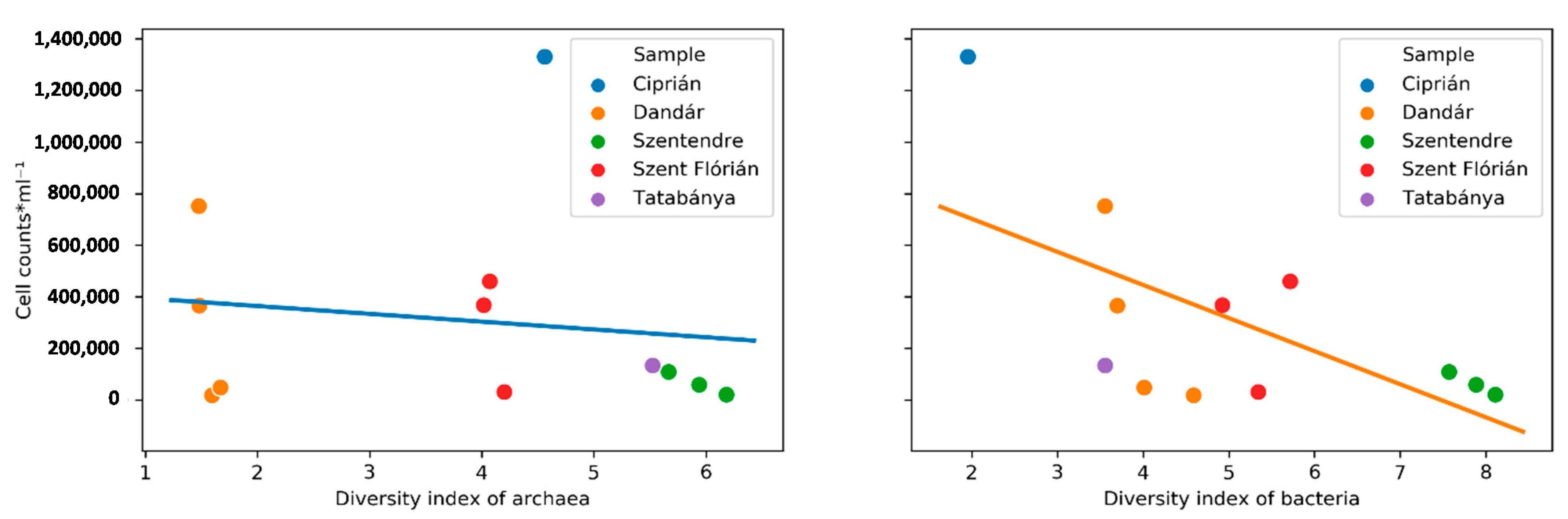
3.2. Microscopic Cell Counts and Diversity Indices of the Samples

The microscopic cell counts of the Ciprián sample were the highest followed by the Dandár and Szent Flórián samples. The latter ones were characterized with high standard deviation values indicating the variability of the cell counts. The cell count values are given in Table 2. Although the Szentendre water sample was characterized by the lowest cell count values based on amplicon sequencing, it had the highest values of diversity indices for both archaea and bacteria. This observation is similar to the Tatabánya sample for the archaea (Table 2). The values of the same samples did not differ significantly in the tested time period. Values of archaeal and bacterial species richness and other diversity indices calculated from 16S rRNA gene amplicon sequencing are given in Supplementary Tables S1 and S2.

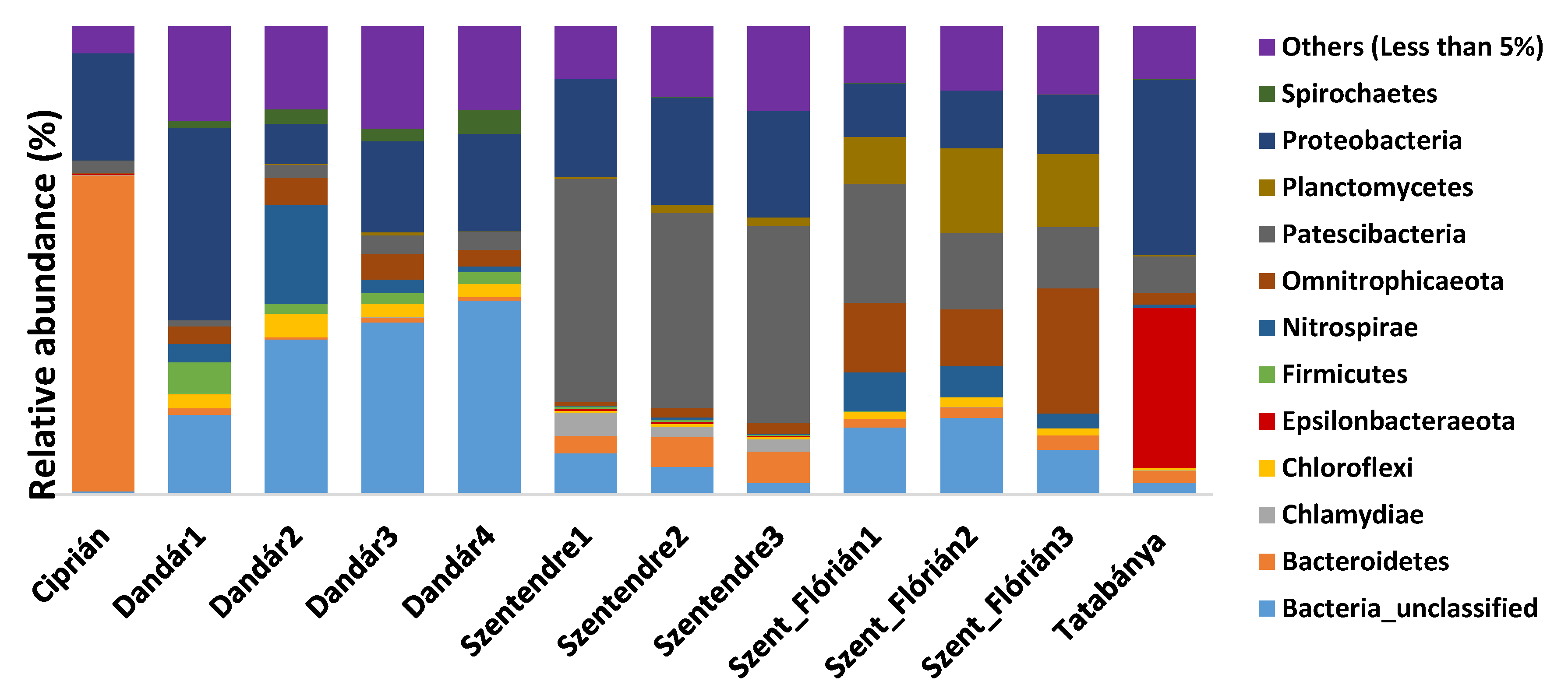
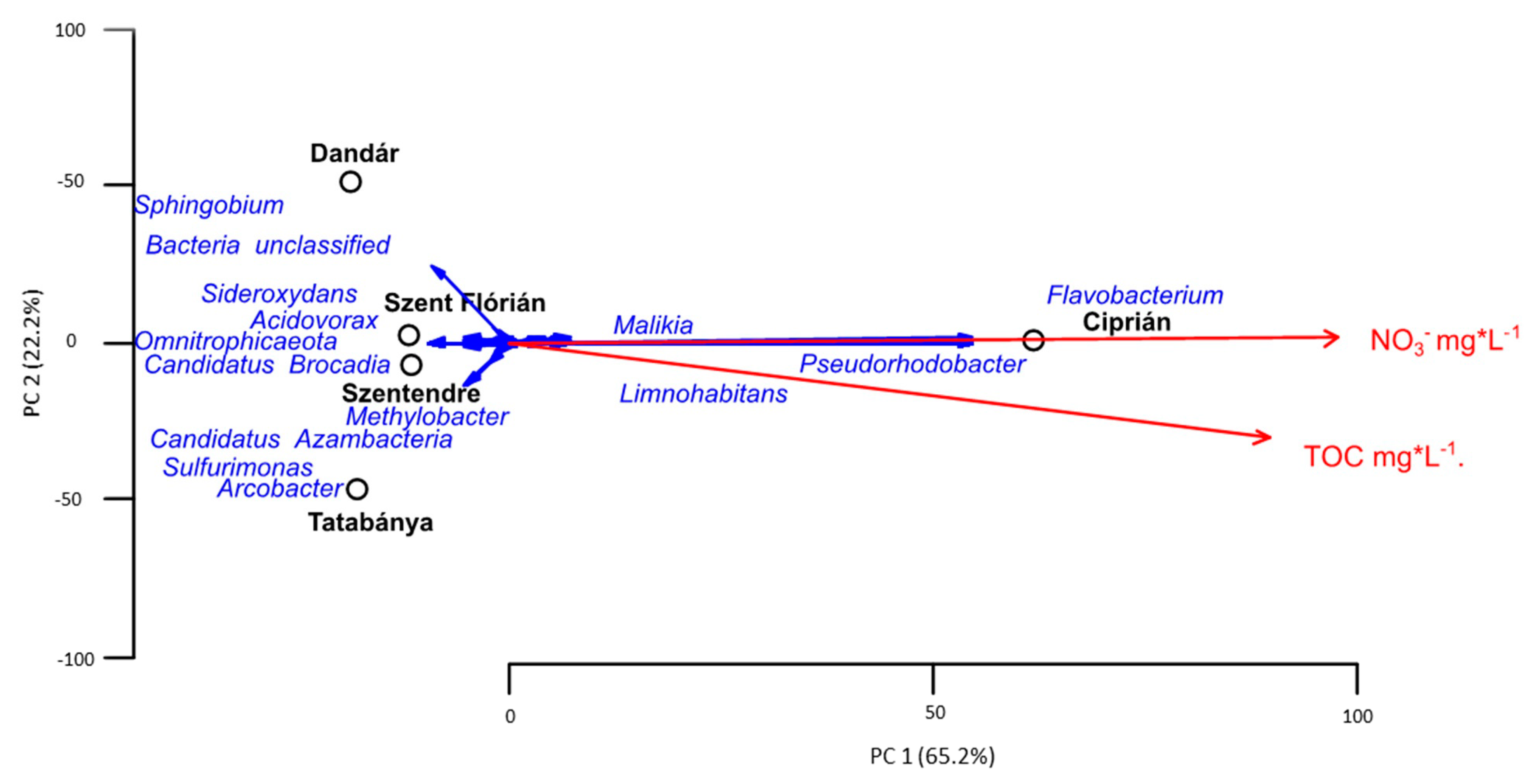
3.3. Bacterial Community Composition of the Different Samples Based on Amplicon Sequencing

Altogether, 55 bacterial phyla were identified by amplicon sequencing. Rarefaction curves of the samples (Supplementary Figure S1) showed that the sequencing depth was sufficient to recover the majority of the bacterial taxa. Patescibacteria and Proteobacteria were present in all the samples. Proteobacteria present in the Ciprián sample showed a high ratio of Malikia (22.29%), Pseudorhodobacter (19.5%), Limnohabitans (12.17%), and unclassified genera of the families Burkholderiaceae (5.13%) and Rhodobacteracea (2.08%). The most abundant sequences in the Dandár water sample belonged to Stenotrophomonas, Pseudomonas, Desulfobacca, Desulfomonile, unclassified Myxococcales and Sphingomonadaceae. In the Szentendre sample within the Proteobacteria phylum sequences of Pseudomonas, Legionella, Aquicella, Cellvibrio, Methylotenera, Rhizobacter, unclassified Burkholderiaceae, Oligoflexus, Haliangium, Bdellovibrio, Peredibacter, Bacteriovorax, Sphingomonas, unclassified Rickettsiales, Amphiplicatus, Brevundimonas and Caulobacter were detected. The Szent Flórián sample was characterized by the presence of unclassified Bacteriovoracaceae, Myxococcales, Sandaracinaceae, Acidiferrobacteraceae, Sulfurifustis and Ferritrophicum. The Tatabánya sample showed a high presence of Desulfocapsa, Desulfurivibrio, Sulfuricella, Sideroxydans, and also the families Gallionellaceae, Hydrogenophilaceae, Methylophilaceae, Rhodocyclaceae, Methylococcaceae and Methylomonaceae. The Ciprián sample was dominated by the phylum Bacteroidetes, being one order of magnitude higher as compared to the Szentendre and Tatabánya samples and approximately 2 orders of magnitude higher as compared to the Szent Flórián and Szentendre samples. Most bacteria belonged to the genus Flavobacterium. In The Dandár sample a high ratio of Chloroflexi was detected, and most of them belonged to Anaerolineae, while also Desantisbacteria, Firestonebacteria, Firmicutes appeared. The most abundant genera within this phylum are Desulfotomaculum and Thermodesulfitimonas, in addition to the presence of Kiritimatiellaeota and Spirochaetes. The Candidate phylum Dependentiae and the phylum Verrucomicrobia characterized the Szentendre sample, and most of the latter belonged to genus Lacunisphaera and the genera of the family Pedosphaeraceae. Rokubacteria and WOR-1 dominated the Szent Flórián samples. Tatabánya had a high number of Epsilonbacteraeota, while this phylum was nearly absent in the other samples; the identified members of this phylum are Arcobacter, unclassified Campylobacterales, Sulfurovum, Sulfuricurvum and Sulfurimonas (Figure 1).

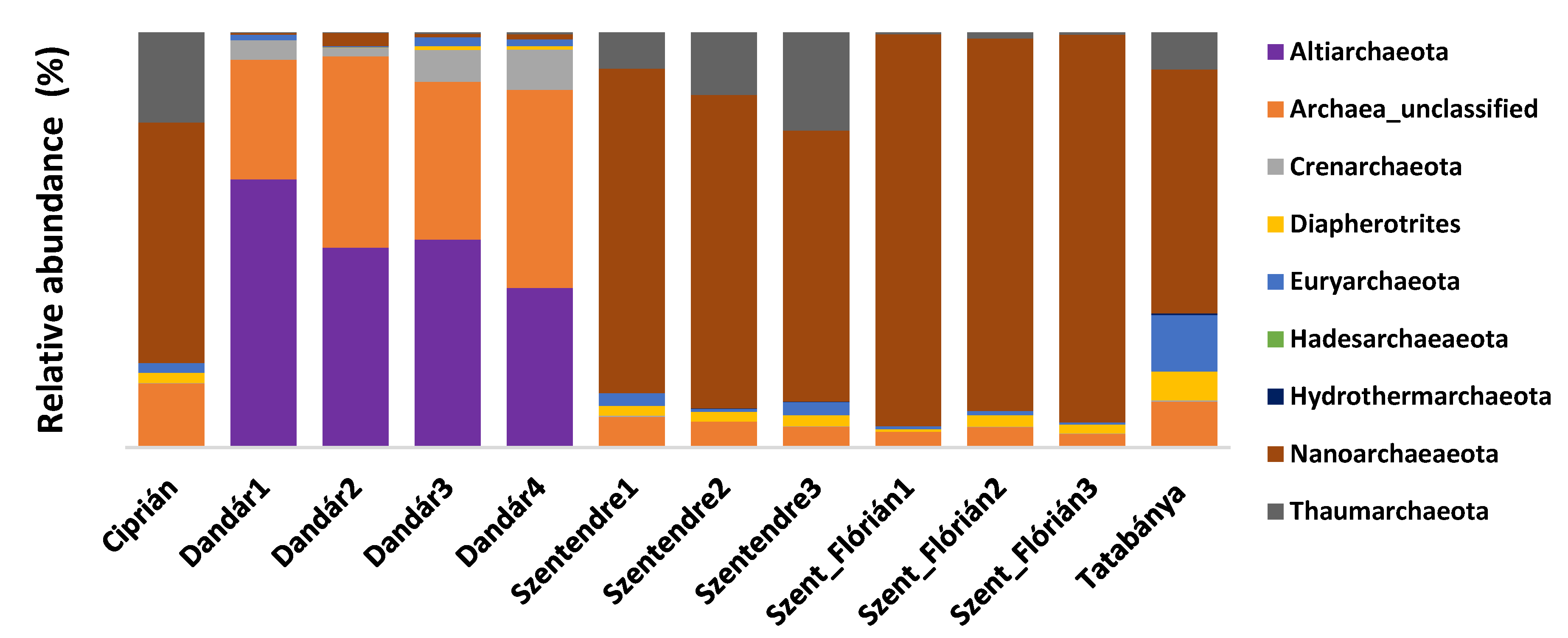
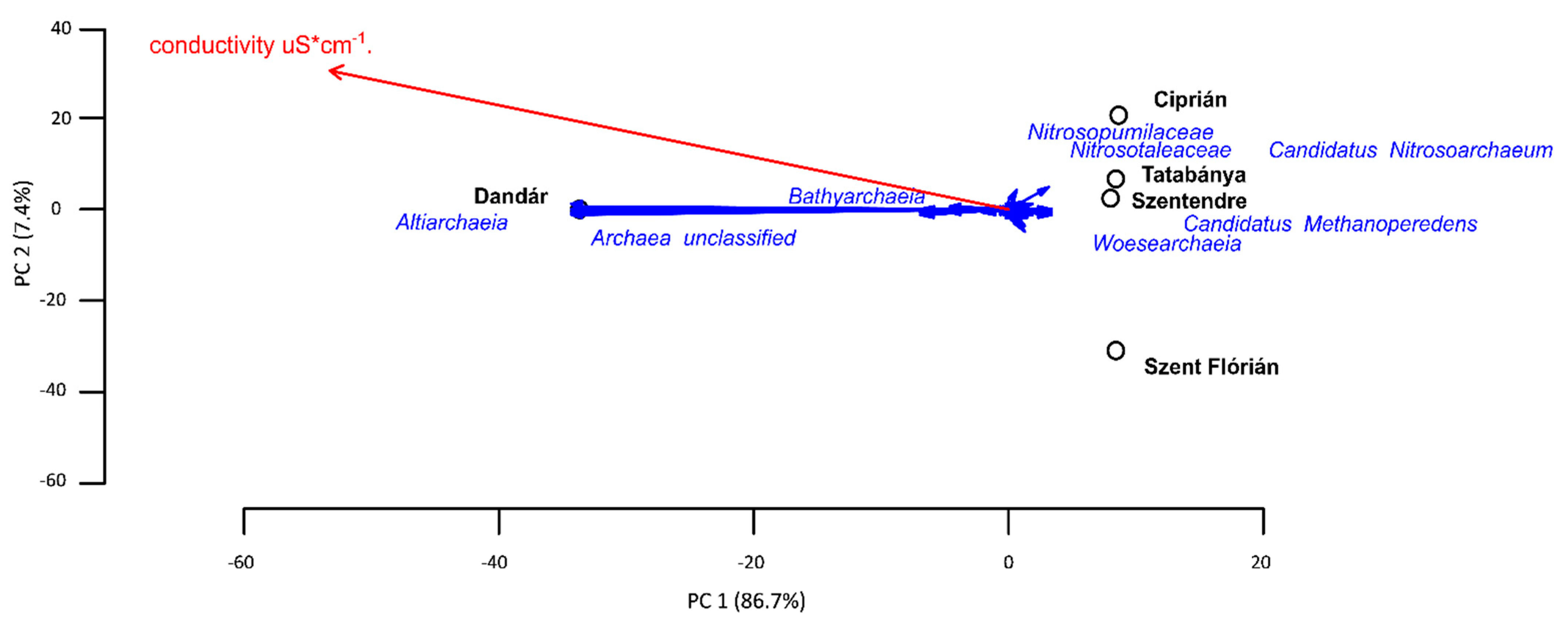
3.4. Archaeal Community Composition of the Different Samples Based on Amplicon Sequencing

Nine archaeal phyla were detected in the water samples. The rarefaction curves of the samples (Supplementary Figure S2) showed that the sequencing depth was sufficient to recover the majority of the archaeal taxa. The samples were characterized by the presence of unclassified archaea and the predominance of Nanoarchaeota, among them Woesearchaeia being the most characteristic. The family Nitrosopumilaceae, (member of Thaumarchaeota) and orders of Nitrosotaleales and Nitrososphaerales appeared in the samples (except Tatabánya); moreover, Marine_Benthic_Group_A was detected in the Szent Flórián and Tatabánya samples. Hydrothermarchaeota phylum was present only in the Szentendre and Tatabánya water samples. Within the Phylum of Euryarchaeota, methanogenic Archaea, e.g., Methanobacterium, Methanoregulaceae were present in both the Ciprián and Tatabánya water samples. Methanospirillaceae and Methanoperedenaceae were present only in the Tatabánya water sample, and Methanomethylophilaceae and Methanosarcinaceae in the Ciprián sample. Thermoplasmata was present in all samples, among them Marine_Benthic_Group_D_and_DHVEG-1 in the case of Tatabánya and Szent Flórián. The order Methanomassiliicoccales was present in the Ciprián, Szent Flórián and Tatabánya samples. Within the Diapherotrites phylum, members of Micrarchaeia were present in all samples together with Iainarchaeia (except in the Dandár water sample). Both Crenarchaeota and Altiarchaeota were characteristic in the Dandár water samples (Figure 2).

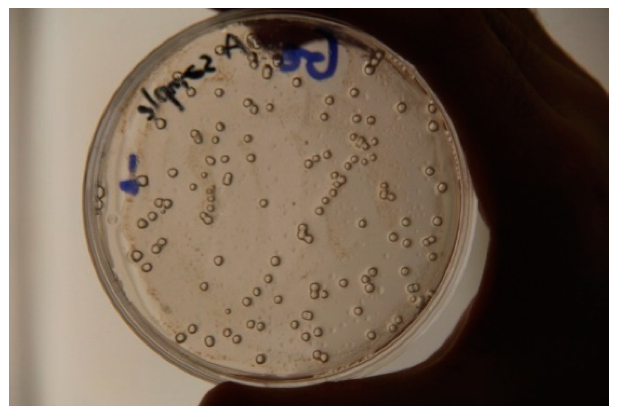
3.5. Results of Cultivation

It is worth mentioning that before isolation, in the media of the Tatabánya water sample, many bubbles were observed (Figure 3), indicating strong gas production of the cultivated bacteria.
In order to reveal the cultivable diversity, 314 bacterial strains were isolated from the samples (their taxonomic position are given in Supplementary Table S3). Based on their 16S rRNA gene sequence similarities most bacterial strains showed between 98 and 100% similarity values to the reference sequences of the type strains of the given species. Nine bacterial strains had lower than 98% similarity to their closest relative, presenting them as novel taxa among the isolated bacteria.
The isolated bacteria were affiliated into four phyla, with most of them being Proteobacteria (59%) followed by Actinobacteria (21%), Firmicutes (17%) and Verrucomicrobia (1%). In the case of amplicon sequencing, the Proteobacteria phylum was dominant, Firmicutes were present in high numbers in the Dandár water sample, and Actinobacteria were represented by less than 5%. The cultivable microbial communities showed similarities between the different samples, e.g., Micrococcus, Pseudomonas, Bacillus and Pseudoxanthomas genera were present in the majority of the samples. The members of the facultative chemolitothrophic genera of Sphingobium, Sphingomonas, Sphingopyxis and the heterotrophic Microbacterium were characteristic only of the Szentendre, Szent Flórián and Tatabánya samples. Many detected taxa known to thrive under nutrient-depleted circumstances, e.g., Acinetobacter, Novosphingobium and Nevskia, were also detected. The distribution of the different cultivated genera is given in Figure 4.

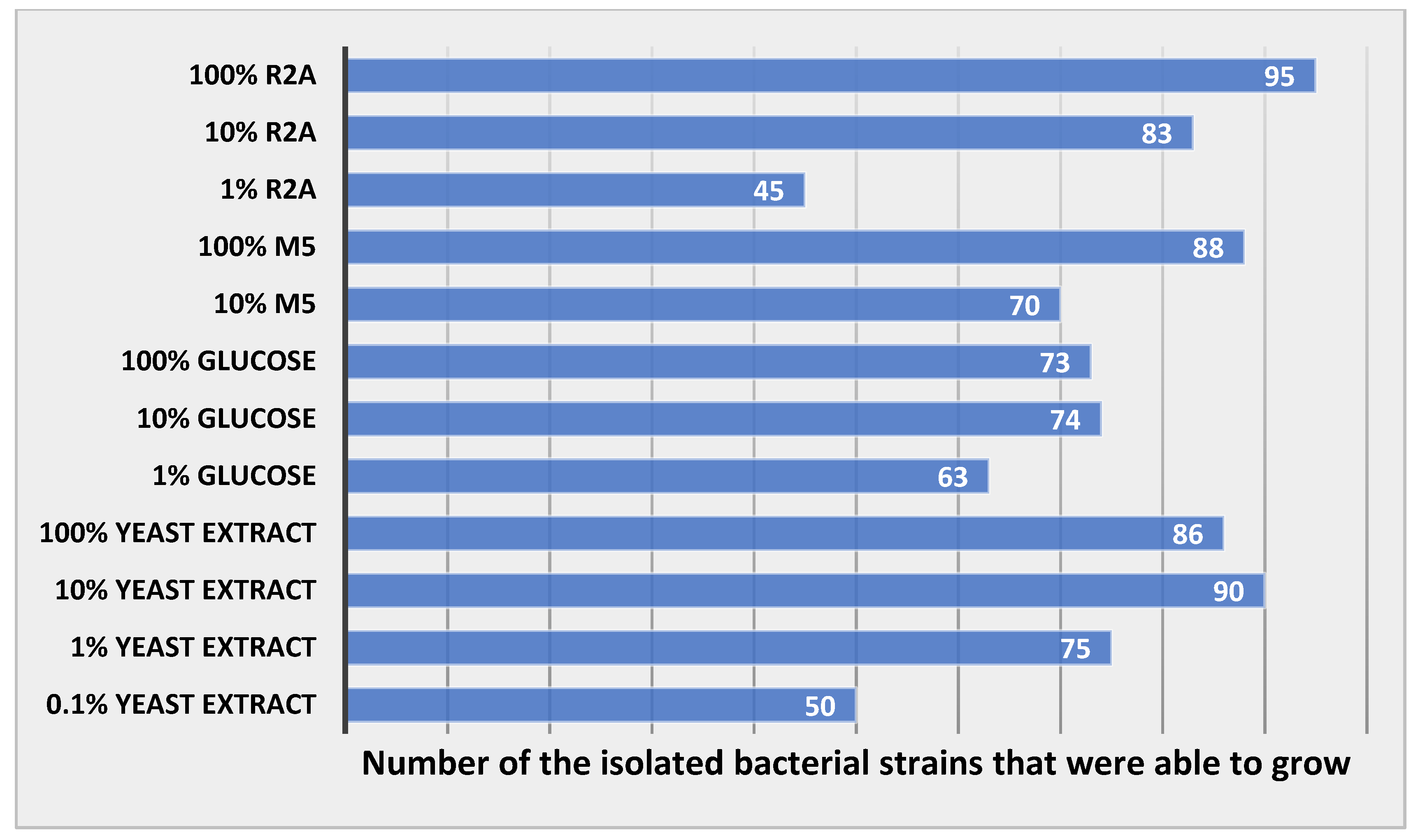
3.6. Results of Bacterial Growth in Different Concentrations of Media

The number of bacterial strains that were able to grow at the different nutrient concentrations was calculated, and results are shown in Figure 5.
24 of the 100 cultivated strains could thrive under all the used media, while the rest of them were absent in one or more media. Only 10 of the tested bacteria were able to grow in one or more oligotrophic media and unable to survive in the 100% yeast extract media (Supplementary Figure S3). The list of these bacteria is given in Table 3, and four of them belong to novel bacterial taxa, as their 16S rRNA gene sequence homology is below 98.8% [36].

4. Discussion

4.1. Influence of the Environmental Factors to the Diversity of Archaeal and Bacterial Communities

The value of the Shannon diversity index of bacteria in the Ciprián water sample was low. However, its cell count value was the highest as compared to all samples. To the contrary, the Szentendre water sample was characterized with the highest diversity index among the samples but its cell count value was low (Figure 6).
The Ciprián sample is characterized by high cell counts, most probably due to the intensive agriculture activity at the top of its catchment area. The nitrate content and TOC value of this sample was also high, indicating the human influence within this region. The negative correlation in cell counts and diversity indices in case of the Szentendre sample can be explained by the location area: In fact, the Stravoda region is located within the Visegrád mountains with many forests; therefore, many soil bacteria with the ability to tolerate low nutrient content could infiltrate into the groundwater and thus increase the diversity within the sample. This assumption is in accordance with the results of Herrmann et al. [37]. The Dandár and Szent Flórián water samples had similar Shanon diversity index values showing low external influence on the waters. These are confined and semi-confined aquifers that protect their groundwaters from external influence. Moreover, previous reports indicated that a high concentration of SO42− in the Dandár water sample significantly influenced the microbial diversity due to the negative interaction of several sulfate-reducing bacteria with other microorganisms [35]. The Tatabánya water sample had a relatively high diversity index in case of archaea, and this can be explained by the high abundance of methanogenic prokaryotes, and they can be connected also to dolomite formation [38] or precipitation in shallow groundwater [39], such as in the case of the Tatabánya well which belongs to a dolomite karst aquifer. To test the effect of environmental factors on the prokaryotic communities, PCA ordination was performed (Figure 6). The calculated PCA components explained 94.1% of the variation in case of archaea and explained 87.4% in case of bacteria. The analysis revealed the impact of chemical characteristics (p < 0.1) on archaea and bacteria OTUs distribution.
It is known that hydrogeological factors influence the microbial processes; at the same time, the metabolism of microorganisms can affect the water quality of hydrogeological systems. In case of the Dandár groundwater, the uptake of ions by the water flow from the host rock results in high conductivity. The PCA ordination of the results show that it moves together with the higher presence of many unclassified archaea and Altiarchaeia (Figure 7). Previous studies did not determine yet the exact electron donor and acceptors for this archaeon; however, it is assumed to be an autotrophic organism [40]. This finding highlights the importance of the ions existing in the water as potential electron donors for the many existing unclassified archaea.
Many OTUs related to the nitrogen cycle characterized the Ciprián water sample and methanogens in the case of the Tatabánya sample, but no significant impact was detected. In case of bacteria, NO3 and the TOC level had an important impact on the bacterial community structure, and these two parameters separated the Ciprián water sample from the others. The most abundant genera in this sample were Flavobacterium and Pseudorhodobacter (Figure 8); among their members some species are able to use nitrate as an electron acceptor (e.g., Pseudomonas denitrificans, Brevundimonas denitrificans).

4.2. Microbial Communities of the Different Samples Based on Amplicon Sequencing

A high percentage of unclassified and uncultured OTUs are commonly seen in nutrient-depleted aquatic environments [41], and this is endorsed by the presence of unclassified Parcubacteria in all samples. The predominance of Patescibacteria is general in groundwaters, often caused by their mobilization from soils and their good survival under oligotrophic conditions [37]. The predominance of Woesearcheota in all the samples can be explained by a syntrophic metabolic model [42], which removes the thermodynamic bottlenecks and enables several metabolic reactions under nutrient-depleted conditions [43]. These results were confirmed by a co-occurrence network analysis [44], and indeed a short distance was shown between many Woesearcheota OTU and both Methanomicrobia and Nitrososphaeria. These results suggest that Woesearchaeota might form a common consortium with methanogens in anaerobic environments. Moreover, Woesearchaeota may have a role in the processes of denitrification, nitrogen fixation, or even dissimilatory nitrite reduction. These findings are in accordance with Liu et al. [45].
The widespread presence of Omnitrophicaeota in the samples is in accordance with previous studies showing their presence in groundwaters and drinking water treatment plants [46].
The dominance of Altiarchaeia within the archaeal community of the Dandár water samples can be explained also by their adaptation to this environment. Based on literature data, Altiarchaeia have evolved specific structural and metabolic features, e.g., developing nanograppling hooks. This anchor allows it to stay stationary on the top of the water despite the water current [47]. Moreover, their presence is common in anaerobic groundwaters; in addition, relatives of Altiarchaeia were found to be widespread in sulfide springs in Europe [48]. Their role is important, often being a carbon dioxide sink [47]. The presence of Sphingomonadaceae and Rhodocyclaceae families in the Dandár water sample can be explained by their capability to degrade many substrates. They can possess a variety of metabolic pathways catalyzing various organic compounds, which is an important feature in oligotrophic environments [49,50]. Unclassified members of Thermodesulfovibrio were isolated earlier from terrestrial hot springs and deep aquifers. They are able to reduce sulfate, thiosulfate or sulfite [51]. The families Pseudomonadaceae, Burkholderiaceae and both the phyla Omnitrophicaeota and Desantisbacteria were described from different aquifers in previous studies [52].
Potential metabolic capabilities of the microbial community of Szentendre and Ciprián water samples:
In the Szentendre and Ciprián samples, an important fraction of ammonia-oxidizing archaea was detected (Nitrosopumilaceae, Nitrosotaleaceae and Nitrososphaeraceae). Compared to ammonia-oxidizing bacteria (AOB), ammonia-oxidizing archaea (AOA) have the ability to inhabit a wide range of extreme environments [53]. This can explain their dominance in nutrient-depleted environments. The sequences assigned to the genus Flavobacterium are shown to be widespread in nature including groundwaters, rivers and oligotrophic lakes [54]. Moreover, many of the Flavobacterium species are able to reduce nitrate to nitrite [55]. This can explain their predominance in the Ciprián water sample, which is characterized by a high NO3 content. Rhodobacteraceae and Rhodocyclaceae can overcome oligotrophic conditions by photoheterotrophic metabolism. Methanoregulaceae, Methanobacteriaceae and Methanosarcinaceae are methanogenic bacteria, and by their metabolism they are able to thrive in nutrient-poor, low ionic-strength environments [56].
Potential metabolic capabilities of the microbial community of Szent Flórián water sample:
The Szent Flórián sample was characterized by distinctive families, the most abundant among them being Candidatus Kaiserbacteria, Candidatus Magasanikbacteria, Candidatus Uhrbacteria, Candidatus Azambacteria and the family Brocadiaceae. Many members of the family Brocardiaceae can be responsible for anaerobic oxidation of ammonium (anammox bacteria). This can suggest that in this environment some ammonia-oxidizing bacteria (AOB) and ammonia-oxidizing archaea (AOA) are present, oxidizing the ammonium to nitrite, while anammox bacteria such as Brocadia would convert what remains from the ammonium. In addition, nitrite reducer bacteria such as Kaiserbacteria [57] would convert what remains from the nitrite to dinitrogen gas [58]. The action of these organisms is very important in these environments where the nitrogen is often in limited concentrations. Members of the taxa Brocadiaceae, Parcubacteria, Peribacteria and Saccharimonadales are characterized by small genomes and a high degree of specialization. These features are often associated with microorganisms performing a limited range of metabolic activities [59,60,61]. The reduced genome size is often related to strong dependent conditions (e.g., interactions between various microbial populations).
Potential metabolic capabilities of the microbial community of Tatabánya water sample:
The Tatabánya sample is characterized by many hydrogenotrophic methanogens, among them Methanobacteriaceae and Methanoperedenaceae. The latter is often found at oxic-anoxic interfaces where they are involved in nitrate-dependent anaerobic oxidation of methane. This reaction links carbon and nitrogen cycles [62]. The presence of Micrarchaeia is reported in several oxygen-poor aquatic environments e.g., shallow groundwater [63], oxygen-minimum zones of Arabian Sea, Bay of Bengal [64], or estuarine water [65]. The group of Marine Benthic Group D and A were found previously in oxygen-depleted water columns [66]. They have the ability to play important roles in the sedimentary carbon cycle [67]. The Deep Sea Euryarchaeotic Group is reported to occur together with anaerobic methanotrophic archaea, and many of them were present in the Tatabánya water sample [68]. Methylorubrum pseudosasae, a methylotrophic bacterium, was even cultivated. The presence of Arcobacteraceae in the Tatabánya sample can be explained by having high survival rate in nutrient-limited groundwater [69]. Some species are capable of autotrophic carbon dioxide fixation via the reverse tricarboxylic acid cycle [70]. In accordance with the archaeal community, the bacterial community is characterized by the presence of members able to metabolize molecular hydrogen as a source of energy (among them Hydrogenophilaceae [71,72] and Sulfurimonas (belonging to Thiovulaceae family [73,74]). Therefore, the experienced gas bubbles are most probably the result of microbial actions, by H2 production of bacteria, or by methane production of archaea. To prove this, further studies would be essential. To reveal precisely the source of gas production at the sampling site, further analysis would also be needed.

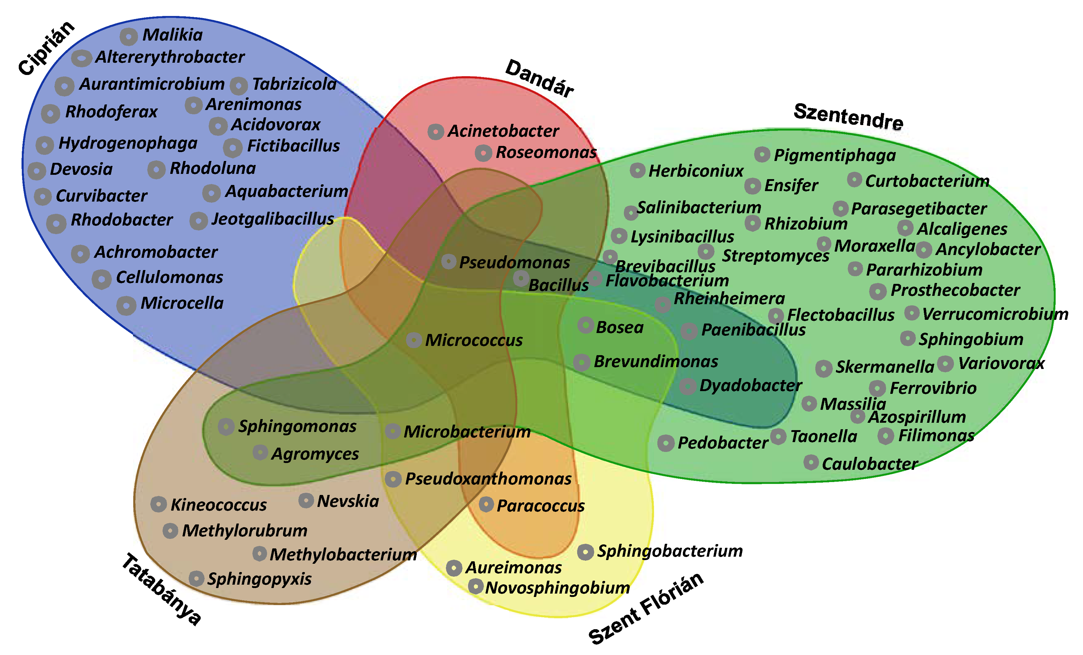
4.3. Cultivable Bacterial Communities of the Samples

Genera of Micrococcus, Pseudomonas, Bacillus and Pseudoxanthomas are widespread in different aquatic environments and they are shown to survive also in nutrient-depleted conditions using different strategies; e.g., most of these bacteria have the ability to form biofilms [75]. Previous literature data showed that many of them have been isolated from an oligotrophic aquifer in West Bengal, and they were characterized by high metabolic flexibility, such as the ability to utilize multiple hydrocarbons and using different electron acceptors [76]. Though Bacillus species are widespread in nature, they are able to produce endospores but often can show extremely slow growth as alternative strategy to survive starvation [77]. Sphingomonas and Brevundimonas species also have the ability to survive in low concentrations of nutrients, as well as to metabolize a wide variety of carbon sources [78,79]. Microbacterium species demonstrated the ability to convert ammonium to nitrogen under aerobic conditions [80]. From the Szentendre water sample, potential nitrogen-fixing bacteria could be isolated, e.g., Herbiconiux [81], Rhizobium [82], Ensifer [83] and also ammonia-oxidizing bacteria, e.g., Prosthecobacter [84]. In oligotrophic environments, nitrogen fixation can be an important feature due to limited nitrogen sources. In addition, some archaeal OTUs were found, which can be responsible for ammonia oxidization (e.g., Nitrosopumilaceae, Nitrosotaleaceae and Nitrososphaeraceae). Rhodobacter azotoformans in the Ciprián water sample is a denitrifying phototrophic bacterium [85], and Fictibacillus is able to perform ammonification and also iron reduction [86]. The Dandár water sample contained members of Acinetobacter. Many species of this genus are able to mobilize inorganic phosphate, and so have a key function for nutrient acquisition in these starved ecosystems [79]. Many of the cultivated species from the Szent Flórián and Tatabánya water samples were isolated previously from groundwaters, suggesting their adaptation to nutrient-depleted environments. The genus Sphingopyxis was represented by different species (S. fribergensis, S. chilensis and S. solisilvae), and it is commonly isolated also from freshwater and marine habitats—many of them are facultatively chemolithotrophs, often producing H2 during their metabolic processes. These bacteria could also be responsible for the observed gas production.

4.4. Growth of the Bacterial Strains in Different Concentrations of Media

It is known that bacteria from low nutrient content environments often lose their ability to grow in rich nutrient content circumstances [87]. In our case, 10 species were not able to grow in the presence of higher nutrient content (Table 3). Flardh et al. [88] suggested that this is the result of the development of high substrate affinities during nutrient limitations. Based on this, our findings contradict the assumption stating that the limiting factor in the bacterial growth is always the nutrient availability. In fact, the limiting factor is the ability of the cell itself to grow. Previous literature’s data show that a sudden addition of high quantities of nutrient to an organism can lead to rapid death via osmotic swelling [89].
Hodgson [90] described Streptomyces as a facultative oligotrophic microorganism, and some species could grow under oligocarbophile conditions. Semenov [91] discovered that members of Prosthecobacteria have an extremely high affinity for different substrates. Curvibacter delicatus, which was characteristic in the Ciprián water sample, was among the bacterial communities that have fouled polyvinylidene fluoride microfiltration membranes, which are used for drinking water treatment [92]. Rheinheimera aquatica was isolated from hot springs from the Jazan region in Saudi Arabia, which is considered as an oligotrophic environment [93]. Both Aquabacterium citratiphilum and Aquabacterium commune were isolated from biofilms of the Berlin drinking water system, where they could resist a severe limitation of low nutrient contents [94]. Different species belonging to the genus Ferrovibrio were isolated from a thermal bath in Budapest, where the water contains only limited organic carbon source [24]. Ferrovibrio species are often related to corrosion in different pipelines, while F. denitrificans can be responsible also for nitrate reduction [95]. No previous studies reported the existence of Malikia spinosa and Salinibacterium hongtaonis in similar environments. All these findings show that some of the cultivated taxa are true oligocarbophile microorganisms, and most probably they all contribute to the survival of the community in the tested aquatic habitats.

5. Conclusions

In conclusion, we can state that our findings confirm the fact that nutrient-depleted aquatic environments are highly colonized by microorganisms which are able to participate actively in different biogeochemical cycles.
This study could reveal the existence of bacteria and archaea involved in the nitrogen cycle, e.g., ammonia-oxidizing bacteria (AOB), e.g., Prosthecobacter—and ammonia-oxidizing archaea (AOA), e.g., Nitrosopumilaceae, nitrate-respiring organisms as well as nitrogen-fixing prokaryotes, e.g., Herbiconiux. Others are involved in the biogeochemical cycle of sulfur such as sulfide/sulfur oxidizers, e.g., Sulfurimonas—which is able to reduce nitrate, and oxidize both sulfur and hydrogen. The members of the family Thiovulaceae and Acidithiobacillus sp. can be involved in sulfur as well as iron cycles, and the detected sulfate-reducing bacteria, e.g., Thermodesulfovibrio, are also key organisms of the sulfur cycle. These bacteria were detected with one or more of the applied methods.
In the tested nutrient-depleted aquatic environments, the existing microorganisms were characterized with different metabolic types: a wide range of chemotrophic (even facultative chemolithotrophic) and facultative phototrophic organisms were found; methanogenic, e.g., Methanobacteriaceae—as well as methanotrophic bacteria, e.g., Methylorubrum—were observed, and also phosphate mobilizer microbes e.g., Bacillus circulans.
The majority of the cultivated bacteria were able to thrive in nutrient-rich conditions; however, an important fraction was unable to survive. This indicates that nutrient availability is not the only factor which influences microbial growth, and that the hydrogeological processes and physical conditions (e.g., temperature) also have a high impact. On the other hand, the amount of substrates (TOC) and their availability are also a crucial point for microbial growth in these nutrient-depleted environments.

Supplementary Materials

The followings are available online at https://www.mdpi.com/article/10.3390/w13111533/s1, Figure S1: Rarefaction curves of bacterial OTUs of the samples based on 16S rRNA gene amplicon sequencing, Figure S2: Rarefaction curves of archaeal OTUs of the samples based on 16S rRNA gene amplicon sequencing, Figure S3: PCA ordination of the bacterial strains growing in 100% yeast extract medium, Table S1: Archaeal species richness and diversity indices calculated from 16S rRNA gene amplicon sequencing data, Table S2: Bacterial species richness and diversity indices calculated from 16S rRNA gene amplicon sequencing, Table S3: Results of taxonomic identification of group representative bacterial strains using 16S rRNA gene sequencing.

Author Contributions

Idea and organization of samplings and all work: E.T. and M.T.; Molecular work and bioinformatical data analysis: M.T., R.F. and G.A.; Cultivation: M.T. and Y.S.; Chemical investigations: L.J.; Project administration: É.Á.; Ecological analysis: E.T. and É.Á. All authors have read and agreed to the published version of the manuscript.

Funding

This research was funded by ELTE Thematic Excellence Program 2020 Supported by the National Research—Development and Innovation Office—TKP2020-IKA-05.

Institutional Review Board Statement

Not applicable.

Informed Consent Statement

Not applicable.

Data Availability Statement

The data used for this study is mentionned in sections: DNA Isolation and Identification of the Isolated Bacterial Strains and DNA Extraction from the Water Samples and Amplicon Sequencing.

Acknowledgments

The authors are thankful to Sára Szuróczki, Tamás Felföldi, Balázs Vajna, Ádám Tóth and Attila Szabó for their technical advice.

Conflicts of Interest

The authors declare no conflict of interest.

References

  1. Cavicchioli, R.; Ostrowski, M.; Fegatella, F.; Goodchild, A.; Guixa-Boixereu, N. Life under Nutrient Limitation in Oligotrophic Marine Environments: An Eco/Physiological Perspective of Sphingopyxis alaskensis (formerly Sphingomonas alaskensis). Microb. Ecol. 2003, 46, 249–256. [Google Scholar] [CrossRef]
  2. Kuznetsov, S.I.; Dubinina, G.A.; Lapteva, N.A. Biology of Oligotrophic Bacteria. Annu. Rev. Microbiol. 1979, 33, 377–387. [Google Scholar] [CrossRef]
  3. Barer, M.; Harwood, C.R. Bacterial Viability and Culturability. Adv. Microb. Physiol. 1999, 41, 93–137. [Google Scholar] [CrossRef] [PubMed]
  4. Lengeler, J.W.; Drews, G.; Schlegel, H.G. Biology of the Prokaryotes; Thieme: New York, NY, USA, 1999. [Google Scholar]
  5. Flynn, T.M.; Sanford, R.A.; Ryu, H.; Bethke, C.M.; Levine, A.D.; Ashbolt, N.J.; Domingo, J.W.S. Functional Microbial Diversity Explains Groundwater Chemistry in a Pristine Aquifer. BMC Microbiol. 2013, 13, 146. [Google Scholar] [CrossRef] [Green Version]
  6. Basler, B.L.; Losick, R. Bacterially speaking. Cell 2006, 125, 237–246. [Google Scholar] [CrossRef] [Green Version]
  7. Legan, J.D.; Owens, J.D. Bacterial competition for methylamine: Computer simulation of a three-strain continuous culture supplied continuously or alternatively with two nutrients. FEMS Microbiol. Ecol. 1988, 53, 307–314. [Google Scholar] [CrossRef]
  8. Ho, A.; Di Lonardo, P.; Bodelier, P.L.E. Revisiting life strategy concepts in environmental microbial ecology. FEMS Microbiol. Ecol. 2017, 93, 1–14. [Google Scholar] [CrossRef] [Green Version]
  9. Andrés, M.; Gil, R.; Latorre, A.; Peretó, J.; Garcillán-Barcia, M.P.; De La Cruz, F. Toward Minimal Bacterial Cells: Evolution vs. Design. FEMS Microbiol. Rev. 2009, 33, 225–235. [Google Scholar]
  10. Dombrowski, N.; Lee, J.-H.; Williams, T.; Offre, P.; Spang, A. Genomic diversity, lifestyles and evolutionary origins of DPANN archaea. FEMS Microbiol. Lett. 2019, 366, 1–12. [Google Scholar] [CrossRef] [PubMed] [Green Version]
  11. Phung, N.T.; Jiyoung, L.; Kui, H.K.; In Seop, C.; Geoffrey, M.G.; Byung, H.K. Analysis of Microbial Diversity in Oligotrophic Microbial Fuel Cells Using 16S RDNA Sequences. FEMS Microbiol. Lett. 2004, 233, 77–82. [Google Scholar] [CrossRef] [PubMed] [Green Version]
  12. Hayakawa, D.H.; Huggett, M.J.; Rappé, M.S. Ecology and Cultivation of Marine Oligotrophic Bacteria. In Extremophiles Handbook; Springer Science and Business Media LLC: Berlin/Heidelberg, Germany, 2011; pp. 1161–1178. [Google Scholar]
  13. Kaeberlein, T.; Lewis, K.; Epstein, S.S. Isolating “uncultivable” microorganisms in pure culture in a simulated natural envi-ronment. Science 2002, 296, 1127–1129. [Google Scholar] [CrossRef] [PubMed] [Green Version]
  14. Janssen, P.H.; Yates, P.S.; Grinton, B.E.; Taylor, P.M.; Sait, M. Improved Culturability of Soil Bacteria and Isolation in Pure Culture of Novel Members of the Divisions Acidobacteria, Actinobacteria, Proteobacteria, and Verrucomicrobia. Appl. Environ. Microbiol. 2002, 68, 2391–2396. [Google Scholar] [CrossRef] [PubMed] [Green Version]
  15. Rappé, M.S.; Connon, S.A.; Vergin, K.L.; Giovannoni, S.J. Cultivation of the ubiquitous SAR11 marine bacterioplankton clade. Nat. Cell Biol. 2002, 418, 630–633. [Google Scholar] [CrossRef]
  16. Braun, B.; Schröder, J.; Knecht, H.; Szewzyk, U. Unraveling the Microbial Community of a Cold Groundwater Catchment System. Water Res. 2016, 07, 113–126. [Google Scholar] [CrossRef] [PubMed]
  17. Erőss, A.; Mádl-Szőnyi, J.; Csoma, A.É. Characteristics of discharge at Rose and Gellért Hills, Budapest, Hungary. Cent. Eur. Geol. 2008, 51, 267–281. [Google Scholar] [CrossRef] [Green Version]
  18. Jobbágy, V.; Kávási, N.; Somlai, J.; Dombovári, P.; Gyöngyösi, C.; Kovács, T. Gross Alpha and Beta Activity Concentrations in Spring Waters in Balaton Upland, Hungary. Radiat. Measur. 2011, 46, 159–163. [Google Scholar] [CrossRef]
  19. Mádl-Szőnyi, J.; Tóth, Á. Basin-scale conceptual groundwater flow model for an unconfined and confined thick carbonate region. Hydrogeol. J. 2015, 23, 1359–1380. [Google Scholar] [CrossRef]
  20. Izápy, G. Magyarország forrásainak katasztere. In Budapest, VITUKI Hydrological Institute; Natural Spring Cadastre of Hungary: Berlin/Heidelberg, Germany, 2002. (In Hungarian) [Google Scholar]
  21. Emese, G.; Tóth, G. In cooperation with SGUDS, Transboundary Geothermal, and Energy Resources. In Report on Komárom—Štúrovo Pilot Area Scenario Modeling; Slovakia, 2013; Available online: http://transenergy-eu.geologie.ac.at/Downloads/outputs/Scenario%20model%20report/WP5_5.3_Komarno-Sturovo%20pilot%20area%20scenario%20modeling.pdf (accessed on 7 November 2004).
  22. Rice, E.W.; Baird, R.B.; Eaton, A.D. Standard Methods for the Examination of Water and Wastewater, 23rd ed.; American Public Health Association, American Water Works Association, Water Environment Federation: Washington, DC, USA, 2017; ISBN 9780875532875. [Google Scholar]
  23. Kéki, Z.; Makk, J.; Barkács, K.; Vajna, B.; Palatinszky, M.; Márialigeti, K.; Tóth, E. Critical point analysis and biocide treatment in a microbiologically contaminated water purification system of a power plant. SN Appl. Sci. 2019, 1, 820. [Google Scholar] [CrossRef] [Green Version]
  24. Szuróczki, S.; Kéki, Z.; Káli, S.; Lippai, A.; Márialigeti, K.; Tóth, E. Microbiological investigations on the water of a thermal bath at Budapest. Acta Microbiol. Immunol. Hung. 2016, 63, 229–241. [Google Scholar] [CrossRef] [PubMed] [Green Version]
  25. Carbonnelle, E.; Mesquita, C.; Bille, E.; Day, N.; Dauphin, B.; Beretti, J.-L.; Ferroni, A.; Gutmann, L.; Nassif, X. MALDI-TOF mass spectrometry tools for bacterial identification in clinical microbiology laboratory. Clin. Biochem. 2011, 44, 104–109. [Google Scholar] [CrossRef]
  26. Kalwasińska, A.; Felföldi, T.; Walczak, M.; Kosobucki, P. Physiology and Molecular Phylogeny of Bacteria Isolated from Alka-line Distillery Lime. Pol. J. Microbiol. 2015, 64, 369–377. [Google Scholar] [CrossRef]
  27. Yoon, S.H.; Sung, M.H.; Soonjae, K.; Jeongmin, L.; Yeseul, K.; Hyungseok, S.; Jongsik, C. Introducing EzBioCloud: A Taxo-nomically United Database of 16S RRNA Gene Sequences and Whole-Genome Assemblies. Int. J. Sys. Evol. Microbiol. 2017, 67, 1613–1617. [Google Scholar] [CrossRef] [PubMed]
  28. Herlemann, D.P.R.; Labrenz, M.; Jürgens, K.; Bertilsson, S.; Waniek, J.J.; Andersson, A.F. Transitions in bacterial communities along the 2000 km salinity gradient of the Baltic Sea. ISME J. 2011, 5, 1571–1579. [Google Scholar] [CrossRef] [PubMed] [Green Version]
  29. Klindworth, A.; Pruesse, E.; Schweer, T.; Peplies, J.; Quast, C.; Horn, M.; Glöckner, F.O. Evaluation of general 16S ribosomal RNA gene PCR primers for classical and next-generation sequencing-based diversity studies. Nucleic Acids Res. 2012, 41, e1. [Google Scholar] [CrossRef] [PubMed]
  30. Schloss, P.D.; Westcott, S.L.; Ryabin, T.; Hall, J.R.; Hartmann, M.; Hollister, E.B.; Lesniewski, R.A.; Oakley, B.B.; Parks, D.H.; Robinson, C.J.; et al. Introducing mothur: Open-Source, Platform-Independent, Community-Supported Software for Describing and Comparing Microbial Communities. Appl. Environ. Microbiol. 2009, 75, 7537–7541. [Google Scholar] [CrossRef] [PubMed] [Green Version]
  31. Quast, C.; Pruesse, E.; Yilmaz, P.; Gerken, J.; Schweer, T.; Yarza, P.; Peplies, J.; Glöckner, F.O. The SILVA Ribosomal RNA Gene Database Project: Improved Data Processing and Web-Based Tools. Nucleic Acids Res. 2013, 41, 590–596. [Google Scholar] [CrossRef]
  32. Edgar, R.C.; Haas, B.J.; Clemente, J.C.; Quince, C.; Knight, R. UCHIME Improves Sensitivity and Speed of Chimera Detection. Bioinformatics 2011, 27, 2194–2200. [Google Scholar] [CrossRef] [PubMed] [Green Version]
  33. Reasoner, D.J.; Geldreich, E.E. A new medium for the enumeration and subculture of bacteria from potable water. Appl. Environ. Microbiol. 1985, 49, 1–7. [Google Scholar] [CrossRef] [PubMed] [Green Version]
  34. Oksanen, J.; Blanchet, F.G.; Kindt, R.; Legendre, P.; Minchin, P.R.; O’Hara, R.; Simpson, G.L.; Solymos, P.; Stevens, M.H.H.; Wagner, H. Ordination Methods, Diversity Analysis and Other Functions for Community and Vegetation Ecologists, 2020, Vegan: Community Ecology Package. Available online: https://cran.r-project.org/package=vegan (accessed on 29 May 2021).
  35. Lemos, L.N.; Fulthorpe, R.R.; Triplett, E.W.; Roesch, L.F. Rethinking microbial diversity analysis in the high throughput sequencing era. J. Microbiol. Methods 2011, 86, 42–51. [Google Scholar] [CrossRef] [Green Version]
  36. Giovannoni, S.J.; Britschgi, T.B.; Moyer, C.L.; Field, K.G. Genetic diversity in Sargasso Sea bacterioplankton. Nat. Cell Biol. 1990, 345, 60–63. [Google Scholar] [CrossRef]
  37. Herrmann, M.; Wegner, C.-E.; Taubert, M.; Geesink, P.; Lehmann, K.; Yan, L.; Lehmann, R.; Totsche, K.U.; Küsel, K. Predominance of Cand. Patescibacteria in Groundwater Is Caused by Their Preferential Mobilization from Soils and Flourishing Under Oligotrophic Conditions. Front. Microbiol. 2019, 10, 1407. [Google Scholar] [CrossRef] [Green Version]
  38. Mazzullo, S. Organogenic Dolomitization in Peritidal to Deep-Sea Sediments: PERSPECTIVES. J. Sediment. Res. 2000, 70, 10–23. [Google Scholar] [CrossRef]
  39. Roberts, J.A.; Kenward, P.A.; Fowle, D.; Goldstein, R.H.; González, L.A.; Moore, D.S. Surface chemistry allows for abiotic precipitation of dolomite at low temperature. Proc. Natl. Acad. Sci. USA 2013, 110, 14540–14545. [Google Scholar] [CrossRef] [Green Version]
  40. Perras, A.K.; Daum, B.; Ziegler, C.; Takahashi, L.K.; Musahid, A.; Wanner, G.; Klingl, A.; Leitinger, G.; Kolb-Lenz, D.; Gribaldo, S.; et al. S-Layers at Second Glance? Altiar-chaeal Grappling Hooks (Hami) Resemble Archaeal S-Layer Proteins in Structure and Sequence. Front. Microbiol. 2015, 6, 543. [Google Scholar] [CrossRef] [PubMed]
  41. Lopez-Fernandez, M.; Åström, M.; Bertilsson, S.; Dopson, M. Depth and Dissolved Organic Carbon Shape Microbial Com-munities in Surface Influenced but Not Ancient Saline Terrestrial Aquifers. Front. Microbiol. 2018, 9, 1–16. [Google Scholar] [CrossRef]
  42. Liu, X.; Li, M.; Castelle, C.J.; Probst, A.J.; Zhou, Z.; Pan, J.; Liu, Y.; Banfield, J.F.; Gu, J.-D. Insights into the ecology, evolution, and metabolism of the widespread Woesearchaeotal lineages. Microbiome 2018, 6, 1–16. [Google Scholar] [CrossRef] [PubMed]
  43. Lau, M.C.Y.; Kieft, T.L.; Kuloyo, O.; Linage-Alvarez, B.; van Heerden, E.; Lindsay, M.R.; Magnabosco, C.; Wang, W.; Wiggins, J.B.; Guo, L.; et al. An oligotrophic deep-subsurface community dependent on syntrophy is dominated by sulfur-driven autotrophic denitrifiers. Proc. Natl. Acad. Sci. USA 2016, 113, E7927–E7936. [Google Scholar] [CrossRef] [Green Version]
  44. Kasper, S.A.; Kirkegaard, R.H.; Karst, S.M.; Albertsen, M. ampvis2: An R package to analyse and visualise 16S rRNA amplicon data. bioRxiv 2018, 299537. [Google Scholar] [CrossRef] [Green Version]
  45. Liu, X.; Wang, Y.; Gu, J.-D. Ecological distribution and potential roles of Woesearchaeota in anaerobic biogeochemical cycling unveiled by genomic analysis. Comput. Struct. Biotechnol. J. 2021, 19, 794–800. [Google Scholar] [CrossRef]
  46. Bruno, A.; Sandionigi, A.; Bernasconi, M.; Panio, A.; Labra, M.; Casiraghi, M. Changes in the Drinking Water Microbiome: Effects of Water Treatments along the Flow of Two Drinking Water Treatment Plants in a Urbanized Area, Milan (Italy). Front. Microbiol. 2018, 9, 2557. [Google Scholar] [CrossRef] [PubMed]
  47. Probst, A.J.; Weinmaier, T.; Raymann, K.; Perras, A.K.; Emerson, J.B.; Rattei, T.; Wanner, G.; Klingl, A.; Berg, I.A.; Yoshinaga, M.Y.; et al. Biology of a widespread uncultivated archaeon that contributes to carbon fixation in the subsurface. Nat. Commun. 2014, 5, 5497. [Google Scholar] [CrossRef] [Green Version]
  48. Rudolph, C.; Moissl, C.; Henneberger, R.; Huber, R. Ecology and microbial structures of archaeal/bacterial strings-of-pearls communities and archaeal relatives thriving in cold sulfidic springs. FEMS Microbiol. Ecol. 2004, 50, 1–11. [Google Scholar] [CrossRef] [PubMed]
  49. Oh, S.; Donggeon, C. Microbial Community Enhances Biodegradation of Bisphenol A through Selection of Sphingomona-daceae. Microb. Ecol. 2019, 77, 631–639. [Google Scholar] [CrossRef] [PubMed]
  50. Táncsics, A.; Szalay, A.R.; Farkas, M.; Benedek, T.; Szoboszlay, S.; Szabó, I.; Lueders, T. Stable isotope probing of hypoxic toluene degradation at the Siklós aquifer reveals prominent role of Rhodocyclaceae. FEMS Microbiol. Ecol. 2018, 94, 1–11. [Google Scholar] [CrossRef] [PubMed]
  51. Frank, Y.A.; Kadnikov, V.V.; Lukina, A.P.; Banks, D.; Beletsky, A.V.; Mardanov, A.V.; Sen’kina, E.I.; Avakyan, M.R.; Karnachuk, O.V.; Ravin, N.V. Characterization and Genome Analysis of the First Facultatively Alkaliphilic Thermodesul-fovibrio Isolated from the Deep Terrestrial Subsurface. Front. Microbiol. 2016, 7, 1–11. [Google Scholar] [CrossRef] [PubMed]
  52. Probst, A.J.; Castelle, C.J.; Singh, A.; Brown, C.T.; Anantharaman, K.; Sharon, I.; Hug, L.A.; Burstein, D.; Emerson, J.B.; Thomas, B.C.; et al. Genomic resolution of a cold subsurface aquifer community provides metabolic insights for novel microbes adapted to high CO2concentrations. Environ. Microbiol. 2017, 19, 459–474. [Google Scholar] [CrossRef] [Green Version]
  53. Zhou, L.; Wang, S.; Zou, Y.; Xia, C.; Zhu, G. Species, Abundance and Function of Ammonia-oxidizing Archaea in Inland Waters across China. Sci. Rep. 2015, 5, 15969. [Google Scholar] [CrossRef] [PubMed]
  54. Yi, H.; Chun, J. Flavobacterium weaverense sp. nov. and Flavobacterium segetis sp. nov., novel psychrophiles isolated from the Antarctic. Int. J. Syst. Evol. Microbiol. 2006, 56, 1239–1244. [Google Scholar] [CrossRef]
  55. Jean-François, B.; Bowman, J.P. Flavobacterium; Bergey’s Manual of Systematics of Archaea and Bacteria: New Jersey, NJ, USA, 2015; pp. 1–75. [Google Scholar]
  56. Bräuer, S.; Cadillo-Quiroz, H.; Kyrpides, N.; Woyke, T.; Goodwin, L.; Detter, C.; Podell, S.; Yavitt, J.B.; Zinder, S.H. Genome of Methanoregula boonei 6A8 reveals adaptations to oligotrophic peatland environments. Microbiology 2015, 161, 1572–1581. [Google Scholar] [CrossRef]
  57. Danczak, R.E.; Johnston, M.D.; Kenah, C.; Slattery, M.; Wrighton, K.C.; Wilkins, M.J. Members of the Candidate Phyla Radiation are functionally differentiated by carbon- and nitrogen-cycling capabilities. Microbiome 2017, 5, 112. [Google Scholar] [CrossRef]
  58. Strous, M.; Van Gerven, E.; Gijs, J.K.; Jetten, M. Effects of Aerobic and Microaerobic Conditions on Anaerobic Ammonium- Oxidizing (Anammox) Sludge. Appl. Environ. Microbiol. 1997, 63, 2446–2448. [Google Scholar] [CrossRef] [PubMed] [Green Version]
  59. Nelson, W.C.; Stegen, J.C. The reduced genomes of Parcubacteria (OD1) contain signatures of a symbiotic lifestyle. Front. Microbiol. 2015, 6, 713. [Google Scholar] [CrossRef] [PubMed] [Green Version]
  60. Anantharaman, K.; Brown, C.T.; Burstein, D.; Castelle, C.J.; Probst, A.J.; Thomas, B.C.; Williams, K.H.; Banfield, J.F. Analysis of Five Complete Genome Sequences for Members of the Class Peribacteria in the Recently Recognized Peregrinibacteria Bac-terial Phylum. Peer J. 2016, 1, 1–14. [Google Scholar]
  61. Lemos, L.N.; Medeiros, J.D.; Dini-Andreote, F.; Fernandes, G.R.; Varani, A.D.M.; Oliveira, G.; Pylro, V.S. Genomic signatures and co-occurrence patterns of the ultra-small Saccharimonadia (phylum CPR/Patescibacteria) suggest a symbiotic lifestyle. Mol. Ecol. 2019, 28, 4259–4271. [Google Scholar] [CrossRef]
  62. Guerrero-Cruz, S.; Cremers, G.; Van Alen, T.A.; Camp, H.O.D.; Jetten, M.S.M.; Rasigraf, O.; Vaksmaa, A. Response of the Anaerobic Methanotroph “CandidatusMethanoperedens nitroreducens” to Oxygen Stress. Appl. Environ. Microbiol. 2018, 84, 1–17. [Google Scholar] [CrossRef] [Green Version]
  63. Gayner, N.J. River Bank Inducement Influence on a Shallow Groundwater Microbial Community and Its Effects on Aquifer Reactivity. Master’s Thesis, University of Wisconsin-Milwaukee, Milwaukee, WI, USA, 2018. [Google Scholar]
  64. Fernandes, G.L.; Shenoy, B.D.; Damare, S.R. Diversity of Bacterial Community in the Oxygen Minimum Zones of Arabian Sea and Bay of Bengal as Deduced by Illumina Sequencing. Front. Microbiol. 2020, 10, 1–14. [Google Scholar] [CrossRef]
  65. Zemskaya, T.I.; Bukin, S.V.; Zakharenko, A.S.; Chernitsyna, S.M.; Shubenkova, O.V. Microbial communities in the estuarine water areas of the rivers in the southeastern part of Lake Baikal. Limnol. Freshw. Biol. 2019, 4, 259–265. [Google Scholar] [CrossRef] [Green Version]
  66. Takai, K.; Moser, D.P.; DeFlaun, M.; Onstott, T.C.; Fredrickson, J.K. Archaeal Diversity in Waters from Deep South African Gold Mines. Appl. Environ. Microbiol. 2001, 67, 5750–5760. [Google Scholar] [CrossRef] [Green Version]
  67. Zhou, Z.; Liu, Y.; Lloyd, K.G.; Pan, J.; Yang, Y.; Gu, J.-D.; Li, M. Genomic and transcriptomic insights into the ecology and metabolism of benthic archaeal cosmopolitan, Thermoprofundales (MBG-D archaea). ISME J. 2019, 13, 885–901. [Google Scholar] [CrossRef] [Green Version]
  68. Inagaki, F.; Marcel, M.M.K.; Tsunogai, U.; Ishibashi, J.I.; Nakamura, K.I.; Treude, T.; Ohkubo, S.; Nakaseama, M.; Gena, K.; Chiba, H.; et al. Microbial Community in a Sediment-Hosted CO2 Lake of the Southern Okinawa Trough Hydrothermal System. In Proceedings of the National Academy of Sciences of the United States of America, Stanford University, Stanford, CA, USA, 21 July 2006; Volume 103, pp. 14164–14169. [Google Scholar]
  69. McElwain, R.D. Survival and recovery characteristics of Arcobacter butzleri in groundwater microcosms. In Graduate Theses, Dissertations, and Problem Reports; West Virginia University Libraries: Morgantown, WV, USA, 2019. [Google Scholar]
  70. Waite, D.W.; Vanwonterghem, I.; Rinke, C.; Parks, D.H.; Zhang, Y.; Takai, K.; Sievert, S.M.; Simon, J.; Campbell, B.J.; Hanson, T.E.; et al. Comparative Genomic Analysis of the Class Epsilonproteobacteria and Proposed Reclassification to Epsilonbacteraeota (phyl. nov.). Front. Microbiol. 2017, 8, 682. [Google Scholar] [CrossRef] [PubMed]
  71. Stöhr, R.; Waberski, A.; Liesack, W.; Völker, H.; Wehmeyer, U.; Thomm, M. Hydrogenophilus Hirschii Sp. Nov., a Novel Thermophilic Hydrogen-Oxidizing β-Proteobacterium Isolated from Yellowstone National Park. Int. J. Syst. Evol. Microbiol. 2001, 51, 481–488. [Google Scholar] [CrossRef]
  72. Vésteinsdóttir, H.; Reynisdóttir, D.B.; Örlygsson, J. Hydrogenophilus islandicus sp. nov., a thermophilic hydrogen-oxidizing bacterium isolated from an Icelandic hot spring. Int. J. Syst. Evol. Microbiol. 2011, 61, 290–294. [Google Scholar] [CrossRef] [PubMed] [Green Version]
  73. Han, Y.; Perner, M. The Role of Hydrogen for Sulfurimonas denitrificans’ Metabolism. PLoS ONE 2014, 9, e106218. [Google Scholar] [CrossRef] [PubMed] [Green Version]
  74. Cai, L.; Shao, M.-F.; Zhang, T. Non-contiguous finished genome sequence and description of Sulfurimonas hongkongensis sp. nov., a strictly anaerobic denitrifying, hydrogen- and sulfur-oxidizing chemolithoautotroph isolated from marine sediment. Stand. Genom. Sci. 2014, 9, 1302–1310. [Google Scholar] [CrossRef] [Green Version]
  75. Marsden, A.E.; Grudzinski, K.; Ondrey, J.M.; DeLoney-Marino, C.R.; Visick, K.L. Impact of Salt and Nutrient Content on Biofilm Formation by Vibrio fischeri. PLoS ONE 2017, 12, e0169521. [Google Scholar] [CrossRef] [PubMed]
  76. Mohapatra, B.; Sar, P.; Kazy, S.K.; Maiti, M.K.; Satyanarayana, T. Taxonomy and physiology of Pseudoxanthomonas arseniciresistens sp. nov., an arsenate and nitrate-reducing novel gammaproteobacterium from arsenic contaminated groundwater, India. PLoS ONE 2018, 13, e0193718. [Google Scholar] [CrossRef] [Green Version]
  77. Gray, D.A.; Dugar, G.; Gamba, P.; Strahl, H.; Jonker, M.J.; Hamoen, L.W. Extreme slow growth as alternative strategy to survive deep starvation in bacteria. Nat. Commun. 2019, 10, 1–12. [Google Scholar] [CrossRef] [Green Version]
  78. Fitri, F.; Cavicchioli, R. Physiological Responses to Starvation in the Marine Oligotrophic Ultramicrobacterium Sphingomonas sp. Strain RB. Appl. Environ. Microbiol. 2000, 66, 2037–2044. [Google Scholar]
  79. Barton, H.; Taylor, N.; Kreate, M.; Springer, A.; Oehrle, S.; Bertog, J. The impact of host rock geochemistry on bacterial community structure in oligotrophic cave environments. Int. J. Speleol. 2007, 36, 93–104. [Google Scholar] [CrossRef] [Green Version]
  80. Zhang, D.; Li, W.; Huang, X.; Qin, W.; Liu, M. Removal of ammonium in surface water at low temperature by a newly isolated Microbacterium sp. strain SFA. Bioresour. Technol. 2013, 137, 147–152. [Google Scholar] [CrossRef] [PubMed]
  81. Puri, A.; Padda, K.P.; Chanway, C.P. Can naturally-occurring endophytic nitrogen-fixing bacteria of hybrid white spruce sustain boreal forest tree growth on extremely nutrient-poor soils? Soil Biol. Biochem. 2020, 140, 107642. [Google Scholar] [CrossRef]
  82. Tilak, K.V.B.R.; Ranganayaki, N.; Manoharachari, C. Synergistic effects of plant-growth promoting rhizobacteria and Rhizobium on nodulation and nitrogen fixation by pigeonpea (Cajanus cajan). Eur. J. Soil Sci. 2006, 57, 67–71. [Google Scholar] [CrossRef]
  83. Rogel, M.A.; Hernandez-Lucas, I.; Kuykendall, L.D.; Balkwill, D.L.; Martínez-Romero, E.; Hernández-Lucas, I. Nitrogen-Fixing Nodules with Ensifer adhaerens Harboring Rhizobium tropici Symbiotic Plasmids. Appl. Environ. Microbiol. 2001, 67, 3264–3268. [Google Scholar] [CrossRef] [PubMed] [Green Version]
  84. Gonzalez-Martinez, A.; Rodriguez-Sanchez, A.; Garcia-Ruiz, M.J.; Palazon, B.M.; Cortes-Lorenzo, C.; Osorio, F.; Vahala, R. Performance and bacterial community dynamics of a CANON bioreactor acclimated from high to low operational temperatures. Chem. Eng. J. 2016, 287, 557–567. [Google Scholar] [CrossRef]
  85. Hiraishi, A.; Muramatsu, K.; Ueda, Y. Molecular genetic analyses of Rhodobacter azotoformans sp. nov. and related species of phototrophic bacteria. Syst. Appl. Microbiol. 1996, 19, 168–177. [Google Scholar] [CrossRef]
  86. Zvb, C. Identification of Heterotrophic Soil Ferrosphere Bacteria and Their Sensitivity to Linuron Pesticide. Sci. Period. Ukr. 2017, 79, 75–87. [Google Scholar]
  87. Schut, F.; Prins, R.A.; Gottschal, J.C. Isolation and characterisation of the marine ultramicrobacterium Sphingomonas sp. strain RB. FEMS Microbiol. Rev. 1997, 20, 363–369. [Google Scholar] [CrossRef]
  88. Cohen, K.P.S.F.; Kjelleberg, S. Ribosomes Exist in Large Excess over the Apparent Demand for Protein Synthesis during Carbon Starvation in Marine Vibrio sp. strain CCUG 15956. J. Bacteriol. 1992, 174, 6780–6788. [Google Scholar]
  89. Barton, H.A.; Taylor, M.R.; Pace, N.R. Molecular Phylogenetic Analysis of a Bacterial Community in an Oligotrophic Cave Environment. Geomicrobiol. J. 2004, 21, 11–20. [Google Scholar] [CrossRef]
  90. Hodgson, D.A. Primary metabolism and its control in streptomycetes: A most unusual group of bacteria. Adv. Microb. Physiol. 2000, 42, 47–238. [Google Scholar] [CrossRef]
  91. Semenov, A.M. Physiological bases of oligotrophy of microorganisms and the concept of microbial community. Microb. Ecol. 1991, 22, 239–247. [Google Scholar] [CrossRef] [PubMed]
  92. Chon, K.; Chang, J.-S.; Oh, H.; Lee, E.; Cho, J. Technical Note: Community of bacteria attached on the PVDF MF membrane surface fouled from drinking water treatment, in Seoul, Korea. Drink. Water Eng. Sci. 2009, 2, 35–39. [Google Scholar] [CrossRef]
  93. Yasir, M.; Arooj, K.; Qureshi, I.K.; Fehmida, B.; Mohd, R.; Sher, B.K.; Esam, I.A. Culturomics-Based Taxonomic Diversity of Bacterial Communities in the Hot Springs of Saudi Arabia. OMICS J. Integr. Biol. 2019, 23, 17–27. [Google Scholar] [CrossRef] [PubMed]
  94. Kalmbach, S.; Manz, W.; Wecke, J.; Szewzyk, U. Aquabacterium gen. nov., with description of Aquabacterium citratiphilum sp. nov., Aquabacterium parvum sp. nov. and Aquabacterium commune sp. nov., three in situ dominant bacterial species from the Berlin drinking water system. Int. J. Syst. Evol. Microbiol. 1999, 49, 769–777. [Google Scholar] [CrossRef] [PubMed] [Green Version]
  95. Sorokina, A.Y.; Chernousova, E.Y.; Dubinina, G.A. Ferrovibrio denitrificans gen. nov., sp. nov., a novel neutrophilic facultative anaerobic Fe(II)-oxidizing bacterium. FEMS Microbiol. Lett. 2012, 335, 19–25. [Google Scholar] [CrossRef] [PubMed] [Green Version]
Figure 1. Distribution of the most abundant bacterial phyla based on 16S rRNA gene amplicon sequencing in the water samples. Phyla contributing to less than 5% of each sample are not described.
Water 13 01533 g001
Figure 2. Distribution of the most abundant archaeal phyla based on 16S rRNA gene amplicon sequencing in the water samples.
Water 13 01533 g002
Figure 3. Bubbles observed after spreading the Tatabánya water sample.
Water 13 01533 g003
Figure 4. Distribution of the cultivated taxa among the water samples.
Water 13 01533 g004
Figure 5. Number of the isolated bacterial strains that could grow in the different nutrient content media.
Water 13 01533 g005
Figure 6. Relationship between the cell counts and Shannon diversity indices of the samples.
Water 13 01533 g006
Figure 7. PCA ordination of the water samples based on archaeal community structure and environmental parameters.
Water 13 01533 g007
Figure 8. PCA ordination of the water samples based on bacterial community structure and environmental parameters.
Water 13 01533 g008
Table 1. Physical and chemical parameters of the sampling sites.
DandárSzentendreSzent FlóriánTatabányaCiprián
T (°C)468.611812.5
pH6.708.097.897.018.10
conductivity (μS/cm)17104543887121205
TOC 1 (mg/L)1.751.971.812.403.40
NO3 (mg/L)<1.56.37.0<0.5160
SO42− (mg/L)600718344156
Fe (mg/L)<0.03<0.01<0.010.2933
Hardness CaO (mg/L)3605.611.2234187
1 Total Organic Carbon.
Table 2. Shanon diversity indices and the cell counts of the water samples based on amplicon sequencing data.
SampleDiversity Index of ArchaeaDiversity Index of BacteriaCell Counts·mL−1
Ciprián4.561.951.33 × 106
Dandár_11.473.557.51 × 105
Dandár_21.483.693.65 × 105
Dandár_31.594.591.74 × 104
Dandár_41.674.014.75 × 104
Szentendre6.188.111.98 × 104
Szentendre5.947.885.82 × 104
Szentendre5.667.571.08 × 105
Szent Flórián_14.205.343.01 × 104
Szent Flórián_24.075.714.59 × 105
Szent Flórián_34.014.923.67 × 105
Tatabánya5.523.551.33 × 105
Table 3. The list of bacteria able to grow only in nutrient-depleted conditions.
Sign of the Bacterial StrainClosest Relative Based on 16S rRNA Gene SequencingSimilarity Values (%)
SG_E_30_P1Salinibacterium hongtaonis96.33
CG_13_IMalikia spinosa99.81
SA_6_IStreptomyces umbrinus99.04
SA_E_31_P2Prosthecobacter algae99.62
CG_19_ICurvibacter delicatus99.53
CG_E_13Dyadobacter sediminis97.23
CG_14_IRheinheimera aquatica97.97
CG_9_I_P1Aquabacterium citratiphilum99.53
CA_10_IAquabacterium commune97.19
SA_E_40Ferrovibrio soli99.15
Publisher’s Note: MDPI stays neutral with regard to jurisdictional claims in published maps and institutional affiliations.

