
A Brief Review of the Structure, Cytotoxicity, Synthesis, and Biodegradation of Microcystins

Biological Science Department, Kent State University, Kent, OH 44242, USA
*
Authors to whom correspondence should be addressed.
Water 2021, 13(16), 2147; https://doi.org/10.3390/w13162147
Submission received: 5 July 2021
/
Revised: 31 July 2021
/
Accepted: 3 August 2021
/
Published: 5 August 2021
(This article belongs to the Special Issue Microbial Ecology in Reservoirs and Lakes)
Abstract
:Harmful cyanobacterial blooms pose an environmental health hazard due to the release of water-soluble cyanotoxins. One of the most prevalent cyanotoxins in nature is microcystins (MCs), a class of cyclic heptapeptide hepatotoxins, and they are produced by several common cyanobacteria in aquatic environments. Once released from cyanobacterial cells, MCs are subjected to physical chemical and biological transformations in natural environments. MCs can also be taken up and accumulated in aquatic organisms and their grazers/predators and induce toxic effects in several organisms, including humans. This brief review aimed to summarize our current understanding on the chemical structure, exposure pathway, cytotoxicity, biosynthesis, and environmental transformation of microcystins.
1. Introduction
Microcystins (MCs) are a class of liver toxins that are toxic to humans and animals, alike [1]. MCs are produced as secondary metabolites by a number of widely distributed freshwater cyanobacteria, including Microcystis, Planktothrix, Anabaena, and Oscillatoria genera [2]. Once synthesized, MCs are stored intracellularly and only released into the water following cell lysis, either by viral infection or during cell senescence [3]. During growth seasons, MCs are often measured at concentrations that exceed the guideline values published by the World Health Organization for safe use for drinking (>1 µg/L) and recreational purposes (>20 µg/L for moderate probability for adverse health effects) in freshwaters across the world, even in some of the largest lakes, like Lake Erie [4] (United States) and Lake Taihu [5] (China).
Due to the wide distribution and important health implications of MCs, related studies have attracted broad interests from scientists of various disciplines, including (bio)chemistry, biology, ecology, and toxicology. Several reviews are available on specific aspects of MC research, including chemical structures, detection [6,7], ecological impacts [8], human health risk [9], mechanism of toxicity [10,11], synthesis [12], degradation/removal pathways [13,14], and more. Among these different aspects, cellular toxicity has been relatively under studied, which has been addressed in this review. However, a review that encompasses the multifaceted features of MC research is lacking, which is necessary to yield a comprehensive understanding of the health effects and ecosystem impact of MCs. This review compiled the MC-related research on aspects which include MCs’ structure, synthesis, toxicity, bioaccumulation, and biological degradation while highlighting the current updates, and it aimed to provide a succinct but more comprehensive view on our current understanding of MCs. This approach attempted to attract a broader readership from researchers of varied backgrounds and to raise interdisciplinary research interests on MC research.
Structure of MCs
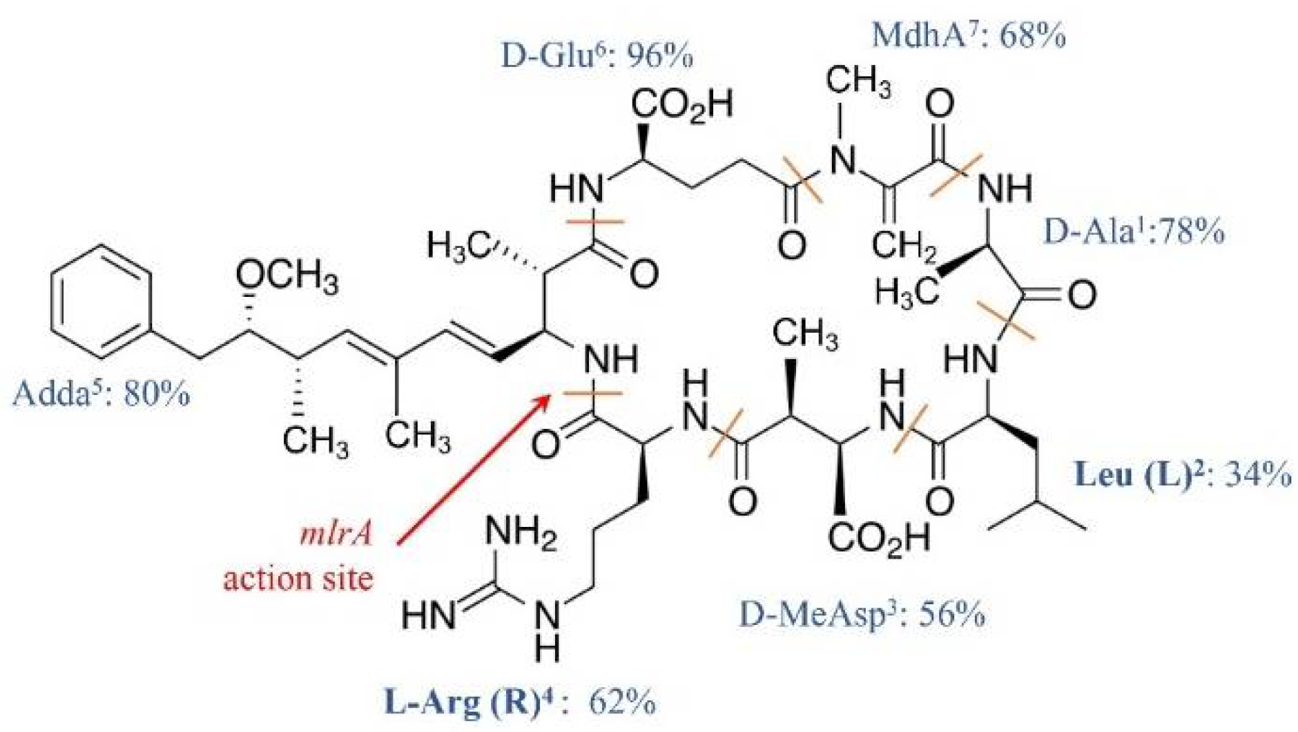
MCs congeners share a common cyclic structure that is formed by seven amino acid residues, cyclo-(D-Ala1)-X2-(D-MeAsp3)-Z4-Adda5-(D-Glu6)-Mdha7 [15] (Figure 1). The percentage variation for the amino acid residue has been depicted in Figure 1. The two L-amino acids of MCs at positions 2 and 4, i.e., X2 and Z4, are the most variable by substitution and account for the most diversity of MC congeners [15]. The structures of the rest of the amino acid residues are largely constant, although variations at each of these positions have been reported (Figure 1). The number of identified MC congeners has been consistently increasing and reached 279 very recently [16]; more are expected to be discovered [17].
Ala1, MeAsp3, and Glu6 were D-amino acids. Adda5, or C20 amino acid (3-amino-9-methoxy-2, 6, 8-trimethyl-10-phenyldeca-4, 6-dienoic acid), and MdhA7 are non-proteinogenic amino acids; they contribute significantly to the toxicity of MCs and are also found in the structure of another cyanotoxin, i.e., nodularin [18].
MC congeners were named after the X2 and Z4 amino acid residues. With decreasing commonalities, MC-Leucine2-Arginine4 (MC-LR), MC-Arginine2-Arginine4 (MC-RR), MC-Leucine2-Phenylalanine4 (MC-LF), and MC-Leucine2-Alanine4 (MC-LA) are the most abundant in natural environments [19,20]. Among these common MCs, MC-LR is the most toxic [21], although it is milder than MC-LF, a far less common MC congener [22]. Due to the universal distribution and high toxicity, MC-LR is widely used as a model for MC studies [21,23].
2. Exposure and Toxicology
2.1. Modes of Exposure and Primary Toxicities
MCs in natural waters can affect humans via various routes, including chronic and accidental ingestion of contaminated drinking or recreational water, inhalation or contact with the nasal mucous membrane, dermal contact with toxins during recreational activities, and consumption of contaminated food irrigated with (vegetables, fruits) or grown in (i.e., fish and shellfish) contaminated water [24] (Figure 2).
Developing a better understanding of the toxicity of MCs will enable us to assess the risk of exposure to these commonly encountered cyanotoxins. The primary cytotoxicity of MCs is the inhibition of Protein Phosphatases 1 and 2a (PP1 and 2a), which leads to several subsequent harmful effects. Acute exposure via ingestion of MCs by humans at concentrations over 10 µg/L can cause various symptoms, including vomiting, diarrhea, abdominal pain, and blistering around the mouth, or it can even ultimately lead to liver failure [25]. However, MCs are rarely ingested directly at acute lethal doses by humans. Chronic and frequent exposure to MCs at low concentrations can ultimately lead to liver failure due to chronic liver cell apoptosis or uncontrolled cell proliferation, leading to primary liver cancer [26,27,28].
2.2. Mechanism of MC Cytotoxicity
Once they have gained exposure to animal cells, MCs first get absorbed into the intestinal tract and then earn entry to the blood stream, where they can be distributed to a range of organs [29]. MCs enter human cells via transmembrane organic anion transporter peptides (OATPs) (Fischer et al., 2005). Receptors of OATPs are abundant on the hepatic cells, making liver a primary target for MCs; about 50–70% of MCs in blood streams can be taken up by the liver [30]. With less abundance, OATPs also exist on cells of other organs, such as kidney and brain cells, making them also susceptible to MC toxicity [30]. In comparison with hepatic cells, non-hepatic cells require a greater MC dose and longer exposure time for cell death to occur [10,31]. When being exposed to the same concentrations of MC-LR (0.8 µM), hepatocytes were found to shrink and lose their viability (dying) within 30 min, while endothelial, fibroblasts, and epithelial cells remained viable for up to 5 days [31].
The final stage of MC intoxication may include cell death due to apoptosis or necrosis27; the former mechanism has been found to be more common [10]. Many studies found that MCs can induce apoptosis via the intrinsic pathway, starting with the production of reactive oxidative species (ROS), which then increase mitochondrial permeability potential (MPP) followed by the induction of caspase 9 and then 3 in that sequence [32,33,34]. A few recent studies have also suggested the importance of extrinsic apoptosis, which starts with the Fas/Fas-L receptor, followed by the induction of caspase 8, followed by the induction of caspase 3 [35,36,37]. MC can also induce apoptosis via activation of the NF-kB pathway (part of the extrinsic pathway) followed by caspase 9 induction [38] in INS-1 cells, which involves both extrinsic and intrinsic pathways. A recent work has shown the induction of caspase 8 before the induction of caspase 9 when we have a combination of extrinsic and intrinsic pathways in MC-treated hepatic cells [37].
3. Production of MCs and Their Regulating Factors
MC synthesis in cyanobacteria is carried out by a gene cluster that possesses eight genes of a total of over 55 kbp DNA (mcyA-J), which encode 48 catalytic reactions [39]. A number of precursors are incorporated during the MC synthesis, including phenylacetate, malonyl-CoA, SAM (S-adenosyl methionine), glutamate, serine, alanine, leucine, D-methyl-iso-aspartate, and arginine [39,40]. The arrangements and sequences of mcy genes in the genome and their products (enzymes/proteins) differ among cyanobacterial species [41] (12); therefore, the prevalence of MC congeners during cyanobacterial blooms is determined by dominant cyanobacterial species [17].
Besides inherent genetic properties, a number of environmental factors can impact MC synthesis. Higher dissolved oxygen in water has been found to increase MC production [42,43,44]. High nitrogen concentrations have been found to limit the amount of MCs produced by Microcystis aeruginosa, while high sulfur and phosphorous supplies behave the opposite [45,46]. Solar irradiance, and UV-B intensities, in particular, has been found to encourage the growth of MC-producing cyanobacteria over non-toxin producing strains [47]. Solar irradiance and nutrient supplies together have also been found to impact not only the amount but also the types of MC congeners produced by Microcystis aeruginosa and Planktothrix agardhii [48,49]. Specifically, these cyanobacteria produced higher amounts of MCs with more toxic variants, like MC-LR, than MC-RR under high solar irradiance and nutrient supply. However, the above observations can be species-specific, as MC productions were not altered by light for M. wesenbergii and Aphanizomenon aphanizomenoides under varied light and nutrients [50].
4. Fate of Extracellular MCs in Environments
Dissolved MCs in natural environments are subjected to both abiotic and biotic transformations.
4.1. Abiotic Transformations
After being released from cyanobacterial cells, a significant fraction (34.8–72.6%) of dissolved MCs in aquatic environments can be physically adsorbed to sediments [51]. Adsorption of MCs to sediment is affected by pH; an increase in adsorption is expected when pH decreases [52]. Organic matter content in soil/sediment was found to enhance MC adsorption to sediment [52,53].
Chemically, the ring-structured MCs are very stable under normal light irradiance and temperatures in natural environments [54]. However, humic acids and pigments (chlorophyll) can serve as photosensitizers to induce photolysis that starts at the site of Adda [55,56]. MC-LR was completely degraded in the presence of photosensitizers in 48 hrs; in contrast, MC removal needed up to weeks if photosensitizers were not supplied [56]. The rate of MC photolysis is affected by pH (acidic pH reduces the degradation rate), temperature (higher temperature promotes degradation) and the wavelength (energy) of the light (UV showed higher lysis than visible light) [54].
4.2. Bioaccumulations
MCs can be taken up and gradually accumulate in aquatic plants and animals (Figure 2) that live in or use CyanoHAB-impacted waters [50]. Depending on locations in the food web (diets), organisms can have different bioaccumulation factors (BF = concentration in tissue vs. in ambient water) of MCs. Primary consumers, like filter feeders and herbivore fish, that graze on MC-producing cyanobacteria can have BF values over a wide range between 436–2409 [57]. Primary producers, like Hydrophytes in Lake Taihu, have also been shown to bioaccumulate MCs with a BF of 200–250 [58]. The bioaccumulation factor of MCs can vary seasonally [59]. Studies on freshwater fishes and mussels have revealed higher BFs in June and July (132.81) than colder months [58]. MCs can be accumulated even in the human body, through consumption of contaminated foods [58]. Bioaccumulation of MCs in humans differs among organs, and the liver typically showed concentrations (767 ng/g) that were different than other organs [60].
4.3. Biodegradation
4.3.1. Pathways and Taxa Diversity
The stable ring structure of MCs protects them from proteases that are commonly found in environments and a group of enzymes that is responsible for degradation of many organic compounds [55,61,62].
Our current knowledge on MC biodegradation is primarily based on studies on bacteria. Currently, one complete MC degradation pathway is known, which is encoded by multiple mlr genes. Four genes, namely mlrA, mlrB, mlrC, and mlrD, are sequentially located in the cluster. mlrD encodes a transporter-like protein, whereas mlrA, B. and C encode enzymes required for the degradation of MCs. Enzyme MlrA (microcystinase) initiates the degradation by breaking up the ring structure of MCs at the peptide bond between Adda and Arg and linearizing the cyclic MC-LR (Figure 1). The linearized MC-LR is 100 times less toxic than the cyclic one [62]. Next, a serine peptidase (MlrB) catalyzes the linearized MC-LR at the Ala~Leu peptide bond and produces a tetrapeptide [63]. Finally, the third enzyme (MlrC) randomly cuts the peptide bonds between amino acid residues, resulting in undetermined peptide fragments and amino acids. Since the initial cleavage is the most critical step in MC-LR degradation, so far, genetic analysis of MC-LR degradation is mostly based on the analysis of mlrA genes [64,65,66]. This mlr-dependent degradation has been found to be regulated by acyl homoserine lactose (AHL)-mediated quorum sensing (QS) [67]. This QS encodes two enzyme systems, namely novR1/novI1 and novR2/novI2, which upregulate microcystin degradation. Identification of these regulators will help control the biological degradation of microcystins. This mlr-dependent degradation has been found to be regulated by acyl homoserine lactose (AHL)-mediated quorum sensing (QS) [67]. This QS encodes two enzyme systems. namely novR1/novI1 and novR2/novI2, which upregulate microcystin degradation. Identification of these regulators will help control the biological degradation of microcystins.
Studies on bacterially mediated MC degradation are dominated by culture-dependent work [62,68]. Nearly a third of current MC-degrading bacterial isolates are affiliated with genera of a single family, i.e., Sphingomonas, Sphingopyxis, and Sphingoscinicella of the Sphingomonaceae family (alphaproteobacteria) [69], with the rest belonging to gammaprotoebacteria and firmicutes. However, many recent culture-dependent and independent studies have consistently suggested that MC-degrading bacteria might be more taxonomically diverse [70,71]. MC-degrading bacteria are now known to be distributed among species (approx. 100) of Microbacterium of Actinobacteria families [72,73]; Bacillus, Brevibacillus, and Lactobacillus of Firmicutes families [74,75,76]; and Pseudomonas, Stenotrophomonas, Acidovorax, and Sphingomonas of alphaproteobacteria, betaproteobacteria, and gammaprotobacteria families [62,77].
Most MC-degrading bacteria are isolated from aerobic environments, but studies have also obtained MC degraders from oxygen-limited environments, such as alphaproteobacteria and gammproteobacteria from drinking water sludge [78], deltaproteobacteria from the mucilage of Microcystis cells [79], and commercially available probiotic bacteria [76], which belong to Proteobacteria (alpha, beta, and gamma), Actinobacteria, and Firmicutes.
Both culture-dependent and-independent studies have revealed that MC-degrading bacteria may or may not carry mlr genes [70,71,72]. Non-mlr bacteria might degrade MCs via the xenobiotic degradation pathway [70]. A recent study has shown the presence of microcystinase, linearized-microcystinase, tetrapeptidease, and PAAase (Phenylacetic acid-ase) as enzymes for the degradation of microcystin by bacteria [80]. This study is the first to have reported the presence of PAAase for complete degradation of microcystin using the mlr degradation pathway. Non-mlr MC-degrading bacterial cultures [62] are taxonomically more diverse than mlr-based MC degraders; they are mostly affiliated with Proteobacteria (80% of the non-mlr based degrading bacteria) (alpha, beta, and gamma classes), Actinobacteria (10%), and Firmicutes (10%), with bacteria containing mlr genes [30] primarily belonging to alpha proteobacteria (60%), with some belonging to Firmicutes and Gammaproteobacteria [68].
4.3.2. Factors Impacting MC Biodegradation
The rate of microbial degradation of MCs is affected by a number of abiotic factors, including temperature, pH, DOC (dissolved organic carbon), and nutrient availability [81]. Temperature at the 30–40 °C range has been found to favor MC biodegradation [71,82,83]. Temperatures that are lower or higher (±10–20 °C) than this range can slow down the degradation of MCs [82].
MC degradation has been observed in a wide range of pH (3–9) for most of the bacterial isolates; whereby some isolates degrade at an alkaline pH of 9 [71,82,83], but rapid degradation was observed at a neutral pH (0.09–0.15 µg/mL/h). Lower rates of MC-degradation were recorded at acidic pH of 3~5 [71,82,84].
Organic nutrients (C.N and P) are commonly found in excess in eutrophic lakes, especially during CyanoHABs [85]. Many studies have revealed that a supply of dissolved organic nutrients may slow down MC degradation [65,81]. However, a few recent studies have found improved or unchanged microcystin degradation by certain bacteria species after additions of an organic C source [71,86].
Studies on MC degradation by mixed cultures are limited. MC-degrading bacterial consortia have been isolated from polluted reservoirs in Taiwan and contained a few bacterial species, such as Sphingomonas spp., Sphingobium spp., and Pseudoxanthomonas spp. However, whether and how each of these bacteria participate in MC degradation are unknown [87]. A recent study using Lake Erie bacterial isolates found that combinations of certain pairs of MC-degrading bacterial isolates could increase the MC degradation rate by up to two-fold (mixed: 0.4 µg/mL/h; singular stains: 0.2 µg/mL/h [71].
5. Conclusions
MCs are commonly encountered toxins in lakes and coastal marine environments that are affected by cyanobacterial harmful algal blooms. So far, greater than 100 variants of MCs have been identified, and MC-LR, MC-RR, MC-LF, and MC-LA are among the most common and toxic. MCs can be exposed to animals and humans via multiple routes (orally, dermally), and their primary target organ is the liver. In liver cells, MCs’ toxicity likely manifests by induction of both extrinsic and intrinsic apoptosis, which dysregulates the cell cycle and induces carcinogenesis in hepatic cells. There still exists a knowledge gap for the identification of the pathway used for the induction of carcinogenic effects. Biosynthesis of MCs by cyanobacteria requires 48 catalytic reactions and is affected by cyanobacterial species and environmental factors such as solar irradiance and nutrient supply. MCs are released into environments during cyanobacterial death, and then, they are transformed mainly by microbial degradation. A diversity of MC-degrading bacteria has been identified across phyla, including alpha-, beta-, and gamma-Proteobacteria; Actinobacteria; and Firmicutes. Bacterial degradation of MCs is carried out by mlr and non-mlr pathways. Enzymes/genes of the mlr pathway have been identified, while much less is known for the non-mlr pathway. Factors, such as pH, temperature, and presence of organic nutrients, can impact bacterially mediated MC degradation in the environment.
Author Contributions
Conceptualization, A.K. and X.M.; methodology, A.K. and X.M.; software, A.K. and X.M.; validation, A.K. and X.M.; formal analysis, A.K. and X.M.; investigation, A.K. and X.M.; resources, A.K. and X.M.; data curation, A.K. and X.M.; writing—original draft preparation, A.K. and X.M.; writing—review and editing, A.K. and X.M.; visualization, A.K. and X.M.; supervision, A.K. and X.M.; project administration, A.K. and X.M.; funding acquisition, A.K. and X.M. Both authors have read and agreed to the published version of the manuscript.
Funding
This work was funded by a Harmful Algal Bloom Research Initiative grant from Ohio Department of Higher Education.
Conflicts of Interest
The authors declare no conflict of interest.
References
- Labine, M.; Minuk, G. Cyanobacterial toxins and liver disease. Can. J. Physiol. Pharmacol. 2009, 87, 773–788. [Google Scholar] [CrossRef]
- Harke, M.J.; Steffen, M.M.; Gobler, C.J.; Otten, T.G.; Wilhelm, S.W.; Wood, S.A. A review of the global ecology, genomics, and biogeography of the toxic cyanobacterium, Microcystis spp. Harmful Algae 2016, 54, 4–20. [Google Scholar] [CrossRef] [PubMed] [Green Version]
- Graham, L.; Loftin, K.; Meyer, M.; Ziegler, A. Cyanotoxin mixtures and taste-and-odor compounds in cyanobacterial blooms from the Midwestern United States. Environ. Sci. Technol. 2010, 19, 7361–7368. [Google Scholar] [CrossRef]
- Michalak, A.; Anderson, E.; Beletsky, D.; Boland, S.; Bosch, N.; Bridgeman, T.; Chaffin, T.; Cho, K.; Confesor, R.; Daloglu, I. Record-setting algal bloom in Lake Erie caused by agricultural and meteorological trends consistent with expected future conditions. Proc. Natl. Acad. Sci. USA 2013, 110, 6448–6452. [Google Scholar] [CrossRef] [Green Version]
- Sakai, H.; Hao, A.; Iseri, Y.; Wang, S.; Kuba, T.; Zhang, Z.; Katayama, H. Occurrence and distribution of microcystins in Lake Taihu, China. Sci. World J. 2013, 2013, 1–7. [Google Scholar] [CrossRef]
- Hawkins, P.R.; Novic, S.; Cox, P.; Neilan, B.A.; Burns, B.P.; Shaw, G.; Saitou, T. A review of analytical methods for assessing the public health risk from microcystin in the aquatic environment. J. Water Supply Res.Technol. 2005, 54, 509–518. [Google Scholar] [CrossRef] [Green Version]
- Sangolkar, L.N.; Maske, S.S.; Chakrabarti, T. Methods for determining microcystins (peptide hepatotoxins) and microcystin-producing cyanobacteria. Water Res. 2006, 40, 3485–3496. [Google Scholar] [CrossRef] [PubMed]
- Machado, J.; Campos, A.; Vasconcelos, V.; Freitas, M. Effects of microcystin-LR and cylindrospermopsin on plant-soil systems: A review of their relevance for agricultural plant quality and public health. Environ. Res. 2017, 153, 191–204. [Google Scholar] [CrossRef] [PubMed] [Green Version]
- Preece, E.P.; Hardy, F.J.; Moore, B.C.; Bryan, M. A review of microcystin detections in estuarine and marine waters: Environmental implications and human health risk. Harmful Algae 2017, 61, 31–45. [Google Scholar] [CrossRef] [Green Version]
- Campos, A.; Vasconcelos, V. Molecular mechanisms of microcystin toxicity in animal cells. Int. J. Mol. Sci. 2010, 11, 268–287. [Google Scholar] [CrossRef] [PubMed] [Green Version]
- Lone, Y.; Kohli, R.; Bhide, M. An overview of the toxic effect of potential human carcinogen MC-LR on testis. Toxicol. Rep. 2015, 2, 289–296. [Google Scholar] [CrossRef] [Green Version]
- Christiansen, G.; Fastner, J.; Erhard, M.; Börner, T.; Dittmann, E. Microcystin biosynthesis in Planktothrix: Genes, evolution, and manipulation. J. Bacteriol. Res. 2003, 185, 564–572. [Google Scholar] [CrossRef] [Green Version]
- Westrick, J.A.; Szlag, D.C.; Southwell, B.J.; Sinclair, J. A review of cyanobacteria and cyanotoxins removal/inactivation in drinking water treatment. Analyt. Bioanalyt. Chem. 2010, 397, 1705–1714. [Google Scholar] [CrossRef] [PubMed]
- Li, J.; Li, R.; Li, J. Current research scenario for microcystins biodegradation—A review on fundamental knowledge, application prospects and challenges. Sci. Total Environ. 2017, 595, 615–632. [Google Scholar] [CrossRef] [PubMed]
- Du, X.; Liu, H.; Yuan, L.; Wang, Y.; Ma, Y.; Wang, R.; Chen, X.; Losiwicz, M.; Guo, H.; Zhang, H. The diversity of cyanobacterial toxins on structural characterization, distribution, and identification: A systematic review. Toxins 2019, 11, 530. [Google Scholar] [CrossRef] [PubMed] [Green Version]
- Bouaicha, N.; Miles, C.; Beach, D.; Labidi, Z.; Djabri, A.; Benayache, N.; Nguyen-Quang, T. Structural diversity, characterization and toxicology of microcystins. Toxins 2019, 11, 714. [Google Scholar] [CrossRef] [Green Version]
- Diez-Quijada, L.; Prieto, A.; Guzman-Guilllen, R.; Jos, A.; Camean, A. Occurrence and toxicity of microcystin congeners other than MC-LR and MC-RR: A review. Food Chem. Toxicol. 2019, 125, 106–132. [Google Scholar] [CrossRef]
- He, X.; Stanford, B.; Adams, C.; Rosenfield, E.; Wert, E. Varied influence of microcystin structural difference on ELISA cross-reactivity and chlorination efficiency of congener mixtures. Water Res. 2017, 126, 515–523. [Google Scholar] [CrossRef] [PubMed]
- Zastepa, A.; Pick, F.; Blais, J. Distribution and flux of microcystin congeners in lake sediments. Lakes Reserv. Res. Manag. 2017, 33, 444–451. [Google Scholar] [CrossRef] [Green Version]
- Zurawell, R.W.; Chen, H.; Burke, J.M. Hepatotoxic cyanobacteria: A review of the biological importance of microcystins in freshwater environments. J. Toxicol. Environ. Health Part B 2005, 8, 1–37. [Google Scholar] [CrossRef] [PubMed]
- Greer, B.; Meneely, J.; Elliot, C. Uptake and accumulation of Microcystin-LR based on exposure through drinking water: An animal model assessing the human health risk. Science 2018, 2018, 4913. [Google Scholar] [CrossRef] [PubMed] [Green Version]
- Ward, C.J.; Codd, G.A. Comparative toxicity of four microcystins of different hydrophobicities to the protozoan, Tetrahymena pyriformis. J. Appl. Microbiol. 1999, 86, 74–82. [Google Scholar]
- Welten, R.; Meneely, J.; Elliott, C. A Comparative Review of the Effect of Microcystin-LR on the Proteome. Exp. Health 2020, 12, 111–129. [Google Scholar] [CrossRef] [Green Version]
- Poste, A.; Hecky, R.; Guildford, S. Evaluating MC Exposure Risk through Fish Consumption. Environ. Sci. Technol. 2011, 45, 5806–5811. [Google Scholar] [CrossRef] [PubMed] [Green Version]
- Blaha, L.; Babica, P.; Marsalek, B. Toxins produced in cyanobacterial water blooms-toxicity and risks. Interdiscip. Toxicol. 2009, 2, 36–41. [Google Scholar] [CrossRef] [Green Version]
- McLellan, N.; Manderville, R. Toxic mechanisms of microcystins in mammals. Toxicol. Res. 2017, 6, 391–405. [Google Scholar] [CrossRef] [Green Version]
- Woolbright, B.; Williams, D.; Ni, H.; Kumer, S.; Schmitt, T.; Kane, B.; Jaeschke, H. Microcystin-LR induced liver injury in mice and in primary human hepatocytes is caused by oncotic necrosis. Toxicon 2017, 125, 99–109. [Google Scholar] [CrossRef] [Green Version]
- Ong, M.M.; Deng, S.; Halim, C.; Cai, W.; Huang, R.; Sethi, G.; Hooi, S.; Kumar, A.; Yap, C. Cytoskeletal proteins in cancer and intracellular stress: A therapeutic perspective. Cancers 2020, 12, 238. [Google Scholar] [CrossRef] [Green Version]
- Meier-Abt, F.; Hammann-Hanni, A.; Stieger, B.; Ballatori, N.; Boyer, J. The organic anion transport polypeptide 1d1 (Oatp1d1) mediates hepatocellular uptake of phalloidin and microcystin into skate liver. Toixcol. Appl. Pharmacol. 2007, 218, 274–279. [Google Scholar] [CrossRef]
- Fischer, W.; Altheimer, S.; Cattori, V.; Meier, P.; Dietrich, D.; Hagenbuch, B. Organic anion transporting polypeptides expressed in liver and brain mediate uptake of microcystin. Toxicol. Appl. Pharmacol. 2005, 203, 257–263. [Google Scholar] [CrossRef] [PubMed] [Green Version]
- McDermott, C.; Nho, C.; Howard, W.; Holton, B. The cyanobacterial toxin, MC-LR, can induce apoptosis in a variety of cell types. Toxicon 1998, 36, 1981–1986. [Google Scholar] [CrossRef]
- Weng, D.; Yan, L.; Yinna, W.; Ying, L.; Pingping, S. The Role of ROS in MC-LR-induced Hepatocyte Apoptosis and Liver Injury in Mice. Toxicology 2007, 232, 15–23. [Google Scholar] [CrossRef] [PubMed]
- Li, Y.; Shao, J.; Wu, X.; Xu, Y.; Li, R. MC production by Microcystis aeruginosa: Direct regulation by multiple environmental factors. FEMS Microbiol. Lett. 2011, 322, 108–114. [Google Scholar]
- Huang, H.; Liu, C.; Fu, X.; Zhang, S.; Xin, Y.; Li, Y.; Xue, L.; Cheng, X.; Zhang, H. Microcystin-LR induced apoptosis in rat sertoli cells via the mitochondrial caspase-dependent pathway: Role of reactive oxygen species. Front. Physiol. 2016, 7, 397–412. [Google Scholar] [CrossRef] [Green Version]
- Zhao, Y.; Li, R.; Xia, W.; Neuzil, J.; Lu, Y.; Zhang, H.; Zhao, X.; Zhang, X.; Sun, C.; Wu, K. Bid integrates intrinsic and extrinsic signaling in apoptosis induced by α-tocopheryl succinate in human gastric carcinoma cells. Cancer Lett. 2010, 288, 42–49. [Google Scholar] [CrossRef] [PubMed]
- Wang, M.; Su, P. The role of the Fas/FasL signaling pathway in environmental toxicant-induced testicular cell apoptosis: An update. Syst. Biol. Reprod. Med. 2017, 64, 93–102. [Google Scholar] [CrossRef]
- Krishnan, A.; Koski, G.; Mou, X. Characterization of microcystin-induced apoptosis in HepG2 hepatoma cells. Toxicon 2019, 173, 20–26. [Google Scholar] [CrossRef]
- Feng, G.; Abdalla, M.; Li, Y.; Bai, Y. NF-kB mediates the induction of Fas receptor and Fas ligand by microcystin-LR in HepG2 cells. Mol. Cell Biochem. 2011, 352, 209–219. [Google Scholar] [CrossRef] [PubMed]
- Tillett, D.; Dittmann, E.; Erhard, M.; von Döhren, H.; Börner, T.; Neilan, B.A. Structural organization of MC biosynthesis in Microcystis aeruginosa PCC7806: An integrated peptide–polyketidesynthetase system. Chem. Biol. 2000, 7, 753–764. [Google Scholar] [CrossRef] [Green Version]
- Dittmann, E.; Neilan, B.; Erhard, M.; Dohren, H.; Borner, T. Insertional mutagenesis of a peptide synthetase gene that is responsible for hepatotoxin production in the cyanobacterium Microcystis aeruginosa PCC 7806. Mol. Microbiol. 1997, 26, 779–787. [Google Scholar] [CrossRef]
- Rouhiainen, L.; Vakkilainen, T.; Siemer, B.L.; Buikema, W.; Haselkorn, R.; Sivonen, K. Genes coding for hepatotoxic heptapeptides (microcystins) in the cyanobacterium Anabaena strain 90. Appl. Environ. Microbiol. 2004, 70, 686–692. [Google Scholar] [CrossRef] [PubMed] [Green Version]
- Wicks, R.; Thiel, P. Environmental factors affecting the production of peptide toxins in floating scums of the cyanobacterium Microcystis aeruginosa in a hypertrophic African Reserve. Environ. Sci. Technol. 1990, 24, 1413–1418. [Google Scholar] [CrossRef]
- Srivastav, A.; Choi, G.; Ahn, C.; Oh, H.; Ravi, A.; Asthana, R. dynamics of microcystin production and quantification of potentially toxigenic microcystis sp. using real-time PCR. Water Res. 2012, 46, 817–827. [Google Scholar]
- Kotak, B.; Lam, A.K.Y.; Prepas, A.E.; Hrudey, S. Role of chemical and physical variables in regulating MC-LR concentration in phytoplankton of eutrophic lakes. Can. J. Fisher Aqu. Sci. 2000, 57, 1584–1593. [Google Scholar] [CrossRef]
- Oh, H.; Le, S.; Jang, M.; Yoon, B. MC Production by Microcystis aeruginosa in a Phosphorus-Limited Chemostat. Appl. Environ. Microbiol. 2000, 66, 176–179. [Google Scholar] [CrossRef] [Green Version]
- Laszakovits, J.; MacKay, A. Removal of cyanotoxins by potassium permanganate: Incorporating competition from natural water constituents. Water Res. 2019, 155, 1–28. [Google Scholar] [CrossRef] [PubMed]
- Ding, X.; Li, X.; Hong-Ying, D.; Ik-Kyo, C.; Jin-Ae, L. Toxic effects of microcystis cell extracts on the reproductive system of male mice. Toxicon 2006, 48, 973–979. [Google Scholar] [CrossRef]
- Tonk, L. The MC composition of the cyanobacterium Planktothrix agardhii changes toward a more toxic variant with increasing light intensity. Appl. Environ. Microbiol. 2005, 71, 5177–5181. [Google Scholar] [CrossRef] [Green Version]
- Hesse, K.; Kohl, J.-G. Effects of light and nutrient supply on growth and MC content of different strains of Microcystis aeruginosa. Appl. Environ. Microbiol. 2001, 104–115. [Google Scholar]
- Xie, L.; Xie, P.; Guo, L.; Li, L.; Miyabara, Y.; Park, H. Organ distribution and bioaccumulation of MCs in freshwater fish at different trophic levels from the eutrophic Lake Chaohu, China. Environ. Toxicol. 2005, 20, 293–300. [Google Scholar] [CrossRef]
- Maghsoudi, E.; Prevost, M.; Vo Duy, S.; Sauve, S.; Dorner, S. Adsorption characteristics of multiple microcystins and cylindrospermopsin on sediment: Implications for toxin monitoring and drinking water treatment. Toxicon 2015, 103, 48–54. [Google Scholar] [CrossRef]
- Wu, X.; Xiao, B.; Li, R.; Wang, C.; Huang, C.; Huang, J.; Wang, Z. Mechanism and factors affecting sorption of microystins onto natural sediments. Environ. Sci. Technol. 2011, 45, 2641–2647. [Google Scholar] [CrossRef]
- Chen, W.; Song, L.; Gan, N.; Li, L. Sorption, degradation and mobility of microcystins in Chinese agricultural soils: Risk assessment for groundwater protection. Environ. Poll. 2016, 144, 752–758. [Google Scholar] [CrossRef] [PubMed]
- Boopathi, T.; Ki, J. Impact of environmental factors on the regulation of cyanotoxin production. Toxins 2014, 6, 1951. [Google Scholar] [CrossRef] [Green Version]
- Tsuji, K.; Naito, S.; Kondo, F.; Ishikawa, N.; Watanabe, M.; Suzuki, M.; Harada, K. Stability of MCs from cyanobacteria: Effect of light on decomposition and isomerization. Environ. Sci. Technol. 1994, 28, 173–177. [Google Scholar] [CrossRef] [PubMed]
- Munuswamy, T.; Hu, Y.; Lee, J. Adsorption and photodegradation of microcystin-LR onto sediments collected from reservoirs and rivers in Taiwan: A laboratory study to investigate the fate, transfer, and degradation of microcystin-LR. Environ. Sci. Pollut. Res. 2012, 19, 2390–2399. [Google Scholar] [CrossRef] [PubMed]
- Berry, J.; Lee, E.; Walton, K.; Wilson, A.; Bernal-Brooks, F. Bioaccumulation of microcystins by fish associated with a persistent cyanobacterial bloom in Lago de Patzcuaro (Michoacan, Mexico). Environ. Toxicol. Chem. 2011, 30, 1621–1628. [Google Scholar] [CrossRef] [PubMed]
- Papadimitriou, T.; Kagalou, I.; Stalikas, C.; Pilidis, G.; Leonardos, I. Assessment of microcystin distribution and biomagnification in tissues of aquatic food web compartments from a shallow lake and evaluation of potential risks to public health. Ecotoxicology 2012, 21, 1155–1166. [Google Scholar] [CrossRef]
- Ferrao-Filho, A.; Kozlowsky-Suzuki, B. Cyanotoxins: Bioaccumulation and effects on aquatic animals. Mar. Drugs 2011, 9, 2729–2772. [Google Scholar] [CrossRef]
- Ni, W.; Zhang, J.; Luo, Y. Microcystin accumulation in bighead carp (Aristichthys nobilis) during a Microcystis-dominated bloom and risk assessment of the dietary intake in a fish pond in China. Environ. Sci. Pollut. Res. 2017, 24, 8894–8902. [Google Scholar] [CrossRef] [PubMed]
- Harada, K.M.F.; Watanabe, K.I.; Harada, W.W.C.; Fujiki, H. Chemistry and Detection of MCs in Toxic MCs; CRC Press: Boca Raton, FL, USA, 1996; pp. 103–143. [Google Scholar]
- Jones, G.J.; Bourne, D.G.; Blakeley, R.L.; Doelle, H. Degradation of the cyanobacterial hepatotoxin microcystin by aquatic bacteria. Nat. Toxins 1994, 2, 228–235. [Google Scholar] [CrossRef]
- Jones, G.; Orr, P. Release and degradation of MC following algicide treatment of a Microcystis aeruginosa bloom in a recreational lake, as determined by HPLC and protein phosphatase inhibition assay. Water Res. 1994, 28, 871–876. [Google Scholar] [CrossRef]
- Ho, L.; Meyn, T.; Keegan, A.; Hoefle, D.; Brookes, J.; Saint, C.; Newcombe, G. Bacterial degradation of microcystin toxins within a biologically active sand filter. Water Res. 2006, 40, 768–774. [Google Scholar] [CrossRef]
- Wang, J.; Wang, C.; Li, Q.; Shen, M.; Bai, P.; Li, J.; Lin, Y.; Gan, N.; Li, T.; Zhao, J. Microcystin-LR Degradation and Gene Regulation of Microcystin-Degrading Novosphingobium sp. THN1 at Different Carbon Concentration. Front. Microbiol. 2019, 2019, 1740–1750. [Google Scholar] [CrossRef] [Green Version]
- Yang, F.; Zhou, Y.; Sun, R.; Wei, H.; Li, Y.; Yin, L.; Pu, Y. Biodegradation of microcystin-LR and-RR by a novel microcystin-degrading bacterium isolated from Lake Taihu. Biodegradation 2014, 25, 447–457. [Google Scholar] [CrossRef]
- Zeng, Y.; Cai, Z.; Zhu, J.; Du, X.; Zhou, J. Two hierarchical LuxR-LuxI type quorum sensing systems in Novosphingobium activate microcystin degradation through transcriptional regulation of the mlr pathway. Water Res. 2020, 183, 1–14. [Google Scholar] [CrossRef] [PubMed]
- Park, H.; Sasaki, Y.; Maruyama, T.; Yanagisawa, E.; Hiraishi, A.; Kato, K. Degradation of the cyanobacterial hepatotoxin MC by a new bacterium isolated from a hypertrophic lake. Environ.Toxicol. 2001, 16, 337–343. [Google Scholar] [CrossRef] [PubMed]
- Massey, I.; Yang, F. A mini review on microcystins and bacterial degradation. Toxins 2020, 12, 268. [Google Scholar] [CrossRef] [PubMed] [Green Version]
- Mou, X.; Lu, X.; Jacob, J.; Heath, R. Metagenomic identification of bacterioplankton taxa and pathways involved in MC degradation in Lake Erie. PLoS ONE 2013, 8, e61890. [Google Scholar] [CrossRef] [Green Version]
- Krishnan, A.; Zhang, Y.; Balaban, M.; Seo, Y.; Mou, X. Taxonomical and genotypical heterogeneity of microcystin degrading bacterioplankton in western Lake Erie. Harmful Algae 2020, 98, 1–10. [Google Scholar] [CrossRef] [PubMed]
- Manage, P.; Edwards, C.; Singh, B.; Lawton, L. Isolation and identification of novel MC-degrading bacteria. Appl. Environ. Microbiol. 2009, 75, 6924–6928. [Google Scholar] [CrossRef] [Green Version]
- Lawton, L.A.; Welgamage, A.; Manage, P.M.; Edwards, C. Novel bacterial strains for the removal of MCs from drinking water. Water Sci. Technol. 2011, 63, 1137–1142. [Google Scholar] [CrossRef] [PubMed]
- Hu, L.; Zhang, F.; Li, C.; Wang, C. Biodegradation of MCs by Bacillus sp. strain EMB. Energy Procedia 2012, 16, 2054–2059. [Google Scholar] [CrossRef] [Green Version]
- Kansole, M.; Lin, T. MC-LR Biodegradation by Bacillus sp.: Reaction Rates and Possible Genes Involved in the Degradation. Water 2016, 8, 508. [Google Scholar] [CrossRef] [Green Version]
- Nybom, S.; Salminen, S.; Meriluoto, J. Removal of MC-LR by strains of metabolically active probiotic bacteria. FEMS Microbiol. Lett. 2007, 270, 27–33. [Google Scholar] [CrossRef] [Green Version]
- Krishnan, A.; Zhang, Y.; Mou, X. Isolation and characterization of microcystin-degrading bacteria from Lake Erie. Bull. Environ. Contamin. Toxicology 2018, 101, 617–623. [Google Scholar]
- Ma, G.; Pei, H.; Hu, W.; Xu, X.; Ma, C.; Li, X. The removal of cyanobacteria and their metabolites through anoxic biodegradation in drinking water sludge. Biores. Technol. 2014, 165, 191–198. [Google Scholar] [CrossRef]
- Maruyama, T.; Kato, K.; Yokoyama, A.; Tanaka, T.; Hiraishi, A.; Park, H.-D. Dynamics of MC-degrading bacteria in mucilage of Microcystis. Microb. Ecol. 2003, 46, 279–288. [Google Scholar] [CrossRef] [Green Version]
- Yang, F.; Huang, F.; Feng, H.; Wei, J.; Massey, I.; Liang, G.; Zhang, F.; Yin, L.; Kacew, S.; Zhang, X.; et al. A complete route for biodegradation of potentially carcinogenic cyanotoxin microcystin-LR in a novel indigenous bacterium. Water Res. 2020, 174, 1–12. [Google Scholar] [CrossRef]
- Phujomjai, Y.; Somdee, A.; Somdee, T. Biodegradation of MC [Dha7] MC-LR by a novel MC-degrading bacterium in an internal airlift loop bioreactor. Water Sci. Technol. 2013, 73, 267–274. [Google Scholar] [CrossRef]
- Yang, F.; Masssey, I.; Guo, J.; Yang, S.; Pu, Y.; Zeng, W.; Tan, H. Microcystin-LR degradation utilizing a novel effective indigenous bacterial community YFMCD1 from Lake Taihu. J. Toxicol. Environ. Health Part. A 2018, 81, 1–10. [Google Scholar] [CrossRef] [PubMed]
- Okano, K.; Shimizu, K.; Kawauchi, Y.; Maseda, H.; Motoo, U.; Zhnag, Z.; Neilan, B.; Suguira, N. Characteristics of a microcystin-degrading bacterium under alkaline environmental conditions. J. Toxicol. 2009, 2009, 1687–1691. [Google Scholar] [CrossRef]
- Somdee, T.; Thunders, M.; Rucks, M.; Allison, M.; Page, R. Degradation of [Dha (7)] MC-LR by a MC degrading bacterium isolated from Lake Rotoiti, New Zealand. ISRN Microbiol. 2013, 2013, 1–8. [Google Scholar] [CrossRef] [PubMed] [Green Version]
- Carmichael, W. The toxins of cyanobacteria. Sci. Am. 1992, 2, 78–86. [Google Scholar] [CrossRef] [PubMed]
- Li, J.; Shimizu, K.; Sakharkar, M.K.; Utsumi, M.; Zhang, Z.; Sugiura, N. Comparative study for the effects of variable nutrient conditions on the biodegradation of microcystin-LR and concurrent dynamics in microcystin-degrading gene abundance. Bioresour. Technol. 2011, 102, 9509–9517. [Google Scholar] [CrossRef]
- Tsao, S.; Wei, D.; Chnag, Y.; Lee, J. Aerobic biodegradation of microcystin-LR by an indigenous bacterial mixed culture isolated in Taiwan. Int. Biodet. Biodegrad. 2017, 2017, 101–108. [Google Scholar] [CrossRef]
Figure 1.
Chemical structure of hepatotoxic microcystin-LR. Numbers 1–7 signify the presence of amino acid residues with their percent of variation. Positions 2 and 4 depict the highly variable L-amino acids. mlrA site of cleavage on the MC-LR cyclic structure has also been depicted.
Figure 1.
Chemical structure of hepatotoxic microcystin-LR. Numbers 1–7 signify the presence of amino acid residues with their percent of variation. Positions 2 and 4 depict the highly variable L-amino acids. mlrA site of cleavage on the MC-LR cyclic structure has also been depicted.

Figure 2.
Schematic representation of MC-uptake, release, and transfer when in the environment. This schematic diagram represents the transfer of MCs from zooplankton to fish and finally humans.
Figure 2.
Schematic representation of MC-uptake, release, and transfer when in the environment. This schematic diagram represents the transfer of MCs from zooplankton to fish and finally humans.

Publisher’s Note: MDPI stays neutral with regard to jurisdictional claims in published maps and institutional affiliations. |
© 2021 by the authors. Licensee MDPI, Basel, Switzerland. This article is an open access article distributed under the terms and conditions of the Creative Commons Attribution (CC BY) license (https://creativecommons.org/licenses/by/4.0/).
Share and Cite
MDPI and ACS Style
Krishnan, A.; Mou, X. A Brief Review of the Structure, Cytotoxicity, Synthesis, and Biodegradation of Microcystins. Water 2021, 13, 2147. https://doi.org/10.3390/w13162147
AMA Style
Krishnan A, Mou X. A Brief Review of the Structure, Cytotoxicity, Synthesis, and Biodegradation of Microcystins. Water. 2021; 13(16):2147. https://doi.org/10.3390/w13162147
Chicago/Turabian StyleKrishnan, Anjali, and Xiaozhen Mou. 2021. "A Brief Review of the Structure, Cytotoxicity, Synthesis, and Biodegradation of Microcystins" Water 13, no. 16: 2147. https://doi.org/10.3390/w13162147
Note that from the first issue of 2016, this journal uses article numbers instead of page numbers. See further details here.