
Synthesis and Characterization of Sr-Doped ZnSe Nanoparticles for Catalytic and Biological Activities


 ,
, 
Abstract
:1. Introduction
2. Materials and Methods
2.1. Chemicals and Reagents
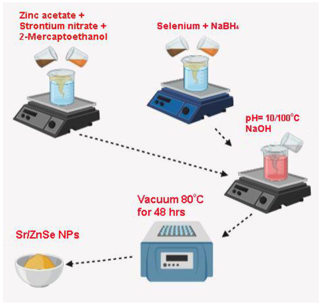
2.2. Synthesis of Sr-Doped Znse Nanoparticles
2.3. Characterization
2.4. Antibacterial Activity
2.5. Photocatalytic Activity
3. Results and Discussion
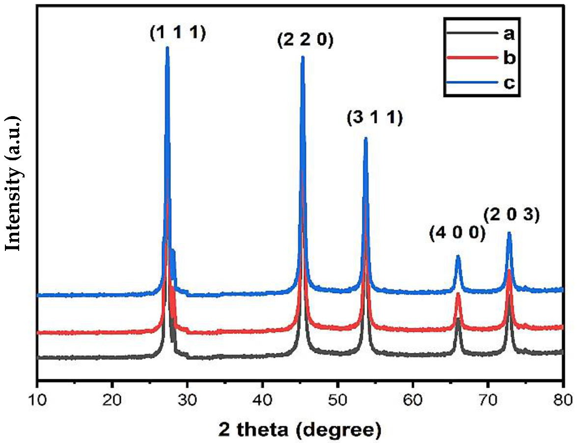
3.1. XRD Analysis
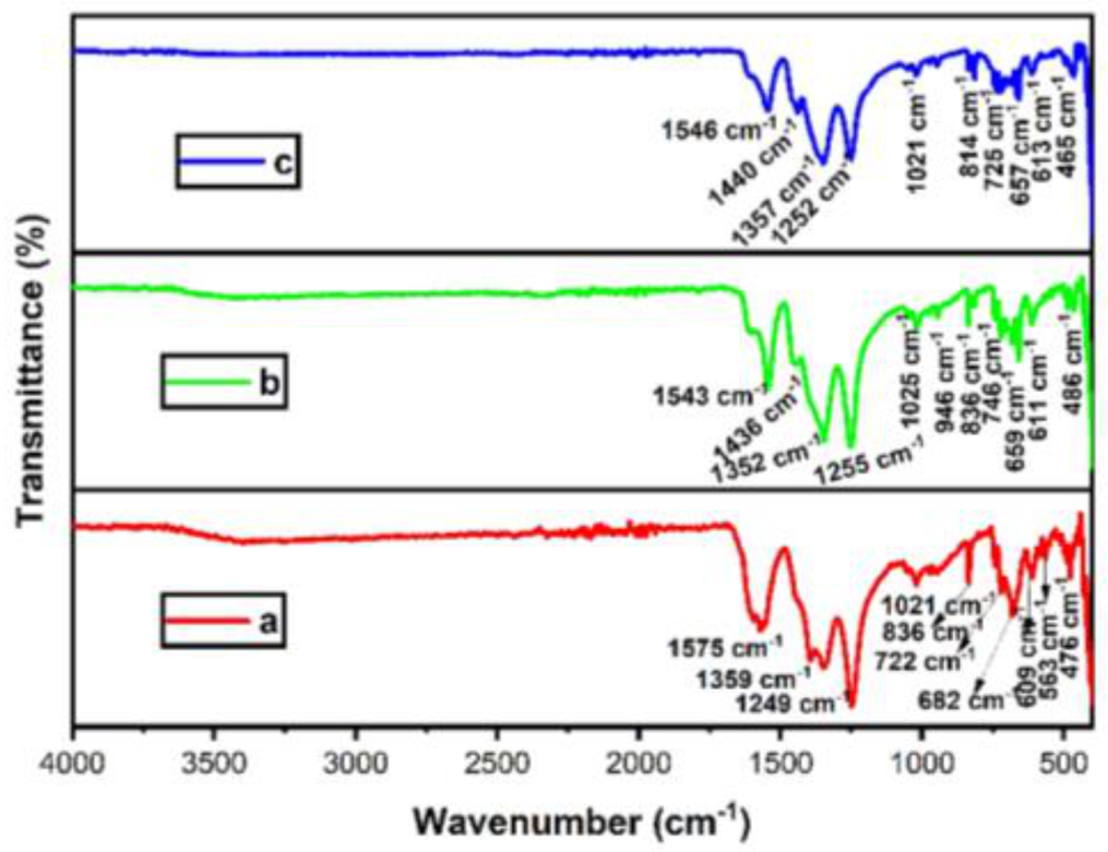
3.2. FTIR Analysis
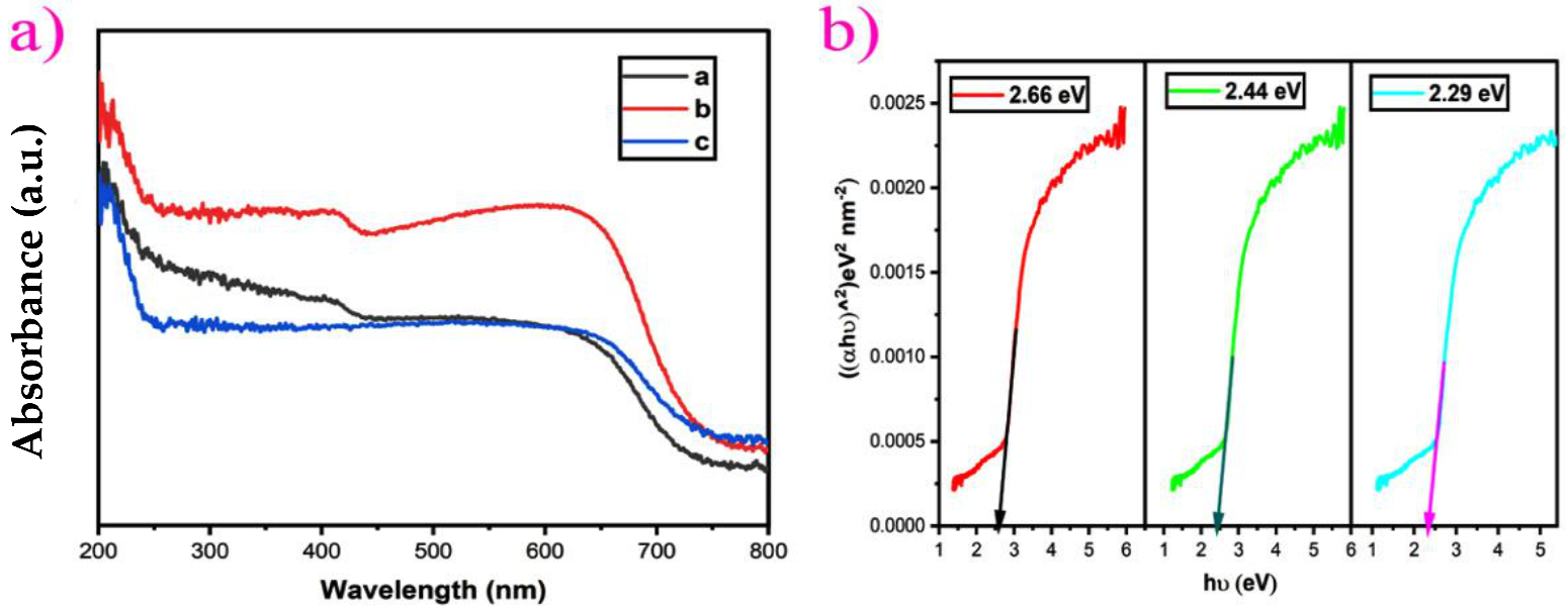
3.3. UV-DRS Analysis
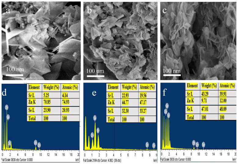
3.4. FE-SEM Analysis
3.5. EDS Analysis
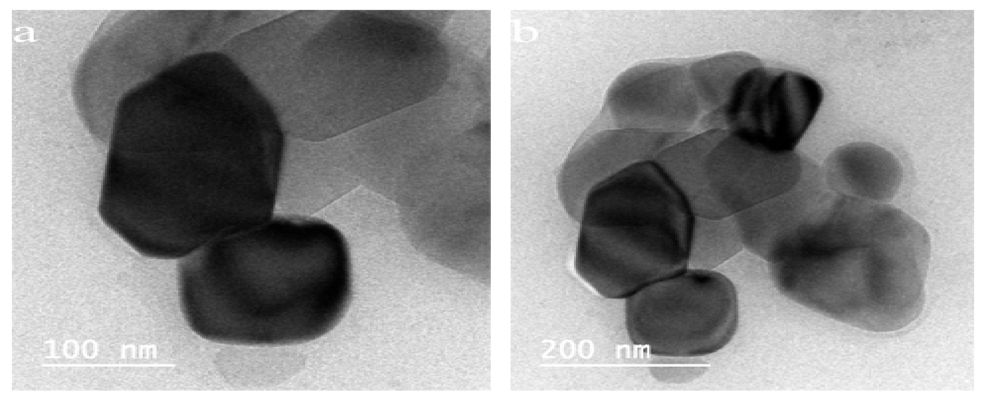
3.6. HR-TEM Analysis
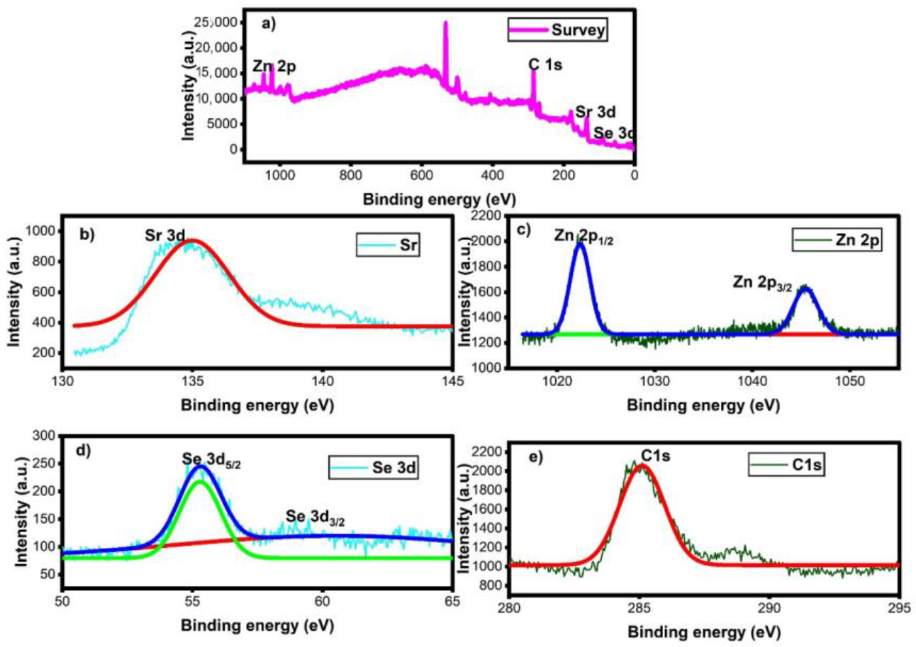
3.7. XPS Analysis
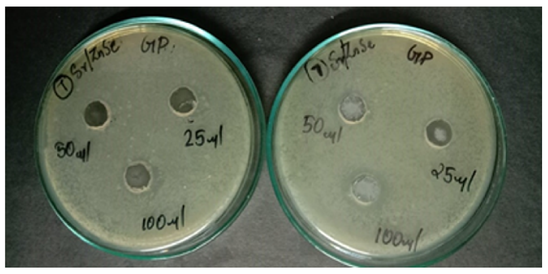
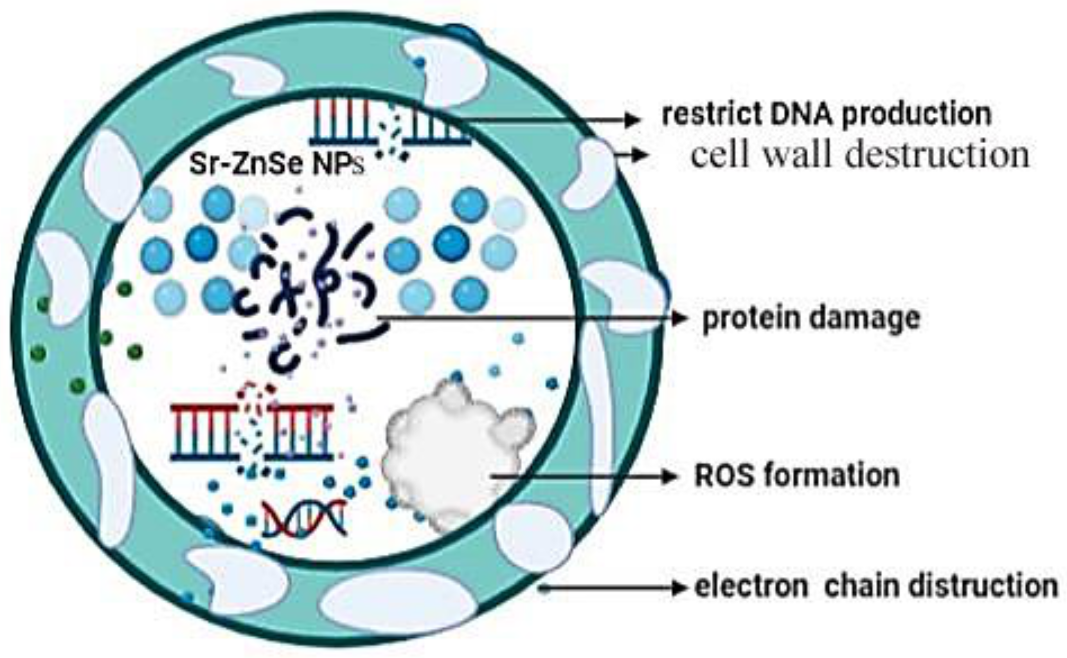
3.8. Antibacterial Activity
Mechanism of Antibacterial Activity
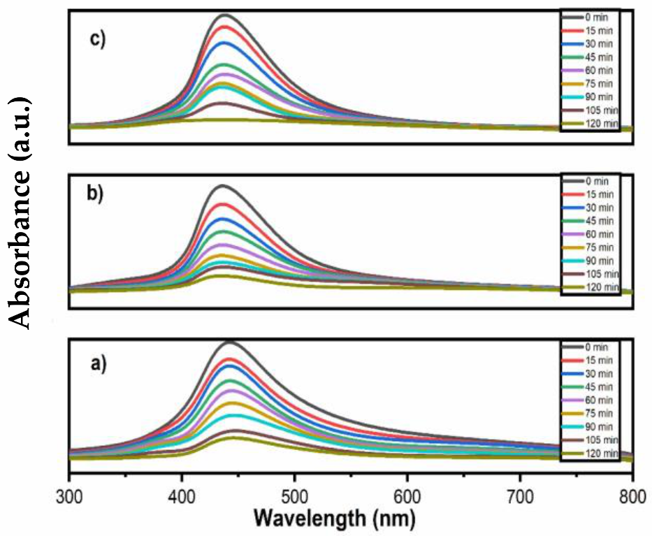
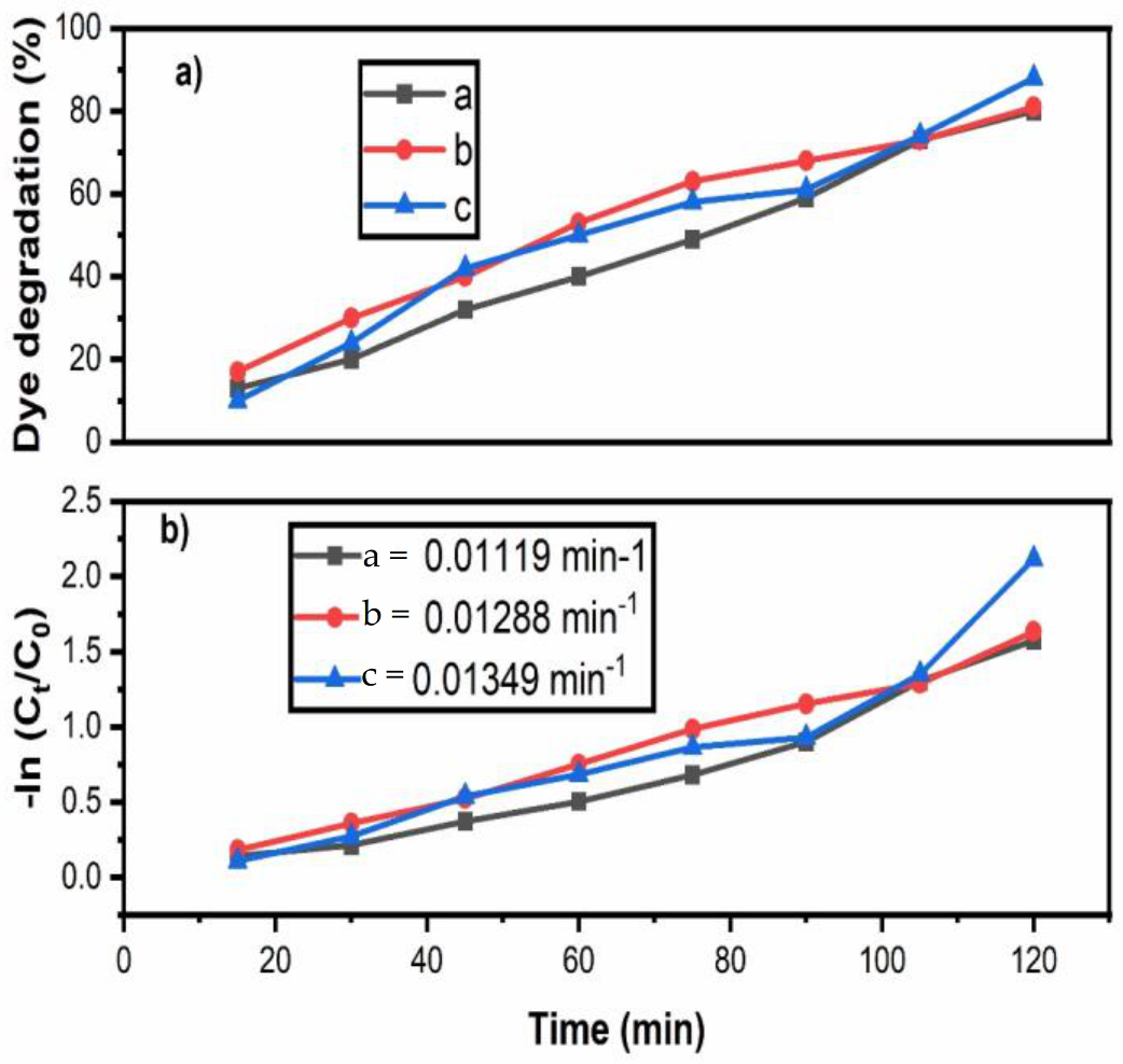
3.9. Photocatalytic Dye Degradation
3.10. Kinetics of Photodegradation
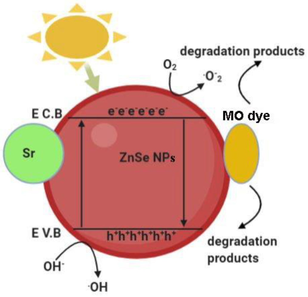
Mechanism of Photodegradation
4. Conclusions
Author Contributions
Funding
Informed Consent Statement
Data Availability Statement
Conflicts of Interest
References
- Aeenjan, F.; Javanbakht, V. Methylene blue removal from aqueous solution by magnetic clinoptilolite/chitosan/EDTA nanocomposite. Res. Chem. Intermed. 2017, 44, 1459–1483. [Google Scholar] [CrossRef]
- Pandey, S.; Fosso-Kankeu, E.; Redelinghuys, J.; Kim, J.; Kang, M. Implication of biofilms in the sustainability of acid mine drainage and metal dispersion near coal tailings. Sci. Total Environ. 2021, 788, 147851. [Google Scholar] [CrossRef]
- Gupta, V.K.; Jain, R.; Nayak, A.; Agarwal, S.; Shrivastava, M. Removal of the hazardous dye—Tartrazine by photodegradation on titanium dioxide surface. Mater. Sci. Eng. C 2011, 31, 1062–1067. [Google Scholar] [CrossRef]
- Erfani, M.; Javanbakht, V. Methylene Blue removal from aqueous solution by a biocomposite synthesized from sodium alginate and wastes of oil extraction from almond peanut. Int. J. Biol. Macromol. 2018, 114, 244–255. [Google Scholar] [CrossRef]
- Mittal, A.; Mittal, J.; Malviya, A.; Gupta, V. Removal and recovery of Chrysoidine Y from aqueous solutions by waste materials. J. Colloid Interface Sci. 2010, 344, 497–507. [Google Scholar] [CrossRef]
- Yagub, M.T.; Sen, T.K.; Afroze, S.; Ang, H. Dye and its removal from aqueous solution by adsorption: A review. Adv. Colloid Interface Sci. 2014, 209, 172–184. [Google Scholar] [CrossRef]
- Bibi, S.; Ahmad, A.; Anjum, M.A.R.; Haleem, A.; Siddiq, M.; Shah, S.S.; Al Kahtani, A. Photocatalytic degradation of malachite green and methylene blue over reduced graphene oxide (rGO) based metal oxides (rGO-Fe3O4/TiO2) nanocomposite under UV-visible light irradiation. J. Environ. Chem. Eng. 2021, 9, 105580. [Google Scholar] [CrossRef]
- Pandey, S.; Do, J.Y.; Kim, J.; Kang, M. Fast and highly efficient catalytic degradation of dyes using κ-carrageenan stabilized silver nanoparticles nanocatalyst. Carbohydr. Polym. 2020, 230, 115597. [Google Scholar] [CrossRef]
- Pandey, S.; Fosso-Kankeu, E.; Spiro, M.; Waanders, F.; Kumar, N.; Ray, S.; Kim, J.; Kang, M. Equilibrium, kinetic, and thermodynamic studies of lead ion adsorption from mine wastewater onto MoS2-clinoptilolite composite. Mater. Today Chem. 2020, 18, 100376. [Google Scholar] [CrossRef]
- Aravind, M.; Ahmad, A.; Ahmad, I.; Amalanathan, M.; Naseem, K.; Mary, S.M.M.; Parvathiraja, C.; Hussain, S.; Algarni, T.S.; Pervaiz, M.; et al. Critical green routing synthesis of silver NPs using jasmine flower extract for biological activities and photocatalytical degradation of methylene blue. J. Environ. Chem. Eng. 2020, 9, 104877. [Google Scholar] [CrossRef]
- Ahmad, A.; Jini, D.; Aravind, M.; Parvathiraja, C.; Ali, R.; Kiyani, M.Z.; Alothman, A. A novel study on synthesis of egg shell based activated carbon for degradation of methylene blue via photocatalysis. Arab. J. Chem. 2020, 13, 8717–8722. [Google Scholar] [CrossRef]
- Younas, U.; Hassan, S.; Ali, F.; Hassan, F.; Saeed, Z.; Pervaiz, M.; Khan, S.; Jannat, F.; Bibi, S.; Sadiqa, A.; et al. Radical Scavenging and Catalytic Activity of Fe-Cu Bimetallic Nanoparticles Synthesized from Ixora finlaysoniana Extract. Coatings 2021, 11, 813. [Google Scholar] [CrossRef]
- Chong, M.N.; Jin, B.; Chow, C.W.K.; Saint, C. Recent developments in photocatalytic water treatment technology: A review. Water Res. 2010, 44, 2997–3027. [Google Scholar] [CrossRef]
- Chakrabarti, S.; Chaudhuri, B.; Bhattacharjee, S.; Das, P.; Dutta, B.K. Degradation mechanism and kinetic model for photocatalytic oxidation of PVC–ZnO composite film in presence of a sensitizing dye and UV radiation. J. Hazard. Mater. 2008, 154, 230–236. [Google Scholar] [CrossRef]
- Lan, Y.; Lu, Y.; Ren, Z. Mini review on photocatalysis of titanium dioxide nanoparticles and their solar applications. Nano Energy 2013, 2, 1031–1045. [Google Scholar] [CrossRef]
- Ong, C.B.; Ng, L.Y.; Mohammad, A.W. A review of ZnO nanoparticles as solar photocatalysts: Synthesis, mechanisms and applications. Renew. Sustain. Energy Rev. 2018, 81, 536–551. [Google Scholar] [CrossRef]
- Mishra, M.K.; Singh, N.; Pandey, V.; Haque, F.Z. Synthesis of SnO2 Nanoparticles and Its Application in Sensing Ammonia Gas Through Photoluminescence. J. Adv. Phys. 2016, 5, 8–12. [Google Scholar] [CrossRef]
- Cheng, L.; Xiang, Q.; Liao, Y.; Zhang, H. CdS-based photocatalysts. Energy Environ. Sci. 2018, 11, 1362–1391. [Google Scholar] [CrossRef]
- Boulkroune, R.; Sebais, M.; Messai, Y.; Bourzami, R.; Schmutz, M.; Blanck, C.; Halimi, O.; Boudine, B. Hydrothermal synthesis of strontium-doped ZnS nanoparticles: Structural, electronic and photocatalytic investigations. Bull. Mater. Sci. 2019, 42, 223. [Google Scholar] [CrossRef] [Green Version]
- Dai, S.; Wang, B.; Li, W.; Wang, L.; Song, X.; Guo, C.; Li, Y.; Liu, F.; Zhu, F.; Wang, Q.; et al. Systemic application of 3-methyladenine markedly inhibited atherosclerotic lesion in ApoE−/− mice by modulating autophagy, foam cell formation and immune-negative molecules. Cell Death Dis. 2016, 7, e2498. [Google Scholar] [CrossRef] [Green Version]
- Patel, B.; Rath, S.; Sarangi, S.N.; Sahu, S. HgS nanoparticles: Structure and optical properties. Appl. Phys. A 2007, 86, 447–450. [Google Scholar] [CrossRef]
- Naseem, K.; Rehman, M.Z.U.; Ahmad, A.; Dubal, D.; Algarni, T.S. Plant Extract Induced Biogenic Preparation of Silver Nanoparticles and Their Potential as Catalyst for Degradation of Toxic Dyes. Coatings 2020, 10, 1235. [Google Scholar] [CrossRef]
- Fallah, Z.; Zare, E.N.; Ghomi, M.; Ahmadijokani, F.; Amini, M.; Tajbakhsh, M.; Arjmand, M.; Sharma, G.; Ali, H.; Ahmad, A.; et al. Toxicity and remediation of pharmaceuticals and pesticides using metal oxides and carbon nanomaterials. Chemosphere 2021, 275, 130055. [Google Scholar] [CrossRef]
- Hu, X.-L.; Shang, Y.; Yan, K.-C.; Sedgwick, A.C.; Gan, H.-Q.; Chen, G.-R.; He, X.-P.; James, T.D.; Chen, D. Low-dimensional nanomaterials for antibacterial applications. J. Mater. Chem. B 2021, 9, 3640–3661. [Google Scholar] [CrossRef]
- Menazea, A.; Ahmed, M. Synthesis and antibacterial activity of graphene oxide decorated by silver and copper oxide nanoparticles. J. Mol. Struct. 2020, 1218, 128536. [Google Scholar] [CrossRef]
- Krishna, A.G.R.; Espenti, C.S.; Reddy, Y.V.R.; Obbu, A.; Satyanarayana, M.V. Green Synthesis of Silver Nanoparticles by Using Sansevieria Roxburghiana, Their Characterization and Antibacterial Activity. J. Inorg. Organomet. Polym. Mater. 2020, 30, 4155–4159. [Google Scholar] [CrossRef]
- Varghese, B.; Kurian, M.; Krishna, S.; Athira, T. Biochemical synthesis of copper nanoparticles using Zingiber officinalis and Curcuma longa: Characterization and antibacterial activity study. Mater. Today Proc. 2020, 25, 302–306. [Google Scholar] [CrossRef]
- Agarwal, H.; Menon, S.; Shanmugam, V.K. Functionalization of zinc oxide nanoparticles using Mucuna pruriens and its antibacterial activity. Surf. Interfaces 2020, 19, 100521. [Google Scholar] [CrossRef]
- Rajbongshi, H.; Kalita, D. Morphology-Dependent Photocatalytic Degradation of Organic Pollutant and Antibacterial Activity with CdS Nanostructures. J. Nanosci. Nanotechnol. 2020, 20, 5885–5895. [Google Scholar] [CrossRef] [PubMed]
- Yang, Y.; Wu, Z.; Yang, R.; Li, Y.; Liu, X.; Zhang, L.; Yu, B. Insights into the mechanism of enhanced photocatalytic dye degradation and antibacterial activity over ternary ZnO/ZnSe/MoSe2 photocatalysts under visible light irradiation. Appl. Surf. Sci. 2021, 539, 148220. [Google Scholar] [CrossRef]
- Beena, V.; Ajitha, S.; Rayar, S.L.; Parvathiraja, C.; Kannan, K.; Palani, G. Enhanced Photocatalytic and Antibacterial Activities of ZnSe Nanoparticles. J. Inorg. Organomet. Polym. Mater. 2021, 4, 1–12. [Google Scholar]
- Gao, Z.; Chen, S.; Li, R.; Lou, Z.; Han, W.; Jiang, K.; Qu, F.; Shen, G. An artificial olfactory system with sensing, memory and self-protection capabilities. Nano Energy 2021, 86, 106078. [Google Scholar] [CrossRef]
- Kageshima, Y.; Gomyo, Y.; Matsuoka, H.; Inuzuka, H.; Suzuki, H.; Abe, R.; Teshima, K.; Domen, K.; Nishikiori, H. Z-Scheme Overall Water Splitting Using ZnxCd1–xSe Particles Coated with Metal Cyanoferrates as Hydrogen Evolution Photocatalysts. ACS Catal. 2021, 11, 8004–8014. [Google Scholar] [CrossRef]
- Liu, Y.; Bai, G.; Lyu, Y.; Hua, Y.; Ye, R.; Zhang, J.; Chen, L.; Xu, S.; Hao, J. Ultrabroadband Tuning and Fine Structure of Emission Spectra in Lanthanide Er-Doped ZnSe Nanosheets for Display and Temperature Sensing. ACS Nano 2020, 14, 16003–16012. [Google Scholar] [CrossRef]
- Khan, M.S.; Shi, L.; Zou, B. Impact of vacancy defects on optoelectronic and magnetic properties of Mn-doped ZnSe. Comput. Mater. Sci. 2020, 174, 109493. [Google Scholar] [CrossRef]
- Sharma, V.; Mehata, M.S. Synthesis of photoactivated highly fluorescent Mn2+-doped ZnSe quantum dots as effective lead sensor in drinking water. Mater. Res. Bull. 2021, 134, 111121. [Google Scholar] [CrossRef]
- Feng, K.; Xue, W.; Hu, X.; Fan, J.; Liu, E. Z-scheme CdSe/ZnSe heterojunction for efficient photocatalytic hydrogen evolution. Colloids Surf. A Physicochem. Eng. Asp. 2021, 622, 126633. [Google Scholar] [CrossRef]
- Tian, P.; Tang, T.; Zhang, J.; Lin, S.; Huang, G.; Zeng, J.; Kong, Z.; Wang, H.; Xi, J.; Ji, Z. High photocatalytic and photoelectrochemical performance of a novel 0D/2D heterojunction photocatalyst constructed by ZnSe nanoparticles and MoSe2 nanoflowers. Ceram. Int. 2020, 46, 13651–13659. [Google Scholar] [CrossRef]
- Kasture, M.B.; Patel, P.; Prabhune, A.A.; Ramana, C.V.; Kulkarni, A.A.; Prasad, B.L.V. Synthesis of silver nanoparticles by sophorolipids: Effect of temperature and sophorolipid structure on the size of particles. J. Chem. Sci. 2008, 120, 515–520. [Google Scholar] [CrossRef]
- Fayaz, A.M.; Balaji, K.; Kalaichelvan, P.; Venkatesan, R. Fungal based synthesis of silver nanoparticles—An effect of temperature on the size of particles. Colloids Surf. B Biointerfaces 2009, 74, 123–126. [Google Scholar] [CrossRef]
- Lee, G. Preparation of silver nanorods through the control of temperature and pH of reaction medium. Mater. Chem. Phys. 2004, 84, 197–204. [Google Scholar] [CrossRef]
- Wang, Q.; Liu, F.T.; Jiang, Q.H.; Chen, X.X.; Wang, D.Z. Luminescence of Cu2+-Doped ZnSe Quantum Dots with Different Parameters. Adv. Mater. Res. 2010, 148–149, 1268–1272. [Google Scholar]
- Thor, H.Y.; Teow, Y.H.; Ho, K.C. Synthesis and characterization of 2-mercaptoethanol-capped manganese-doped zinc sulfide quantum dots-embedded molecularly-imprinted membranes. Part. Sci. Technol. 2021, 1–8. [Google Scholar] [CrossRef]
- Ganjali, M.R.; Al-Naqshabandi, M.A.; Larijani, B.; Badiei, A.; Vatanpour, V.; Rajabi, H.R.; Rezania, H.; Paziresh, S.; Mahmodi, G.; Kim, S.-J.; et al. Improvement of dye and protein filtration efficiency using modified PES membrane with 2-mercaptoethanol capped zinc sulfide quantum dots. Chem. Eng. Res. Des. 2021, 168, 109–121. [Google Scholar] [CrossRef]
- Yarahmadi, M.; Maleki-Ghaleh, H.; Mehr, M.E.; Dargahi, Z.; Rasouli, F.; Siadati, M.H. Synthesis and characterization of Sr-doped ZnO nanoparticles for photocatalytic applications. J. Alloy Compd. 2021, 853, 157000. [Google Scholar] [CrossRef]
- Khan, S.; Shah, S.; Anjum, M.; Khan, M.; Janjua, N. Electro-Oxidation of Ammonia over Copper Oxide Impregnated γ-Al2O3 Nanocatalysts. Coatings 2021, 11, 313. [Google Scholar] [CrossRef]
- Ban, Y.; Prates, L.L.; Feng, X.; Khan, N.A.; Yu, P. Novel Use of Ultra-Resolution Synchrotron Vibrational Micropectroscopy (SR-FT/vIMS) to Assess Carinata and Canola oilseed tissues within Cellular and Subcellular Dimensions. Spectrochim. Acta Part A Mol. Biomol. Spectrosc. 2021, 246, 118934. [Google Scholar] [CrossRef]
- Hack, J.H.; Dombrowski, J.P.; Ma, X.; Chen, Y.; Lewis, N.H.C.; Carpenter, W.B.; Li, C.; Voth, G.A.; Kung, H.H.; Tokmakoff, A. Structural Characterization of Protonated Water Clusters Confined in HZSM-5 Zeolites. J. Am. Chem. Soc. 2021, 143, 10203–10213. [Google Scholar] [CrossRef]
- Ali, R.H.; Ageorges, H.; Nasr, S.; Ben Salem, E. Zinc and strontium co-substituted hydroxyfluorapatite: Synthesis, sintering and mechanical properties. Mater. Res. Bull. 2019, 112, 84–94. [Google Scholar] [CrossRef]
- Dewangan, P.; Bisen, D.P.; Brahme, N.; Sharma, S.; Tamrakar, R.K.; Sahu, I.P. Thermoluminescence glow curve for UV induced Sr3MgSi2O8 phosphor with its structural characterization. J. Mater. Sci. Mater. Electron. 2018, 30, 771–777. [Google Scholar] [CrossRef]
- Shavel, A.; Gaponik, N.; Eychmüller, A. Efficient UV-Blue Photoluminescing Thiol-Stabilized Water-Soluble Alloyed ZnSe(S) Nanocrystals. J. Phys. Chem. B 2004, 108, 5905–5908. [Google Scholar] [CrossRef]
- Quinlan, F.T.; Kuther, J.; Tremel, W.; Knoll, W.; Risbud, S.; Stroeve, P. Reverse Micelle Synthesis and Characterization of ZnSe Nanoparticles. Langmuir 2000, 16, 4049–4051. [Google Scholar] [CrossRef]
- Laajalehto, K.; Kartio, I.; Suoninen, E. XPS and SR-XPS techniques applied to sulphide mineral surfaces. Int. J. Miner. Process. 1997, 51, 163–170. [Google Scholar] [CrossRef]
- Calzaferri, G.; Imhof, R. In situ attenuated total reflection FTIR investigations of H2O, HSiCl3 and Co2(CO)8 on ZnSe in the range 600–4000 cm−1. Spectrochim. Acta Part A Mol. Biomol. Spectrosc. 1996, 52, 23–28. [Google Scholar] [CrossRef]
- Kotsis, K.; Staemmler, V. Ab initio calculations of the O1s XPS spectra of ZnO and Zn oxo compounds. Phys. Chem. Chem. Phys. 2006, 8, 1490–1498. [Google Scholar] [CrossRef]
- Canava, B.; Vigneron, J.; Etcheberry, A.; Guillemoles, J.-F.; Lincot, D. High resolution XPS studies of Se chemistry of a Cu(In, Ga)Se2 surface. Appl. Surf. Sci. 2002, 202, 8–14. [Google Scholar] [CrossRef]
- Yao, T.; Zhao, Q.; Qiao, Z.; Peng, F.; Wang, H.; Yu, H.; Chi, C.; Yang, J. Chemical Synthesis, Structural Characterization, Optical Properties, and Photocatalytic Activity of Ultrathin ZnSe Nanorods. Chem. Eur. J. 2011, 17, 8663–8670. [Google Scholar] [CrossRef]
- Lin, X.; Huang, F.; Wang, W.; Wang, Y.; Xia, Y.; Shi, J. Photocatalytic activities of M2Sb2O7 (M=Ca, Sr) for degrading methyl orange. Appl. Catal. A Gen. 2006, 313, 218–223. [Google Scholar] [CrossRef]
- Ahmad, I.; Jamal, M.; Iftikhar, M.; Ahmad, A.; Hussain, S.; Asghar, H.; Saeed, M.; Yousaf, A.; Karri, R.; Al-Kadhi, N.; et al. Lanthanum-Zinc Binary Oxide Nanocomposite with Promising Heterogeneous Catalysis Performance for the Active Conversion of 4-Nitrophenol into 4-Aminophenol. Coatings 2021, 11, 537. [Google Scholar] [CrossRef]


| Sr-Doped ZnSe NP’s | B. subtilis | E. coli |
|---|---|---|
| 25 µL | 0.5 mm | 0.8 mm |
| 50 µL | 2 mm | 1.5 mm |
| 100 µL | 5 mm | 5.1 mm |
Publisher’s Note: MDPI stays neutral with regard to jurisdictional claims in published maps and institutional affiliations. |
© 2021 by the authors. Licensee MDPI, Basel, Switzerland. This article is an open access article distributed under the terms and conditions of the Creative Commons Attribution (CC BY) license (https://creativecommons.org/licenses/by/4.0/).
Share and Cite
Beena, V.; Rayar, S.L.; Ajitha, S.; Ahmad, A.; Albaqami, M.D.; Alsabar, F.A.A.; Sillanpää, M. Synthesis and Characterization of Sr-Doped ZnSe Nanoparticles for Catalytic and Biological Activities. Water 2021, 13, 2189. https://doi.org/10.3390/w13162189
Beena V, Rayar SL, Ajitha S, Ahmad A, Albaqami MD, Alsabar FAA, Sillanpää M. Synthesis and Characterization of Sr-Doped ZnSe Nanoparticles for Catalytic and Biological Activities. Water. 2021; 13(16):2189. https://doi.org/10.3390/w13162189
Chicago/Turabian StyleBeena, V., S. L. Rayar, S. Ajitha, Awais Ahmad, Munirah D. Albaqami, Fatmah Ahmed Ali Alsabar, and Mika Sillanpää. 2021. "Synthesis and Characterization of Sr-Doped ZnSe Nanoparticles for Catalytic and Biological Activities" Water 13, no. 16: 2189. https://doi.org/10.3390/w13162189