Article

The Role of Aquatic Ecosystems (River Tua, Portugal) as Reservoirs of Multidrug-Resistant Aeromonas spp.

1
CITAB—Centre for the Research and Technology of Agro-Environmental and Biological Sciences, University of Trás-os-Montes and Alto Douro, 5001-801 Vila Real, Portugal
2
CIMO—Centro de Investigação de Montanha, ESA-Polytechnic Institute of Bragança, Campus de Santa Apolónia, 5300-253 Bragança, Portugal
3
CIIMAR—Interdisciplinary Center for Marine and Environmental Research, Terminal de Cruzeiros de Leixões, 4450-208 Matosinhos, Portugal
*
Author to whom correspondence should be addressed.
Water 2021, 13(5), 698; https://doi.org/10.3390/w13050698
Submission received: 14 January 2021 / Revised: 25 February 2021 / Accepted: 28 February 2021 / Published: 5 March 2021
(This article belongs to the Special Issue Freshwater Biophysical Ecosystem Health)

Abstract

:
The inappropriate use of antibiotics, one of the causes of the high incidence of antimicrobial-resistant bacteria isolated from aquatic ecosystems, represents a risk for aquatic organisms and the welfare of humans. This study aimed to determine the antimicrobial resistance rates among riverine Aeromonas spp., taken as representative of the autochthonous microbiota, to evaluate the level of antibacterial resistance in the Tua River (Douro basin). The prevalence and degree of antibiotic resistance was examined using motile aeromonads as a potential indicator of antimicrobial susceptibility for the aquatic environment. Water samples were collected from the middle sector of the river, which is most impacted area by several anthropogenic pressures. Water samples were plated on an Aeromonas-selective agar, with and without antibiotics. The activity of 19 antibiotics was studied against 30 isolates of Aeromonas spp. using the standard agar dilution susceptibility test. Antibiotic resistance rates were fosfomycin (FOS) 83.33%, nalidixic acid (NA) 60%, cefotaxime (CTX) 40%, gentamicin (CN) 26.67%, tobramycin (TOB) 26.67%, cotrimoxazole (SXT) 26.67%, chloramphenicol (C) 16.67%, and tetracycline (TE) 13.33%. Some of the nalidixic acid-resistant strains were susceptible to fluoroquinolones. Multiple resistance was also observed (83.33%). The environmental ubiquity, the natural susceptibility to antimicrobials and the zoonotic potential of Aeromonas spp. make them optimal candidates for studying antimicrobial resistance (AMR) in aquatic ecosystems. Aquatic environments may provide an ideal setting for the acquisition and dissemination of antibiotic resistance because anthropogenic activities frequently impact them. The potential risk of multi- and pan-resistant bacteria transmission between animals and humans should be considered in a “One Health—One World” concept.

1. Introduction

Extensive use of water and anthropogenic activities contribute to water body pollution. Agricultural, urban, and animal waste, often characterized by numerous toxic and carcinogenic chemicals, pathogenic bacteria, and antibiotics, as well as antibiotic resistance genes (ARGs), loaded with microflora, can contaminate water and enter the food chain, posing a considerable danger to public health [1,2].
Antibiotic resistance is rising to dangerously high levels worldwide. New resistance mechanisms are emerging and spreading globally, threatening our ability to treat common infectious diseases in humans and animals [3]. ARGs are found in the clinical and natural environments and are linked to antibiotic-resistant bacteria (ARB) [4,5,6,7,8,9]. Antibiotics, ARBs, and ARGs are released over time into the environment from hospitals, human wastewater, fish farms, livestock facilities and sewage treatment plants, which is being considered as a major public health concern [2,10,11].
Bacteria in the environment are frequently exposed to selective pressures from all types of sources (e.g., healthcare, intensive livestock husbandry, agricultural practices, and manure application) that promote ARG transfer. Anthropogenic pressures, mainly the overuse of antibiotics in both humans and animals, promote the emergence of resistant bacteria, as well as of new resistance genes in natural environments [5,7]. Consequently, the environment itself is a hotspot and route of dissemination for antibiotic resistance, which exhibits significant ecological and human health concerns worldwide [5].
Aeromonas spp. are ubiquitous bacteria, primarily recovered from aquatic ecosystems. They have been isolated from wastewater [12], natural water such as rivers, lakes and estuaries [13,14], aquacultures [15,16,17], urban drinking water [18], and in association with numerous autochthonous aquatic organisms in these environments [19]. However, aeromonads are also etiologic agents of fish diseases and are now recognized as emerging pathogens in humans [20,21,22]. Of the 36 species described so far, several are known as pathogens of cold-blooded animals (e.g., fish and amphibians), and interest in this genus has increased because three of them, namely Aeromonas hydrophila, Aeromonas caviae and Aeromonas dhakensis, present zoonotic potential [19,23].
Infectious diseases, both human and animal, are closely related through the environment in the One World—One Medicine—One Health concept, in order to deal with the growing problem of antibiotic resistance. Although antimicrobial resistance (AMR) is prevalent in the environment, even in pristine areas untouched by human pressures [24], there is insufficient knowledge about the primary source of AMR infections in the environment [25]. Nonetheless, recent research has demonstrated an increased risk of multidrug AMR bacterial carriage in water-associated species [26]. The inappropriate use of antibiotics and their overuse throughout history is one of the causes for the high incidence of antimicrobial-resistant bacteria isolated from aquatic ecosystems [27]. Aeromonas spp. can acquire antimicrobial resistance mechanisms, with the potential to spread via horizontal gene transfer, so they could be a good candidate as an indicator to follow antimicrobial resistance dissemination in water [28,29]. In this sense, studies have recently emerged based on the role of Aeromonas spp. as bioindicators or sentinels for AMR [27,30]. Therefore, this study aimed to evaluate antimicrobial resistance among riverine Aeromonas spp., taken as representative of the autochthonous microbiota, to assess the level of antibacterial resistance in the Tua River (Douro Basin), and the potential risk that it represents.

2. Material and Methods
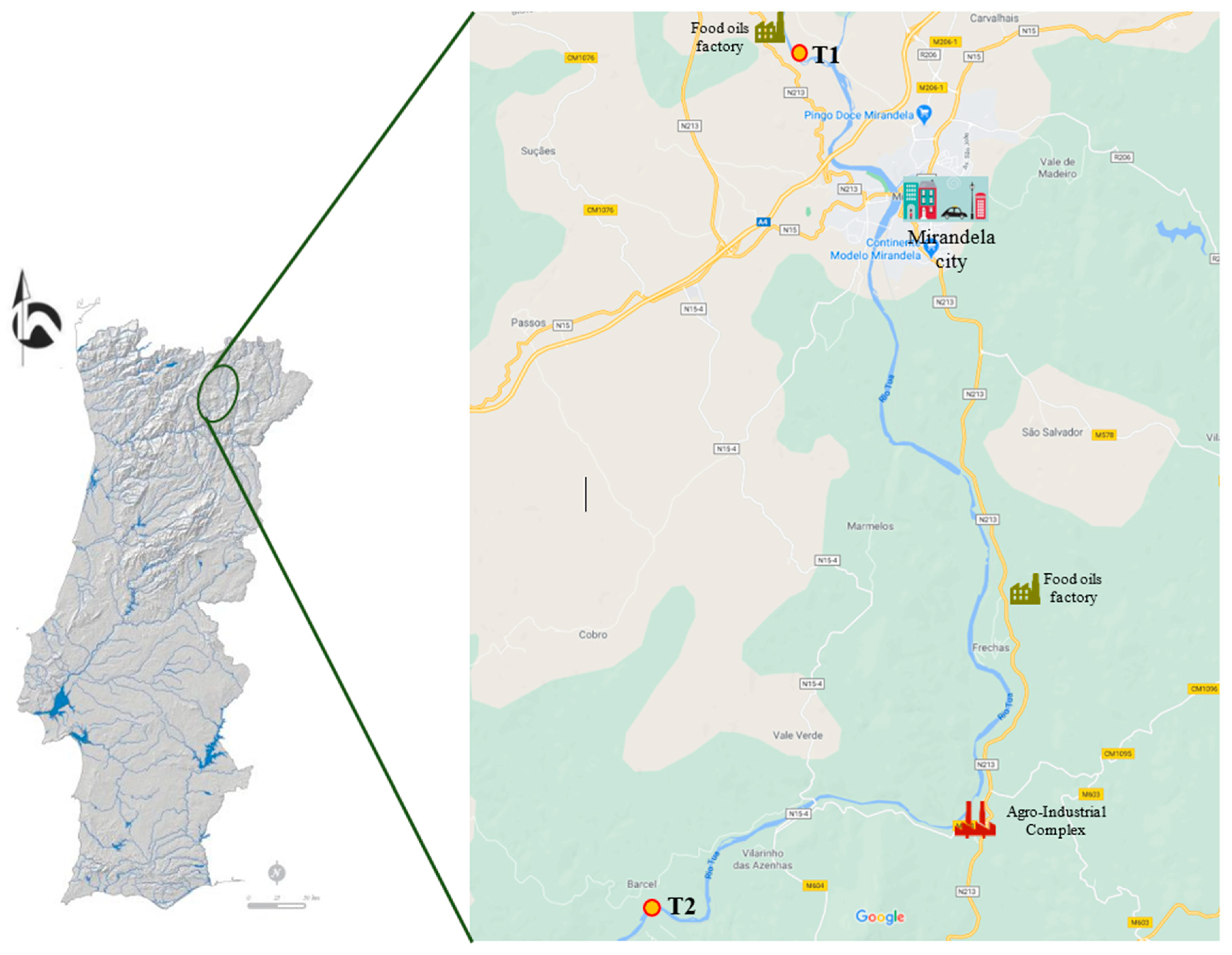
2.1. Study Area

The Tua River is one of the Douro River’s main tributaries (affluent of the right bank) and results from the merging of the Tuela and Rabaçal Rivers, both born in Spain. The Tua River is formed only in Portuguese territory, 4 km upstream from the city of Mirandela, where it starts its journey, until it meets the Douro River (approximately 40 km in length) (Figure 1).
The Tua River basin’s hydrological regime presents significant interannual variability, with an average annual runoff of 988.1 hm3. Intra-annual variability also occurs, with high values in winter and low values in summer (on average it varies between 4 hm3 in August and 277 hm3 in January). The basin area in Portugal is 3,122.80 km2 and in Spain is 690.742 km2, making a total of 3,813.540 km2. The Tua basin is subject to relatively high organic loads, especially the Tua River, coming from agriculture, urban agglomerations, and industrial activities (factories producing and processing food oils, and others that have set up at an agro-industrial complex—wool, nuts, and a regional slaughterhouse), resulting in high biological oxygen demand (BOD), chemical oxygen demand (COD) and total suspended solids (TSS). In this river basin, there is only one small town (Valpaços) upstream from the T1 site (approximately 12 km), with 16,882 inhabitants. The T2 site is influenced by the city of Valpaços (Rabaçal River), Vinhais (9066 inhabitants, Tuela River) and Mirandela (23,850 inhabitants, Tua River).
Phenomena of eutrophication and oil stains over large areas of its waters have been recurrent since 2017 (Portuguese Environment Agency—APA). The fires of 2013 and 2016 also left 5000 tons of hazardous waste from the agro-industrial complex. Additionally, the presence of several hydroelectric dams results in loss of connectivity for aquatic communities and inflow regulation, which, especially in the summer, can lead to the intensification of eutrophication phenomena.

2.2. Sample Processing and Isolation

The water samples were collected at two sites located in the Tua River basin (Rabaçal River T1: 41°30′45.821” N; 7°12’32.92” W, and Tua River T2: 41°24’18.69” N; 7°9’38.93” W), in two seasons, the summer and autumn of 2018 (Figure 1). Two replicates of water were collected in 1 L sterile glass bottles, stored in a cold container, and transported to the Laboratory of Medical Microbiology in Trás-os-Montes e Alto Douro University (UTAD), Vila Real, Portugal. For each replicate sample, three volumes of each were analyzed. The detection and quantification of bioindicators was performed by the filter membrane method. Briefly, 100 mL of water samples were filtered with nitrocellulose membrane filters (0.45 µm pore size) (Millipore, Watford, UK). The filter was put on solid culture media with selective and differential for growth and development of Aeromonas spp. and Pseudomonas spp. (Glutamate Starch Phenol red (GSP) agar, Oxoid Thermo Scientific, Oxoid, UK), supplemented with 2 µg/mL of Imipenem (GSP–IMP) to select for potential carbapenemases producers. For each sampling site (T1 and T2) membranes were also placed on plates without antibiotic in order to estimate the proportion of resistant Aeromonas. Plates were incubated at 30 °C (GSP plates) for 18 to 24 h. From each plate, up to two to five colonies were identified based on colony morphology on GSP and GSP–IMP agar, with Aeromonas spp. presenting a yellow colour and Pseudomonas spp. presenting a red colour. Yellow colonies were selected and purified for further confirmation. Once confirmed, they were stored in aliquots of Brain Heart Infusion (BHI) medium with 17% (v/v) glycerol at −80 °C.

2.3. Identification of Isolates

All the presumptive Aeromonas isolates were identified by classical biochemical methods (indole, Voges–Proskauer, methyl red, citrate reactions, gelatin liquefaction, nitrate reduction, urease test, glucose oxidation and carbohydrate fermentations were determined), Gram-negative staining, the presence of normally positive cytochrome oxidase, catalase reaction, and growth in nutritive broth at 0% to grow in the presence of vibriostatic factor O/129 [23,31]. Additionally, the commercial identification system API 20NE (bioMérieux, https://www.biomerieux.com/ (accessed on 15 January 2021)) was used. Strains were maintained on Tryptone Soya Agar (TSA) (Thermo Scientific, Oxoid, UK).

2.4. Antimicrobial Susceptibility Testing

Susceptibility to antimicrobial agents was performed by the disk-diffusion technique of Kirby–Bauer on Mueller–Hinton agar plates (Oxoid Basingstoke, Oxoid, UK) with inocula adjusted to an optical density of 0.5 McFarland standard units, according to the Performance Standards for Antimicrobial Susceptibility Testing. Aeromonas isolates were tested against 19 antibiotics according to the Clinical Laboratory Standards Institute guidelines [32,33]. The following disks (Oxoid Basingstoke, Oxoid, UK) were used. β-lactam antibiotics tested included penicillins (aminopenicillins, carboxypenicillins and ureidopenicillins), cephalosporins (1st and 3rd generations), monobactams and carbapenems, namely amoxicillin (AML 10 μg), amoxicillin/clavulanic acid (AMC 30 μg), piperacillin (PRL 100 μg), piperacillin/tazobactam (TZP 110 μg), ticarcillin (TIC 75 μg), ticarcillin/clavulanic acid (TIM 85 μg), cephalothin (KF 30μg), cefotaxime (CTX 30 μg), aztreonam (ATM 30 μg), and imipenem (IMP 10 μg). Additionally, another 6 classes of antibiotics were tested: quinolones (nalidixic acid—NA 30 μg, ciprofloxacin—CIP 5 μg), aminoglycosides (gentamicin—CN 10 μg, kanamycin—K 30 μg, tobramycin—TOB 10 μg), tetracyclines (tetracycline—TE 30 μg), fosfomycin (FOS 50 μg), amphenicols (chloramphenicol—C 30 μg), and the combination sulfamethoxazole/trimethoprim (SXT 25 μg). Isolates were classified as sensitive (S) or resistant (R) based on size of the zone of bacteria growth inhibition, according to the Clinical and Laboratory Standard Institute (CLSI) recommendations, after 24 ± 2 h incubation at 30 °C [33]. Multidrug-resistance (MDR) is defined as acquired non-susceptibility to at least one agent in three or more antimicrobial categories [34]. Escherichia coli ATCC 25922 was used as a quality control strain.

2.5. Statistical Analysis

Differences between antimicrobial resistance rates of Aeromonas spp. isolates from different river locations (sites T1 and T2), and the frequency of resistance isolates among different seasons (summer and autumn) were evaluated through Tukey’s test using IBM SPSS statistics v.22.0 software (SPSS Inc., Chicago, IL, USA). Statistical calculations were based on a confidence level of ≥95%; p-value < 0.05 was considered statistically significant.

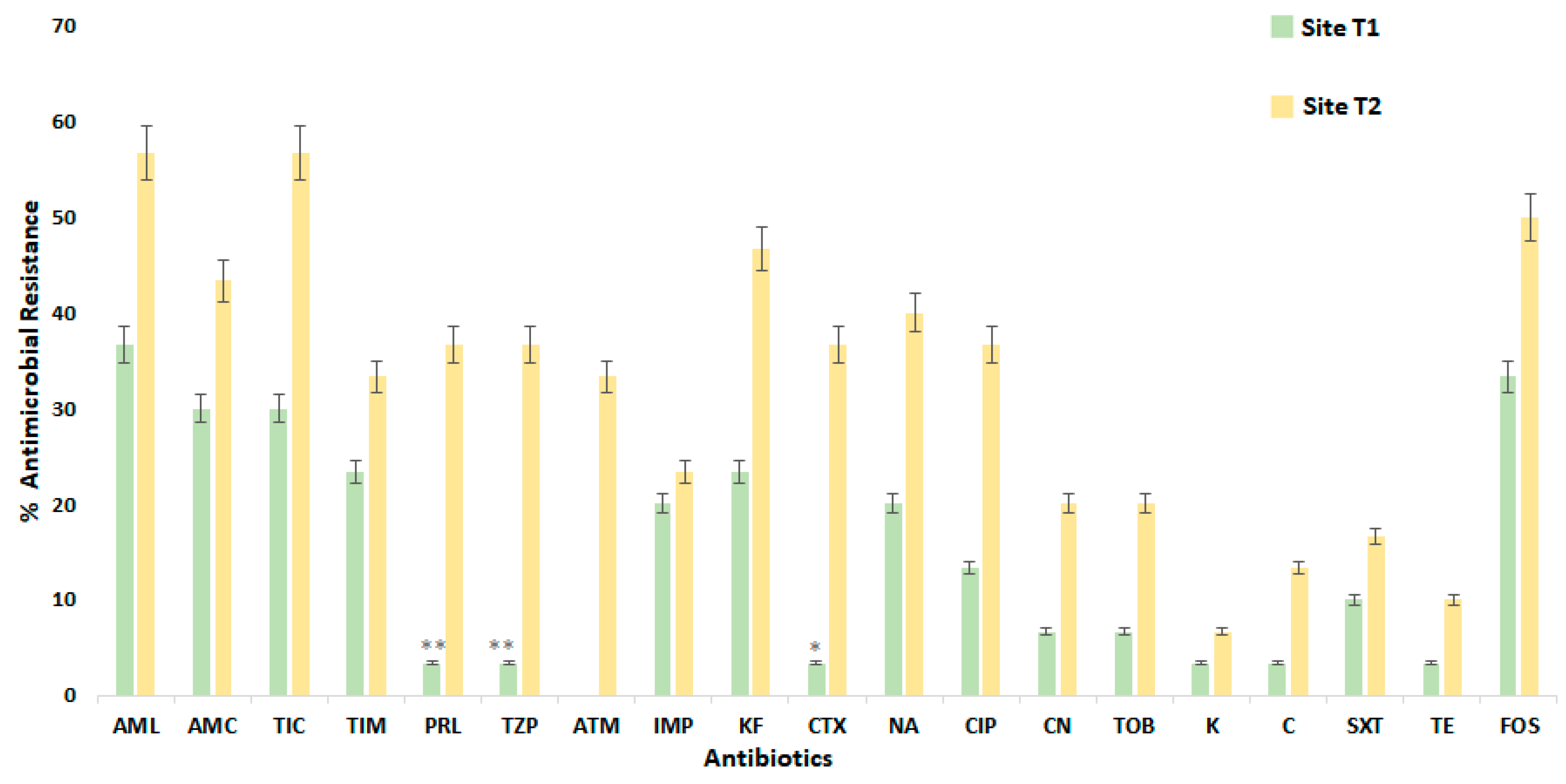
3. Results and Discussion

Although the Aeromonas genus is autochthonous in the aquatic environment, and easy to detect in any water type, they have received increasing attention as opportunistic pathogens [2,35,36,37]. Aeromonas spp. are important pathogens of fish and can cause diseases in humans, as well as in wild animals [19,37].
In the present study, 30 Aeromonas isolates were collected from the sampling survey in the Tua River basin, in summer and in autumn seasons (2018). Specifically, 13 strains were isolated from the site T1 and 17 from the downstream site T2 (Figure 1). All isolates were tested for resistance profile to a panel of 19 antibiotics, namely 20 Aeromonas isolates obtained from GSP agar plates, and 10 Aeromonas isolates from GSP–IMP agar plates. The proportions of antimicrobial resistance of 30 Aeromonas strains are detailed in Figure 2.
Overall, the antibiotic resistance for aeromonads was high and was as follows: fosfomycin (FOS) 83.33% (25/30); nalidixic acid (NA) 60% (18/30); cefotaxime (CTX) 40% (12/30); gentamicin (CN) 26.67% (8/30); tobramycin (TOB) 26.67% (8/30); cotrimoxazole (SXT) 26.67% (8/30); chloramphenicol (C) 16.67% (5/30); and tetracycline (TE) 13.33% (4/30). It is interesting to note that the highest incidence of resistance of Aeromonas spp. isolates was to β-lactam antibiotics, namely to amoxicillin (AML) (93.33%) and to ticarcillin (TIC) (83.33%) (Figure 2). These findings are consistent with those of a previous study which demonstrated that Aeromonas spp. are endogenously resistant to β-lactams, therefore representing a potential risk to public health. β-lactam antibiotics are the most broadly used antibiotics worldwide since they have a broad spectrum of antibacterial activity [28,38,39,40,41,42]. This resistance occurs due to the presence of an unstable β-lactam ring in the structure of β-lactam antibiotics, which are susceptible to bacterial hydrolysis by chromosomal β-lactamases produced by Aeromonas that are easily eliminated [43].
Nevertheless, the combination of an aminopenicillin and a carboxipenicillin with a β-lactamases inhibitor was effective in reducing resistance, as shown by the decrease in the proportion of resistant strains: 93.33% (amoxicillin) versus 73.33% (amoxicillin/clavulanic acid); and 83.33% (ticarcillin) versus 56.67% (ticarcillin/clavulanic acid) (Figure 2). Some of the nalidixic acid-resistant strains were also susceptible to fluoroquinolones (20%). Indeed, the resistance profile of β-lactams occurs because enzymes, named β-lactamases, produced by a large variety of Gram-negative and -positive bacteria are capable of hydrolyzing the β-lactam ring of antibiotics such as penicillins, cephalosporins and aztreonam [44]. Thus, the combination of aminopenicillin with clavulanic acid increases the effectiveness (reduces the resistance profile) by inhibiting the β-lactamases, as evidenced in other studies [45].
Resistance to a first-generation cephalosporin (cephalothin) was measured in 70% of the isolates and for cefotaxime, a third-generation cephalosporin, resistance was measured in 40%. Aztreonam, a monobactam antibiotic, was more effective against these bacteria, with only 33.33% of the isolates being resistant. It should be noted that the occurrence of resistance to imipenem, an antibiotic belonging to the carbapenem group, was observed in 43.33% of isolates. Among aminoglycosides, resistant strains were between 10% (kanamycin) to 26.67% (gentamicin and tobramycin). Although the susceptibility of Aeromonas strains to fluoroquinolones has been observed in other studies [19,46], in the present study, resistance up to 50% to ciprofloxacin was observed. Likewise, more than 70% of the Aeromonas isolates were found to be susceptible to chloramphenicol, trimethoprim-sulfamethoxazole and tetracycline (Figure 2).
The proportions of antimicrobial-resistant Aeromonas spp. isolated from the T1 (n = 13; nine grown in GSP and four in GSP–IMP) and T2 (n = 17; 11 grown in GSP and six in GSP–IMP) river locations are summarized in Figure 3. Generally, the pattern of antimicrobial-resistant Aeromonas spp. showed that high values were observed on site T2. Indeed, the T2 site was the one where the most resistant isolates were observed (32.46%), when compared with the T1 site (14.39%). Likewise, the percentages of fosfomycin resistance were markedly higher among isolates derived from the T2 site than among those obtained from the T1 site. It must be pointed out that from antibiotics employed in clinical practice, for human and veterinary medicine fosfomycin is classified as a “critically important antimicrobial” and a “highly important antimicrobial”, respectively [47,48]. It is noteworthy that it is precisely this site (T2) that presents the greatest anthropogenic pressure, namely, related to the production and refining of food oils and the agro-industrial complex, and all with the urban effluent discharged by the Mirandela city. The T2 site was also heavily affected by the fires of 2013 and 2016 in the agro-industrial complex that left 5000 tons of hazardous waste that continues to be released into the aquatic ecosystem whenever rainfall occurs.
Rivers are considered reservoirs of MDR aeromonads, since they receive water from a wide range of environments, like wastewater treatment plants (WWTPs), industrial effluents, agricultural activities, hospital sewage or animal production effluents [19,37]. The incidence of Aeromonas spp. in wastewater was observed to be high, providing an ideal setting for the acquisition and dissemination of antibiotic resistance mechanisms [29,30,39,40].
Some differences were observed in site T1, between Aeromonas spp. isolates at summer (four strains grown in GSP and two grown in GSP–IMP) and autumn (five grown in GSP and two grown in GSP–IMP) seasons, although they were not statistically significant (Figure 4). In the summer season, a lower percentage of antimicrobial resistance to β-lactams was observed in site T1 (19.25 ± 15.93%) than site T2 (31.17 ± 5.60%) (Figure 4). On the other hand, in site T1 none of the isolates were resistant to aminoglycosides (K) and amphenicos (C) in autumn season (Figure 4). Conversely, in T2 (in summer, four strains grown in GSP and three grown in GSP–IMP; in autumn, seven strains grown in GSP and three grown in GSP–IMP) this absence of resistance was observed in summer. In this work, the percentage of CIP unsusceptible Aeromonas spp. strains was generally lower in comparison with previous studies [29,49,50].
Comparing two sampling periods (summer vs. autumn), it was observed that the resistance to antibiotics increased in autumn, being 1.3-fold higher (Figure 4). However, it should be noted that the analyzed summer period corresponded to the end of spring/early summer when the flow rates were still relatively high after a very rainy April, a relatively dry May and a very rainy June (Portuguese Institute of the Sea and the Atmosphere—IPMA). In contrast, September (the month of autumn sampling) was classified as extremely hot and extremely dry. According to the IPMA, this month was the warmest since 1931 and the second driest in the last 30 years. Thus, and as would be expected, the absence of precipitation, with a direct consequence in the drastic reduction in flows, was the main factor that contributed to a greater concentration of pollution in the river (less capacity for self-purification). Therefore, a high percentage of resistant bacteria could be found, increasing the risk to public health due to the direct use of water in its different applications (human and animal consumption, irrigation, bathing). To highlight this, it is precisely in the months of July, August and part of the month of September that the use of this essential resource and pollution are felt most intensely, due to a seasonal increase in the emigrant population, which is more pronounced in the northern and central regions of Portugal, often doubling the resident population.
However, Knapp et al. [51] showed that it is precisely in periods of greater precipitation that the potential for human exposure to ARB increases, since it is just in these seasons that the ARGs are found to be more equally distributed, both in the watercourse and in the water column. A wide variety of human activities are generally associated with strong impacts on the environment. The stretches of rivers that run through cities are often used as receiving bodies for treated and untreated urban wastewater worldwide [52,53,54]. Some of these rivers, contaminated by man-made sewers and animal effluents, are among the most extreme examples of ecosystems disturbed by anthropogenic activities. General treatment processes to improve water quality in WWTPs, using primary and secondary or biological treatments, are not very efficient in reducing Aeromonas spp. concentration [9,55,56]. Nevertheless, using tertiary treatments, such as chemical (ozone, chlorination), physical (ultraviolet radiation), and natural tertiary treatments such as lagooning, these bacteria can be completely eliminated [57,58,59]. Thus, it would be important to carry out further studies to assess antibiotic resistance variation with the seasons because of the results obtained here.
The results of disk-diffusion phenotypical resistance tests showed a high number of MDR aeromonads among all isolated Aeromonas spp. strains (Figure 5). To further gain knowledge on the MDR in the aquatic environmental bacteria, 25 out of 30 Aeromonas isolates showed an MDR profile to 19 antibiotics, where 83.33% of the strains were resistant to more than three antibiotic classes, indicating a high level of multi-resistance in the Tua River basin (Figure 5). Previous studies [18,60,61,62,63,64] reported β-lactams and quinolone-resistant isolates of Aeromonas spp. recovered from freshwater, fishes, and humans. To our knowledge, this was the first report that showed multidrug-resistant Aeromonas spp. isolated from the Tua River, representing a potential risk to the population since an appreciable part of the population residing in the Tua River Basin still uses fish and river mussels in their daily diet. Foodborne diseases (FD) are a priority in public health issues around the world due to their incidence and mortality [64]. The genus Aeromonas is regarded not only as an important disease-causing pathogen of cold-blooded species but also as the etiologic agent responsible for a variety of infectious complications in both immunocompetent and immunocompromised persons [23]. Carbapenems are often used as “last-line agents” or “antibiotics of last resort” for the treatment of severe infections due to multidrug-resistant hospital-acquired bacteria [50]. However, the appearance of MDR seriously threatens this class of lifesaving antimicrobials.
The presence of multidrug-resistant bacteria in water has resulted from the indiscriminate use of antibiotics in the last decades, which has exerted a selective pressure on bacteria from the environment, together with effluent treatment systems that are inefficient in the antibiotic removal process [65,66,67,68]. Antibiotics and resistant bacteria are entering our local waterways and have the potential to influence biotic processes. Another important point is associated with the transfer of resistant genes from not-pathogenic bacteria to pathogenic bacteria, and on to humans interacting with the aquatic environment [69,70,71].

4. Conclusions

From the results of this study, it was clear that there was a pool of MDR aeromonads in the Tua River basin. These results, together with the resistance patterns of Aeromonas spp. to antibiotic tests, suggest that Aeromonas spp. can be effective bioindicator organisms for monitoring antimicrobial resistance in rivers. This knowledge is essential to manage and mitigate potential risks to human health, emphasizing the need for predicting and preventing the spread of antibiotic-resistant pathogenic aeromonads. A continuous monitoring surveillance in aquatic systems is imperative, considering the interactions between the key elements (geographical, ecological, human activities and the food-agricultural components) within the “One Health—One World” approach.
In view of the results obtained in this study, it would be important to assess, in the long term, the seasonal variation in antibiotic resistance. Further studies are also needed to investigate aquatic animals for the possible presence of antibiotic resistant bacteria, as well as to understand the role of ARG and the mobile genetic element relatedness of these isolates.

Author Contributions

In the overall context of this paper, the contributions are as follows: A.T., S.V. and M.J.S. made the sampling collection. S.G., C.F., S.M. and M.J.S. isolated the bacteria and performed the laboratory measurements. C.F., S.V. and M.J.S. made substantial contributions to conception and design and drafted the manuscript. S.G., C.F., S.V. and M.J.S. edited the draft. E.C. and A.T. provided additional information, local insights, and supplementary references. All authors provided critical feedback and helped shape the research, analysis, and manuscript. All authors have read and agreed to the published version of the manuscript.

Funding

This research was supported by the Portuguese Foundation for Science and Technology (FCT) and the Operational Competitiveness Programme (COMPETE), under the projects UIDB/04033/2020 (CITAB-UTAD) and the UIDB/00690/2020 (CIMO-IPB).

Institutional Review Board Statement

Not applicable.

Informed Consent Statement

Not applicable.

Data Availability Statement

Not applicable.

Conflicts of Interest

The authors declare that the research was conducted in the absence of any commercial or financial relationships that could be construed as a potential conflict of interest.

Abbreviations

AMCAmoxicillin/clavulanic acid
AMRAntimicrobial resistance
AMLAmoxicillin
APAEnvironment Portuguese Agency
ARBAntibiotic resistant bacteria
ARGAntibiotic resistance genes
ATMAztreonam
BHIBrain Hearth Infusion
BODBiological oxygen demand
CChloramphenicol
CIPCiprofloxacin
CLSIClinical Laboratory Standard Institute
CNGentamicin
CODChemical oxygen demand
CTXCefotaxime
FDFoodborne Diseases
ESBLExtended-Spectrum β-lactamases
FOSFosfomycin
GSPGlutamate Starch Phenol
GSP–IMPGlutamate Starch Phenol imipenem
IMPImipenem
IPMAPortuguese Institute of the Sea and the Atmosphere
KKanamycin
KFCephalothin
MDRMultidrug-resistant
NANalidixic acid
PRLPiperacillin
SXTCotrimoxazole
SXTTrimethoprim/sulfamethoxazole
T1Site T1 (Chelas)
T2Site T2 (Barcel)
TETetracycline
TICTicarcillin
TIMTicarcillin/clavulanic acid
TOBTobramycin
TSATryptone Soya Agar
TSSTotal suspended solids
TZPPiperacillin/tazobactam
WWTPWastewater treatment plant

References

  1. Páll, E.; Niculae, M.; Kiss, T.; Şandru, C.D.; Spînu, M. Human impact on the microbiological water quality of the rivers. J. Med. Microbiol. 2013, 62, 1635–1640. [Google Scholar] [CrossRef]
  2. Felis, E.; Kalka, J.; Sochacki, A.; Kowalska, K.; Bajkacz, S.; Harnisz, M.; Korzeniewska, E. Antimicrobial pharmaceuticals in the aquatic environment—Occurrence and environmental implications. Eur. J. Pharmacol. 2020, 866, 172813. [Google Scholar] [CrossRef]
  3. World Health Organization (WHO). Antimicrobial Resistance: Global Report on Surveillance. 2018. Available online: https://www.who.int/medicines/areas/rational_use/who-amr-amc-report20181109.pdf (accessed on 3 February 2021).
  4. Baquero, F.; Martínez, J.; Cantón, R. Antibiotics and antibiotic resistance in water environments. Curr. Opin. Biotechnol. 2008, 19, 260–265. [Google Scholar] [CrossRef] [PubMed]
  5. Wright, G.D. Antibiotic resistance in the environment: A link to the clinic? Curr. Opin. Microbiol. 2010, 13, 589–594. [Google Scholar] [CrossRef] [PubMed]
  6. Suzuki, S.; Ogo, M.; Koike, T.; Takada, H.; Newman, B. Sulfonamide and tetracycline resistance genes in total- and culturable-bacterial assemblages in South African aquatic environments. Front. Microbiol. 2015, 6, 796. [Google Scholar] [CrossRef] [PubMed]
  7. Kraemer, S.A.; Ramachandran, A.; Perron, G.G. Antibiotic Pollution in the Environment: From Microbial Ecology to Public Policy. Microorganisms 2019, 7, 180. [Google Scholar] [CrossRef] [Green Version]
  8. Vipin, C.; Saptami, K.; Fida, F.; Mujeeburahiman, M.; Rao, S.S.; Athmika Arun, A.B.; Rekha, P.D. Potential synergistic activity of quercetin with antibiotics against multidrug-resistant clinical strains of Pseudomonas aeruginosa. PLoS ONE 2020, 15, e0241304. [Google Scholar] [CrossRef]
  9. He, Y.; Yuan, Q.; Mathieu, J.; Stadler, L.; Senehi, N.; Sun, R.; Alvarez, P.J.J. Antibiotic resistance genes from livestock waste: Occurrence, dissemination, and treatment. NPJ Clean Water 2020, 3, 4. [Google Scholar] [CrossRef] [Green Version]
  10. Michael, I.; Rizzo, L.; McArdell, C.S.; Manaia, C.M.; Merlin, C.; Schwartz, T.; Dagot, C.; Fatta-Kassinos, D. Urban wastewater treatment plants as hotspots for the release of antibiotics in the environment: A review. Water Res. 2013, 47, 957–995. [Google Scholar] [CrossRef] [Green Version]
  11. Ghaderpour, A.; Ho, W.S.; Chew, L.L.; Bong, C.W.; Chong, V.C.; Thong, K.L.; Chai, L.C. Diverse and abundant multi-drug resistant E. coli in Matang mangrove estuaries. Malaysia Front. Microbiol. 2015, 10. [Google Scholar] [CrossRef] [Green Version]
  12. Figueira, V.; Vaz-Moreira, I.; Silva, M.; Manaia, C.M. Diversity and antibiotic resistance of Aeromonas spp. in drinking and waste water treatment plants. Water Res. 2011, 45, 5599–5611. [Google Scholar] [CrossRef] [PubMed]
  13. Henriques, I.S.; Fonseca, F.; Alves, A.; Saavedra, M.J.; Correia, A. Occurrence and diversity of integrons and β-lactamase genes among ampicillin-resistant isolates from estuarine waters. Res. Microbiol. 2006, 157, 938–947. [Google Scholar] [CrossRef]
  14. Picão, R.C.; Poirel, L.; Demarta, A.; Silva, C.S.F.; Corvaglia, A.R.; Petrini, O.; Nordmann, P. Plasmid-mediated quinolone resistance in Aeromonas allosaccharophila recovered from a Swiss lake. J. Antimicrob. Chemother. 2008, 62, 948–950. [Google Scholar] [CrossRef] [Green Version]
  15. Schmidt, A.S.; Bruun, M.S.; Dalsgaard, I.; Larsen, J.L. Incidence, distribution, and spread of tetracycline resistance determinants and integron-associated antibiotic resistance genes among motile aeromonads from a fish farming environment. Appl. Environ. Microbiol. 2001, 67, 5675–5682. [Google Scholar] [CrossRef] [PubMed] [Green Version]
  16. Saavedra, M.J.; Guedes-Novais, S.; Alves, A.; Rema, P.; Tacão, M.; Correia, A.; Martínez-Murcia, A. Resistance to beta-lactam antibiotics in Aeromonas hydrophila isolated from rainbow trout (Oncorhynchus mykiss). Int. Microbiol. 2004, 7, 207–211. [Google Scholar]
  17. Sørum, H. Antimicrobial Drug Resistance in Fish Pathogens. In Antimicrobial Resistance in Bacteria of Animal Origin; Aarestrup, F.M., Ed.; American Society for Microbiology Press: Washington, DC, USA, 2006; pp. 213–238. [Google Scholar]
  18. Carvalho, M.J.; Martínez-Murcia, A.; Esteves, A.C.; Correia, A.; Saavedra, M.J. Phylogenetic diversity, antibiotic resistance and virulence traits of Aeromonas spp. from untreated waters for human consumption. Int. J. Food Microbiol. 2012, 159, 230–239. [Google Scholar] [CrossRef]
  19. Fernández-Bravo, A.; Figueras, M.J. An Update on the genus Aeromonas: Taxonomy, epidemiology, and pathogenicity. Microorganisms 2020, 8, 129. [Google Scholar] [CrossRef] [Green Version]
  20. Figueras, M.J.; Alperi, A.; Saavedra, M.J.; Ko, W.C.; Gonzalo, N.; Navarro, M.; Martínez-Murcia, A.J. Clinical relevance of the recently described species Aeromonas aquariorum. J. Clin. Microbiol. 2009, 47, 3742–3746. [Google Scholar] [CrossRef] [Green Version]
  21. Beaz-Hidalgo, R.; Martínez-Murcia, A.; Figueras, M.J. Reclassification of Aeromonas hydrophila subsp. dhakensis Huys and Aeromonas aquariorum Martínez-Murcia et al. 2008 as Aeromonas dhakensis sp. nov. comb nov. and emendation of the species Aeromonas hydrophila. Syst. Appl. Microbiol. 2013, 36, 171–176. [Google Scholar] [CrossRef] [PubMed]
  22. Chen, P.L.; Lamy, B.; Ko, W.C. Aeromonas dhakensis, an increasingly recognized human pathogen. Front. Microbiol. 2016, 7, 793. [Google Scholar] [CrossRef]
  23. Janda, J.M.; Abbott, S.L. The genus Aeromonas: Taxonomy, pathogenicity, and infection. Clin. Microbiol. Rev. 2010, 23, 35–73. [Google Scholar] [CrossRef] [PubMed] [Green Version]
  24. Prestinaci, F.; Pezzotti, P.; Pantosti, A. Antimicrobial resistance: A global multifaceted phenomenon. Pathog. Glob Health 2015, 109, 309–318. [Google Scholar] [CrossRef] [PubMed] [Green Version]
  25. European Court of Auditors. Addressing Antimicrobial Resistance: Progress in the Animal Sector, But this Health Threat Remains A Challenge for the EU. Special Report Nº 21.41. 2019. Available online: http://europeanmemoranda.cabinetoffice.gov.uk/memorandum/court-of-auditors-special-report-no-2019-21-addressing-antimicrobial-resistance-progress-in-the-animal (accessed on 24 February 2021).
  26. Jobbins, S.E.; Alexander, K.A. Whence they came—Antibiotic-resistant Escherichia coli in African wildlife. J. Wildl. Dis. 2015, 51, 811–820. [Google Scholar] [CrossRef] [PubMed]
  27. Furness, L.E.; Campbell, A.; Zhang, L.; Gaze, W.H.; Robbie, A.; McDonald, R.A. Wild small mammals as sentinels for the environmental transmission of antimicrobial resistance. Environ. Res. 2017, 154, 28–34. [Google Scholar] [CrossRef]
  28. Usui, M.; Tagaki, C.; Fukuda, A.; Okubo, T.; Boonla, C.; Suzuki, S. Use of Aeromonas spp. as general indicators of antimicrobial susceptibility among bacteria in aquatic environments in Thailand. Front. Microbiol. 2016, 7, 710. [Google Scholar] [CrossRef] [PubMed] [Green Version]
  29. Varela, A.R.; Nunes, O.C.; Manaia, C.M. Quinolone resistant Aeromonas spp. as carriers and potential tracers of acquired antibiotic resistance in hospital and municipal wastewater. Sci. Total Environ. 2016, 15, 665–671. [Google Scholar] [CrossRef] [PubMed]
  30. Baron, S.; Granier, S.A.; Larvor, E.; Jouy, E.; Cineux, M.; Wilhelm, A.; Gassilloud, B.; Le Bouquin, S.; Kempf, I.; Chauvin, C. Aeromonas diversity and antimicrobial susceptibility in freshwater-an attempt to set generic epidemiological cut-off values. Front. Microbiol. 2017, 8, 503. [Google Scholar] [CrossRef] [Green Version]
  31. Abbott, S.L.; Cheung, W.K.W.; Janda, J.M. The genus Aeromonas: Biochemical characteristics, atypical reactions, and phenotypic identification schemes. J. Clin. Microbiol. 2003, 41, 2348–2357. [Google Scholar] [CrossRef] [PubMed] [Green Version]
  32. Clinical and Laboratory Standards Institute (CLSI). Performance Standards for Antimicrobial Susceptibility Testing, 30th ed.; Clinical and Laboratory Standards Institute (CLSI): Wayne, PA, USA, 2020. [Google Scholar]
  33. Clinical and Laboratory Standards Institute (CLSI). Methods for Antimicrobial Dilution and Disk Susceptibility Testing of Infrequently Isolated or Fastidious Bacteria, 3rd ed.; CLSI Guideline M45; Clinical and Laboratory Standards Institute: Wayne, PA, USA, 2016. [Google Scholar]
  34. Magiorakos, A.P.; Srinivasan, A.; Carey, R.B.; Carmeli, Y.; Falagas, M.E.; Giske, C.G.; Harbarth, S.; Hindler, J.F.; Kahlmeter, G.; Olsson-Liljequist, B.; et al. Multidrug-resistant, extensively drug-resistant and pandrug-resistant bacteria: An international expert proposal for interim standard definitions for acquired resistance. Clin. Microbiol. Infect. 2011, 268–282. [Google Scholar] [CrossRef] [PubMed] [Green Version]
  35. Ko, W.C.; Yu, K.W.; Liu, C.Y.; Huang, C.T.; Leu, H.S.; Chuang, Y.C. Increasing antibiotic resistance in clinical isolates of Aeromonas strains in Taiwan. Antimicrob. Agents Chemother. 1996, 40, 1260–1262. [Google Scholar] [CrossRef] [PubMed] [Green Version]
  36. Saengsitthisak, B.; Chaisri, W.; Punyapornwithaya, V.; Mektrirat, R.; Klayraung, S.; Bernard, J.K.; Pikulkaew, S. Occurrence and antimicrobial susceptibility profiles of multidrug-resistant Aeromonads isolated from freshwater ornamental fish in chiang Mai Province. Pathogens 2020, 9, 973. [Google Scholar] [CrossRef] [PubMed]
  37. Dias, C.; Borges, A.; Saavedra, M.J.; Simões, M. Biofilm formation and multidrug-resistant Aeromonas spp. from wild animals. J. Glob. Antimicrob. Resist. 2018, 12, 227–234. [Google Scholar] [CrossRef] [PubMed]
  38. Lupo, A.; Coyne, S.; Berendonk, T.U. Origin and evolution of antibiotic resistance: The common mechanisms of emergence and spread in water bodies. Front. Microbiol. 2012, 3, 18. [Google Scholar] [CrossRef] [Green Version]
  39. Holmes, P.; Nicolls, L.M.; Sartory, D.P. The ecology of mesophilic Aeromonas in the aquatic environment. In The Genus Aeromonas; John Wiley & Sons Ltd.: West Sussex, UK, 1996; pp. 127–150. [Google Scholar]
  40. McLellan, S.L.; Huse, S.M.; Mueller-Spitz, S.R.; Andreishcheva, E.N.; Sogin, M.L. Diversity and population structure of sewage-derived microorganisms in wastewater treatment plant influent. Environ. Microbiol. 2010, 12, 378–392. [Google Scholar] [CrossRef] [Green Version]
  41. Dias, C.; Mota, V.; Martinez-Murcia, A.; Saavedra, M.J. Antimicrobial resistance patterns of Aeromonas spp. isolated from ornamental fish. J. Aquac. Res. Dev. 2012, 3, 3. [Google Scholar] [CrossRef] [Green Version]
  42. Dias, C.; Serra, C.R.; Simões, L.C.; Simões, M.; Martinez-Murcia, A.; Saavedra, M.J. Extended-spectrum β-lactamase and carbapenemase-producing Aeromonas species in wild animals from Portugal. Vet. Rec. 2014, 24, 532. [Google Scholar] [CrossRef] [PubMed] [Green Version]
  43. Goňi-Urriza, U.; Capdepuy, M.C.; Arpin, C.; Raymond, N.; Caumette, P.; Quentin, C. Impact of an urban effluent on antibiotic resistance of riverine Enterobacteriaceae and Aeromonas spp. Appl. Environ. Microbiol. 2000, 66, 125–132. [Google Scholar] [CrossRef] [Green Version]
  44. Rawat, D.; Nair, D. Extended-spectrum β-lactamases in Gram Negative Bacteria. J. Glob. Infect. Dis. 2010, 2, 263–274. [Google Scholar] [CrossRef] [PubMed]
  45. Chen, J.; Shang, X.; Hu, F.; Lao, X.; Gao, X.; Zheng, H.; Yao, W. β-Lactamase Inhibitors: An Update. Mini-Rev. Med. Chem. 2013, 13, 1846–1861. [Google Scholar] [CrossRef]
  46. Codjoe, F.S.; Donkor, E.S. Carbapenem resistance: A review. Med. Sci. 2017, 6, 1. [Google Scholar] [CrossRef] [Green Version]
  47. World Health Organization. Critically Important Antimicrobials for Human Medicine, 6th ed.; World Health Organization: Geneva, Switzerland, 2019. [Google Scholar]
  48. World Organization for Animal Health. OIE List of Antimicrobial Agents of Veterinary Importance; World Organization for Animal Health: Paris, France, 2018. [Google Scholar]
  49. Piotrowska, M.; Przygodzinska, D.; Matyjewicz, K.; Popowska, M. Occurrence and variety of β-Lactamase genes among Aeromonas spp. isolated from urban wastewater treatment plant. Front. Microbiol. 2017, 8, 863. [Google Scholar] [CrossRef] [Green Version]
  50. Picão, R.C.; Cardoso, J.P.; Campana, E.H.; Nicoletti, A.G.; Petrolin, F.V.; Assis, D.M.; Juliano, L.; Gales, A.C. The route of antimicrobial resistance from the hospital effluent to the environment: Focus on the occurrence of KPC-producing Aeromonas spp. and Enterobacteriaceae in sewage. Diagn. Microbiol. Infect. Dis. 2013, 76, 80–85. [Google Scholar] [CrossRef] [PubMed] [Green Version]
  51. Knapp, C.W.; Lima, L.; Olivares-Rieumont, S.; Bowen, E.; Werner, D.; Graham, D.W. Seasonal variations in antibiotic resistance gene transport in the almendares river, havana, cuba. Front. Microbiol. 2012, 3, 396. [Google Scholar] [CrossRef] [Green Version]
  52. Decker, E.H.; Kerkhoff, A.J.; Moses, M.E. Global patterns of city size Distributions and their fundamental drivers. PLoS ONE 2007, 2, e934. [Google Scholar] [CrossRef] [PubMed]
  53. United Nations (UN). Revision of World Urbanization Prospects. 2009. Available online: www.unpopulation.org (accessed on 21 January 2021).
  54. Abraham, W.-R. Megacities as sources for pathogenic bacteria in rivers and their fate downstream. Int. J. Microbiol. 2011, 2011, 1–13. [Google Scholar] [CrossRef] [PubMed]
  55. Salvat, M.J.F.; Ashbolt, N. Aeromonas in Global Water Pathogen Project; University of Alberta: Edmonton, AB, Canada, 2019. [Google Scholar]
  56. He, L.; Du, P.; Chen, Y.; Lu, H.; Cheng, X.; Chang, B.; Wang, Z. Advances in microbial fuel cells for wastewater treatment. Renew. Sustain. Energy Rev. 2017, 71, 388–403. [Google Scholar] [CrossRef]
  57. Hassani, L.; Imziln, B.; Boussaid, A.; Gauthier, M.J. Seasonal incidence of and antibiotic resistance among Aeromonas species isolated from domestic wastewater before and after treatment in stabilization ponds. Microb. Ecol. 1992, 23, 227–237. [Google Scholar] [CrossRef]
  58. Latif-Eugenín, F.; Beaz-Hidalgo, R.; Figueras, M.J. Evaluation of different conditions and culture media for the recovery of Aeromonas spp. from water and shellfish samples. J. Appl. Microbiol. 2016, 121, 883–891. [Google Scholar] [CrossRef] [PubMed]
  59. Latif-Eugenin, F.; Hidalgo, R.B.; Silvera, C.; Fernandez-Cassi, X.; Figueras, M.J. Chlorinated and ultraviolet radiation-treated reclaimed irrigation water is the source of Aeromonas found in vegetables used for human consumption. Environ. Res. 2017, 154, 190–195. [Google Scholar] [CrossRef] [PubMed]
  60. Daskalov, H. The importance of Aeromonas hydrophila in food safety. Food Control. 2006, 17, 474–483. [Google Scholar] [CrossRef]
  61. Lijon, M.B.; Khatun, M.M.; Islam, A.; Khatun, M.M.; Islam, M.A. Detection of multidrug resistance Aeromonas hydrophila in farm raised fresh water prawns. J. Adv. Vet. Anim. Res. 2015, 2, 469–474. [Google Scholar] [CrossRef]
  62. Martínez-Murcia, A.; Beaz-Hidalgo, R.; Navarro, A.; Carvalho, M.J.; Aravena-Román, M.; Correia, A.; Figueras, M.J.; Saavedra, M.J. Aeromonas lusitana sp. nov., isolated from untreated water and vegetables. Curr. Microbiol. 2016, 72, 795–803. [Google Scholar] [CrossRef] [PubMed]
  63. Patil, H.J.; Benet-Perelberg, A.; Naor, A.; Smirnov, M.; Ofek, T.; Nasser, A.; Minz, D.; Cytryn, E. Evidence of increased antibiotic resistance in phylogenetically-diverse Aeromonas isolates from semi-intensive fish ponds treated with antibiotics. Front. Microbiol. 2016, 7, 1875. [Google Scholar] [CrossRef] [Green Version]
  64. Borella, L.; Salogni, C.; Vitale, N.; Scali, F.; Moretti, V.M.; Pasquali, P.; Alborali, G.L. Motile aeromonads from farmed and wild freshwater fish in northern Italy: An evaluation of antimicrobial activity and multidrug resistance during 2013 and 2016. Acta Vet. Scand. 2020, 23, 6. [Google Scholar] [CrossRef]
  65. Bhunia, A.K. Opportunistic and Emerging Foodborne Pathogens: Aeromonas hydrophila, Plesiomonas shigelloides, Cronobacter sakazakii, and Brucella abortus. In Foodborne Microbial Pathogens; Food Science Text Series; Springer: New York, NY, USA, 2018. [Google Scholar]
  66. Hirsch, R.; Ternes, T.; Haberer, K.; Kratz, K.L. Occurrence of antibiotics in the aquatic environment. Sci. Total Environ. 1999, 12, 109–118. [Google Scholar] [CrossRef]
  67. Huang, J.J.; Hu, H.Y.; Lu, S.Q.; Li, Y.; Tang, F.; Lu, Y.; Wei, B. Monitoring and evaluation of antibiotic-resistant bacteria at a municipal wastewater treatment plant in China. Environ. Int. 2012, 42, 31–36. [Google Scholar] [CrossRef]
  68. Vaz-Moreira, I.; Nunes, O.C.; Manaia, C.M. Bacterial diversity and antibiotic resistance in water habitats: Searching the links with the human microbiome. FEMS Microbiol. Rev. 2014, 38, 761–778. [Google Scholar] [CrossRef] [PubMed]
  69. Costanzo, S.D.; Murby, J.; Bates, J. Ecosystem response to antibiotics entering the aquatic environment. Mar. Pollut. Bull. 2005, 51, 218–223. [Google Scholar] [CrossRef]
  70. Bengtsson-Palme, J.; Kristiansson, E.; Larsson, D.G.J. Environmental factors influencing the development and spread of antibiotic resistance. FEMS Microbiol. Rev. 2018, 42, 68–80. [Google Scholar] [CrossRef]
  71. Lima, T.; Domingues, S.; Da Silva, G.J. Manure as a Potential Hotspot for Antibiotic Resistance Dissemination by Horizontal Gene Transfer Events. Vet. Sci. 2020, 7, 110. [Google Scholar] [CrossRef]
Figure 1. Map of the study area and the location of the two sampling sites (T1 and T2) in the Tua River basin. The water samples were collected in two different seasons (summer and autumn).
Water 13 00698 g001
Figure 2. Susceptibility and resistance profiles (%) of Aeromonas spp. (n = 30) isolates to 19 antibiotics. Antibiotic abbreviations: AML—amoxicillin; AMC—amoxicillin/clavulanic acid; TIC—ticarcillin; TIM—ticarcillin/clavulanic acid; PRL—piperacillin; TZP—piperacillin/tazobactam; ATM—aztreonam; IPM—imipenem; KF—cephalothin; CTX—cefotaxime; NA—nalidixic acid; CIP—ciprofloxacin; CN—gentamicin; TOB—tobramycin; K—kanamycin; C—chloramphenicol; SXT—trimethoprim/sulfamethoxazole; TET—tetracycline; and FOS—fosfomycin. Squares of different colors represent the seven families to which the 19 antibiotics belong: ten β- lactams (AML, AMC, TIC, TIM, PRL, TZP, ATM, IMP, KF, CTX), two quinolones (NA, CIP), three aminoglycosides (CN, TOB, K), one amphenicol (C), one sulfonamide (SXT), one tetracycline (TE) and fosfomycin (FOS).
Water 13 00698 g002
Figure 3. Percentage of antimicrobial-resistant bacteria along the river: Site T1 (green) and Site T2 (yellow). Values were expressed as the mean of resistant Aeromonas spp. ± standard deviation, for the same antimicrobial. * and ** indicate significant differences between different locations (sites T1 and T2) within the same antimicrobial (ANOVA followed by Tukey’s test at p < 0.05 and p < 0.01, respectively). Antibiotic abbreviations: AML—amoxicillin; AMC—amoxicillin/clavulanic acid; TIC—ticarcillin; TIM—ticarcillin/clavulanic acid; PRL—piperacillin; TZP—piperacillin/tazobactam; ATM—aztreonam; IPM—imipenem; KF—cephalothin; CTX—cefotaxime; NA—nalidixic acid; CIP—ciprofloxacin; CN—gentamicin; TOB—tobramycin; K—kanamycin; C—chloramphenicol; SXT—trimethoprim/sulfamethoxazole; TET—tetracycline; and FOS—fosfomycin.
Water 13 00698 g003
Figure 4. Percentage of antimicrobial-resistant bacteria for each sampling site (T1 and T2) by seasons (summer—blue color vs. autumn—orange color). Values were expressed as ths mean of resistant Aeromonas spp. ± standard deviation, for the same antimicrobial. No lettering indicates non-significant differences at a 95% confidence level. Antibiotic abbreviations: AML—amoxicillin; AMC—amoxicillin/clavulanic acid; TIC—ticarcillin; TIM—ticarcillin/clavulanic acid; PRL—piperacillin; TZP—piperacillin/tazobactam; ATM—aztreonam; IPM—imipenem; KF—cephalothin; CTX—cefotaxime; NA—nalidixic acid; CIP—ciprofloxacin; CN—gentamicin; TOB—tobramycin; K—kanamycin; C—chloramphenicol; SXT—trimethoprim/sulfamethoxazole; TET—tetracycline; and FOS—fosfomycin.
Water 13 00698 g004
Figure 5. Multidrug-resistance (MDR) in the Tua River, where 25 out of the 30 Aeromonas isolates (red bars) were resistant to three or more antibiotic classes. The grey bars indicate no MDR isolates (5 out of the 30). The green line represents the number of antibiotic classes of resistance.
Water 13 00698 g005
Publisher’s Note: MDPI stays neutral with regard to jurisdictional claims in published maps and institutional affiliations.

