
Evaluation of the Ecological Potential of Microorganisms for Purifying Water with High Iron Content
by
 , and
, and
Ekaterina Pleshakova
1,*, 
Clement Ngun
1,* ,
,
Mikhail Reshetnikov
1,2 and
Maxim Viktorovich Larionov
3 1
Department of Biochemistry and Biophysics, Saratov State University, 410012 Saratov, Russia
2
LUKOIL-Engineering LLC, Kogalymnipineft Branch in Tyumen, Center for the Study of Core and Reservoir Fluids, 628486 Kogalym, Russia
3
Federal State Budgetary Educational Institution of Higher Education, Russian Timiryazev State Agrarian University, 127550 Moscow, Russia
*
Authors to whom correspondence should be addressed.
Water 2021, 13(7), 901; https://doi.org/10.3390/w13070901
Submission received: 25 February 2021
/
Revised: 20 March 2021
/
Accepted: 22 March 2021
/
Published: 26 March 2021
(This article belongs to the Special Issue Environmental Biogeochemistry of Trace Elements in Contaminated Soils and Water)
Abstract
:The ability of a number of microorganisms isolated from highly magnetic soil of the city Mednogorsk to oxidize Fe (II) under conditions of periodic cultivation in a liquid medium was studied. Among the studied microorganisms, two microbial isolates with maximum growth characteristics and iron-oxidizing activity were selected and identified: Bacillus megaterium 69.3 and B. megaterium 69.5. Individual levels of metal resistance of the isolates were determined: maximum tolerated concentration (MTC) for Fe (II) of the isolates B. megaterium 69.3 and B. megaterium 69.5 was 1200 mg L−1, minimum inhibitory concentration (MIC) was 1800 mg L−1. Both microbial isolates actively oxidized Fe (II) by reducing its high concentration in the medium (1.19 g L−1) by 33 and 39% during 14 days of culturing. Total increase in the biomass of B. megaterium 69.3 and B. megaterium 69.5 after 14 days of culturing was 15.3 and 14.7 g L−1; the active parts of the biomass increased 8.7- and 6.9-fold compared to the inoculum dose, respectively. These microbial isolates could be used in future in the biotechnological process of water purification with increased/high levels of Fe (II).
1. Introduction
The problem of water pollution with heavy metals (HMs) is quite acute all over the world, especially in urbanized regions. Among heavy metals, iron and its compounds are considered one of the major pollutants of urban surface waters. Anthropogenic activities serve as a gateway through which iron compounds enter ecosystems: via wastewater and sludge from metallurgical, chemical, petrochemical, and pharmaceutical industries, paint and varnish, textile production, and due to metal corrosion [1]. Data from scientific analysis have shown a violation of iron content quality standards in surface water bodies in the Russian Federation. Iron concentrations 2–2.5-fold higher than the maximum permissible concentration (MPC) have been observed in urban reservoirs of Dzerzhinsky town (Moscow region, Russia) [2]. In the water and silt of mine water-receiving rivers of the Kizelovsky coal basin (Perm Region, Russia), a multiple excess of the MPC for iron was revealed [3]. High concentrations of iron were also found in the rivers of taiga zones of Western Siberia: Bolshoy Yugan, Tara, etc. [4].
In Russia, urban surface water serves as the main source of drinking water. The MPC of total iron in drinking water, according to the Sanitary Rules and Norms, Russia (SanPiN), is 0.3 mg L−1 [5]. Excess iron in drinking water is known to have a negative effect on human health. The negative health implications include dermatitis, allergic reactions, an increase in the size of the liver, a gradual change in the morphological composition of the blood, weight loss, high fatigue, weakness, violations of the normal heart rhythm, memory impairment, frequent stomach disorders, inflammation of the digestive system, and problems with the thyroid gland [6]. When water with high iron content is consumed regularly, this element accumulates in the kidney, liver, heart, lungs, intestines, and pancreas. In adulthood (at about 50 years), increased iron content in drinking water can lead to pathologies of the central nervous system, development of diabetes, and arthrosis. An extreme manifestation of the negative effect of excess iron intake is hemochromatosis, a lesion of the hematopoietic system, liver, and spleen.
In order to make drinking water safe for humans, iron compounds and other HMs are usually removed by various chemical and physico-chemical methods, such as aeration, using strong oxidizing agents, coagulants, and sorbents [7,8]. The disadvantages of these methods include the incomplete removal of HMs, high energy costs, high operational costs, operational difficulties, the formation of toxic sludge, and low efficiency at low concentrations of HMs [9,10]. Sand filters are often used in water treatment plants, but they are not capable of absorbing the soluble forms of iron which dominate urban surface waters. Therefore, removing iron compounds from urban surface waters is quite a complex task that requires a comprehensive solution.
Biological methods of HM removal, such as biosorption and/or bioaccumulation, can be a good alternative to physico-chemical methods [11,12]. One of the modern trends in the water treatment industry is the use of biotechnological methods for the treatment of drinking and wastewater using microorganisms [13,14].
Microorganisms can directly and indirectly oxidize or reduce iron [15,16,17]. Direct oxidation involves the enzymatic conversion of iron into an oxidized form to produce energy. Indirect oxidation involves localized changes in pH and Eh conditions as a result of metabolism, which then promote mineral deposition or solubilization followed by metal mobilization. In addition, microorganisms can accumulate iron by passive physico-chemical sorption of Fe (III) and Fe (II) ions on the surface of negatively charged microbial cells and extracellular polymer substances [18,19], which can accelerate the residual deposition of iron.
Microorganisms which possess the ability to oxidize and concentrate iron on their cell surface under aerobic conditions in a neutral environment have a wide distribution area [20]. They are divided into freshwater and marine species. The most common type of iron-oxidizing bacteria in closed water bodies is the genus Siderocapsa and Arthrobacter. These microorganisms in urban surface water play a major role in the accumulation of residual iron from complex compounds of iron humates. Microorganisms of the genus Gallionella, which precipitate iron oxides, are most often seen in the form of deposits in soil [21]. In bacteria of the genus Leptothrix, a stalk is formed that performs a protective role against free radicals of oxygen formed during iron oxidation [19]. The predominant species in lakes is Metallogenium personatum. Leptothrix, Siderocapsa, and Gallionella are the morphotypes of iron-oxidizing bacteria that dominate natural associations of sedimentary sand filters of treatment facilities; Sphaerotilus, Metallogenium, Hyphomicrobium, and Micromonospora are less common. Currently, the range of bacteria capable of oxidizing iron is expanding due to active research in this area. However, the search for the most promising bacterial strains to be used in the biotechnology of water purification from high iron content remains relevant today.
In connection with the above, the aim of this study was to analyze the ability of microorganisms isolated from highly magnetic soils of the city of Mednogorsk, to oxidize Fe (II) during periodic cultivation in a liquid medium. Based on the conducted screening studies, microbial isolates with maximum growth characteristics and iron-oxidizing activity were selected and identified.
2. Materials and Methods
2.1. Site Description and Isolation of Microorganisms
Neutrophilic iron-oxidizing microorganisms isolated [22] from soil microbiocenoses of the city of Mednogorsk (Orenburg region, Russia) (N 51°24′46″, E 57°35′41″) were the objects of research in this study. Soils of the city of Mednogorsk (urbanozems) are constantly being polluted by industrial emissions from the copper-sulfuric plant. The pollutants include copper, iron, manganese, and sulfur compounds. The soil samples were obtained at a depth of 5 to 10 cm. The soil samples from which iron-oxidizing microorganisms were isolated were characterized by a maximum degree of technogenic load and an extremely high level of magnetic susceptibility, which indicated an increased content of magnetic minerals in the soil, primarily minerals of iron [23]. Neutrophilic iron-oxidizing microorganisms were isolated on selective agar media with composition, g L−1: peptone, 1.0; sucrose, 2.0; citric acid, 10.0; FeSO4·7H2O, 5.9; (NH4)2SO4, 0.5; NaNO3, 0.5; K2HPO4, 0.5; MgSO4·7H2O, 0.5 and at pH 7.0 [24], using the dilution plate technique method. The microbial isolates were stored at 4 °C in stock cultures of 6% agarized selective medium with sterile Vaseline oil and regular restocking.
2.2. Assessment of Individual Level of Microbial Metal Resistance
To determine the MTC and MIC of Fe (II), daily cultures of microorganisms were cultured in a liquid selective medium with Fe (II) at a concentration of 0–1800 mg L−1 in replicates [25,26]. Test tubes inoculated with the studied microorganisms were incubated in a thermostat at 28 °C. After 2 days, microbial growth was visually evaluated. The concentration of metal in the medium of the last test tube where microbial growth was visible was considered to be the MTC, while MIC was considered to be the metal concentration in the first test tube, where there was complete microbial growth inhibition.
2.3. Determination of Iron-Oxidizing Activity and Growth Indicators of Microorganisms
To evaluate for iron-oxidizing activity, the microbial isolates were cultured in 50 mL of liquid selective medium in 250 mL Erlenmeyer flasks under aeration conditions in a PSU-10i table shaker-incubator (BioSan) at 160 rpm and at a temperature of 22–24 °C for 14 days. The initial concentration of Fe (II) in the medium was 1.19 g L−1. One-day-old cultures of the isolates washed off from the selective agar medium with sterile saline solution were used as the seed culture. The optical density of the initial seed dose was 1.0 units at λ = 540 nm. A blank liquid selective medium without microorganisms was used as the abiotic control. Each isolate was studied in three replicates.
Microbial growth parameters were determined after 7 and 14 days of cultivation using two methods [27]: (i) weight-estimation of the biomass growth by raw weight (g L−1); and (ii) analyzing the absolute biomass growth by measuring the optical density of the liquid culture on a LEKI SS2107UV spectrophotometer (MEDIORA OY, Helsinki, Finland) at λ = 540 nm. For cultures with high densities, samples were diluted appropriately. Absolute increase in biomass was expressed in units of the optical density of the microbial cell suspension. The specific growth rates of the bacterial cultures (µ) were calculated from data obtained from the biomass concentration according to the formula:
where X0 and X1 are the biomass values corresponding to the growth times T0 and T1.
Mass concentration of total iron in the culture medium was determined according to the method based on the qualitative reaction of iron with sulfosalicylic acid in an alkaline medium [28]. Mass concentration of iron was recorded as a change in optical density, which was measured using a LEKI SS2107UV spectrophotometer (MEDIORA OY, Helsinki, Finland) at λ = 400–430 nm. After recording the data, the mass concentration of iron was determined using a pre-constructed calibration graph.
2.4. Identification of the Bacterial Isolates
Two bacterial isolates were identified based on results obtained from studying a complex of cultural, morphological, physiological, and biochemical traits using Bergey’s Manual of Determinative Bacteriology [29] and the principles of molecular typing by 16S rRNA sequencing. The sequencing analysis was performed at the All-Russia Research Institute of Agricultural Biotechnology (Moscow, Russia).
2.5. Statistical Analysis
All experiments were carried out in several replicates. The obtained data were processed using statistical methods, based on the preliminary checking of samples for regularity in their distribution according to the Kolmogorov–Smirnov criterion and their volume. Comparing mean values was performed using Fisher’s exact test and the indicator of the least significant difference (p ≤ 0.05) in the univariate analysis of variants (ANOVA). For data processing and analysis, we used STATISTICA 13.0 package (TIBCO Software Inc. 2017, Statsoft Russia, Moscow, Russia).
3. Results and Discussion
In the course of the experiments, microbial isolates obtained from soil with high magnetism due to strong technogenic influence from industrial emissions of the copper-sulfuric plant in the city of Mednogorsk were tested for their resistance to the impact of Fe (II) concentrations by visually observing for microbial growth in a liquid selective medium containing FeSO4·7H2O. For two microbial strains (69.3 and 69.5), the following indicators were calculated: MTC and MIC Fe (II). It was shown that when the concentration of Fe (II) was 3, 30, 300, 900 and 1200 mg L−1 in the culture medium, microbial strains 69.3 and 69.5 showed good growth. At a concentration of 1800 mg L−1 of Fe (II), these strains showed weak growth. Thus, based on experimental data, it was determined that the MTC Fe (II) for microbial strains 69.3 and 69.5 was 1200 mg L−1; MIC was 1800 mg L−1.
For the quantitative analysis of iron-oxidizing activity of the microorganisms isolated from highly magnetic soil of the city of Mednogorsk, eight microbial strains were analyzed under conditions of periodic cultivation in a liquid medium with Fe (II). Concentration of Fe (II) in the culture medium was 1.19 g L−1, this concentration corresponded to the concentration of Fe (II) in the selective medium used for the isolation of these microbial isolates from the soil. The choice of the Fe (II) concentration was also based on the results of the MTC and MIC analysis on microbial strains 69.3 and 69.5.
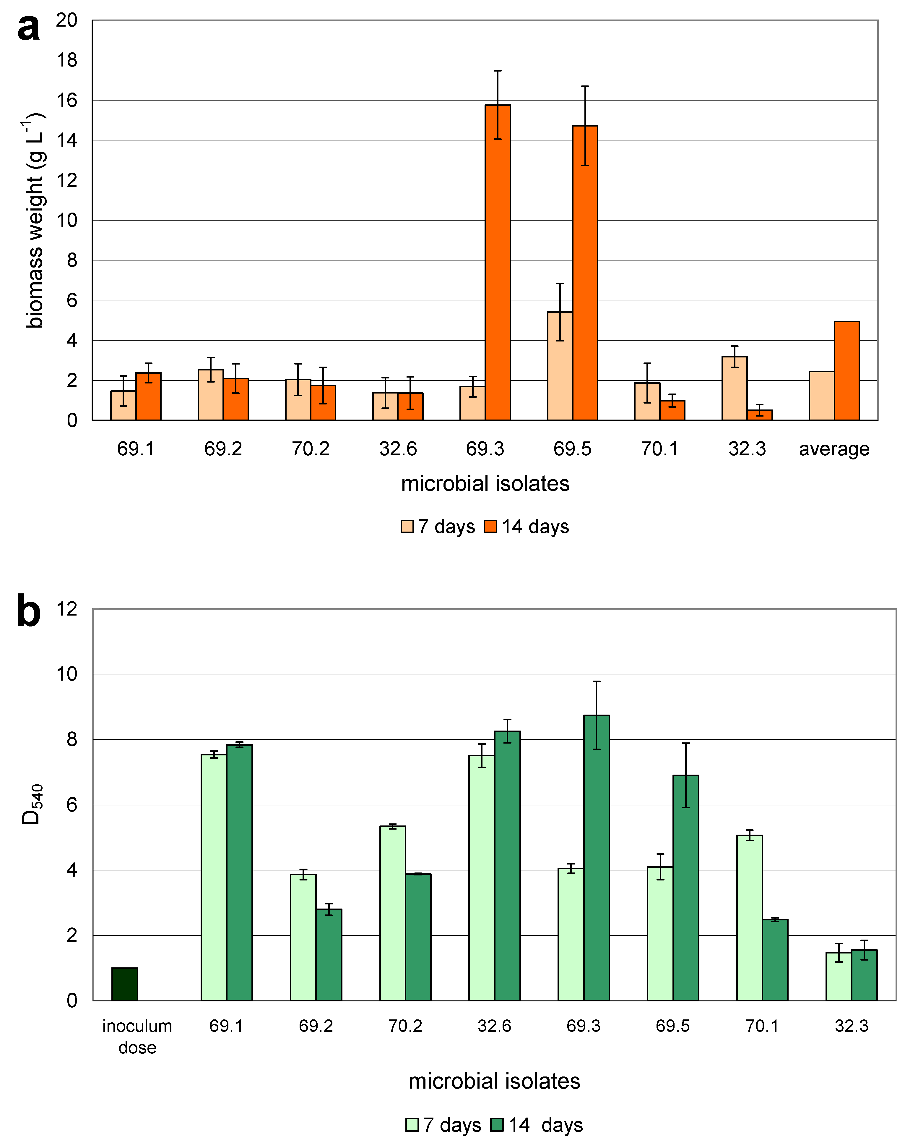
Increase in biomass was evaluated by weighing and photometric methods after 7 and 14 days of cultivation (Figure 1). The weighing method was used to determine the total increase in biomass, which depends on the increase in the number of cells and on changes in the size of individual bacterial cells. A photometric method was used to determine changes in the active part of the biomass.
In Figure 1a, it can be seen that the greatest increase in biomass after seven days of cultivation was demonstrated by microbial isolates with laboratory numbers 69.5 and 32.3. The weight of raw biomass was 5.4 and 3.2 g L−1, respectively. The weight of other studied isolates was less intensive. The smallest increase in biomass after seven days was observed in isolates 32.6 and 69.1. It is noteworthy that after seven days of culturing isolate 69.3, the weight of its raw biomass was 1.5-fold less than the average value of the eight studied isolates, and after 14 days, it was 3.2-fold higher than the average value. After 14 days of cultivation, the weight of the raw biomass of isolate 69.3 was the highest, amounting to 15.3 g L−1. A similar value was also observed in the microbial strain 69.5–14.7 g L−1.
It should be noted that, in contrast to isolate 69.3, the biomass weight of isolate 69.5 after seven days of the experiment was 2.2-fold higher and after 14 days was three-fold higher than the average value of the eight studied isolates. A slight increase in the biomass of microbial isolate 69.1 was observed after 14 days of cultivation, while in the other studied microorganisms, the growth indicators decreased, indicating the end of active growth.
Figure 1b shows the results obtained from the evaluation of optical densities of the liquid cultures of the eight studied microbial isolates after 7 and 14 days of cultivation. After 7 days, isolates 69.1, 32.6, 70.1, and 70.2 grew most actively, which differed from the results obtained using the weight method for estimating biomass growth. After 14 days of cultivation, a slight increase in the optical density was observed in isolates 69.1 and 32.6, which did not contradict data obtained from the weight method. Active increase in the biomass of isolates 69.3 and 69.5 was observed after 14 days of cultivation, as was shown when assessing the increase in biomass by raw weight method.
After seven days of the experiment, the biomass of isolates 69.3 and 69.5 increased 2.2- and 1.7-fold, respectively, while in isolates 69.2, 70.1, 70.2 and 32.3, biomass growth stopped after 14 days of cultivation. Thus, when determining the total increase in biomass by the weight method, which allows for the determination of both the size and the number of microbial cells, the maximum growth rates were found in isolates 69.3 and 69.5. According to results obtained from determining the increase in the active part of the biomass using the photometric method, similar microbial isolates were identified: 69.3 and 69.5, and, in addition to them, isolates 69.1 and 32.6.
Results from the evaluation of total Fe (II) removal by iron-oxidizing microorganisms in a liquid medium is shown in Figure 2. Fe (II) removal was calculated as a percentage based on the abiotic control (selective medium without an isolate). As shown by the experiment, after 7 and 14 days of culturing, there was no loss of Fe (II) in the control medium. According to the results obtained, after 7 days of cultivation, Fe (II) removal in the liquid medium was 8–10% in most of the studied isolates; after 14 days, it was 11–15 %. Such low values were most likely due to the very high concentration of Fe (II) in the culture medium. Considering the fact that the microbial isolates grew well in such an environment indicates their unique ecological properties. Among the studied isolates, microbial isolates 69.3 and 69.5 were noticeably distinguished, based on the Fe (II) removal results in a liquid culture medium, which is consistent with results obtained when analyzing their growth indicators (by weight and photometric method). Fe (II) removal in the culture medium with isolate 69.3 was 21% and 33% for 7 and 14 days, respectively, while in the culture medium with isolate 69.5 it was 34% and 39%, respectively.
Based on the data obtained during the experiments, we calculated one of the kinetic characteristics of culture growth, the specific growth rate µ. It is known that the growth rate of microorganisms is determined by the physiological characteristics of the strain, the conditions of cultivation, the nature of the substrate being utilized, and its concentration in the medium. When selecting promising isolates for practical use, this kinetic characteristic is of great importance [30]. According to results of the weight method, the maximum specific growth rate for isolate 69.5 was 1.5–5.3-fold higher than in other investigated isolates after seven days of cultivation (Table 1). Isolates 69.2 and 32.3 also showed high indices. According to photometric analysis, the maximum specific growth rate after seven days of cultivation was noticed in the following strains: 69.1, 32.6, and 70.2. In general, there were no strong variations in the values of the calculated indicators for the studied microbial isolates, with the exception of isolate 32.3.
Comparing the calculated specific growth rates of the studied microbial isolates, it was observed that after 14 days of cultivation, a number of the studied isolates were in the attenuation phase or their growth rate had decreased significantly, with the exception of isolates 69.3 and 69.5, which were characterized by maximum values of µ. According to results from the weight analysis method, after 14 days of culturing, the µ value of isolate 69.3 increased almost five-fold compared to the previous measurement obtained after seven days of culturing, and for isolate 69.5 it increased 1.3-fold. The results obtained indicate the possible use of a number of the investigated isolates in the biotechnological process of water purification from excess iron.
For further research, we selected the two microbial isolates with maximum growth characteristics and iron-oxidizing activity: 69.3 and 69.5. In order to identify the selected isolates, their cultural, morphological, physiological, and biochemical characteristics were studied, and the results are presented in Table 2. The investigated microorganisms were analyzed for their 16S rRNA nucleotide sequence. Based on a comparative analysis of the studied cultural, morphological, physiological, and biochemical characteristics and in accordance with the criteria for bacterial differentiation proposed in Bergey’s Manual of Determinative Bacteriology [29], the principles of molecular typing of prokaryotic cells, the selected microbial isolates were identified as Bacillus megaterium 69.3 and B. megaterium 69.5. The 16S rDNA homology of the two isolates showed that these two organisms may be different strains of B. megaterium with some differences in their genome. The 16S rRNA sequences of the isolates Bacillus megaterium 69.3 and B. megaterium 69.5 were submitted to the NCBI GenBank with the registration numbers MK764545 and MK764687.
Bacilli found in fresh water are mainly of soil origin and represent typical soil organisms, such as B. megaterium, B. cereus, B. firmus, and B. pumilus [31,32]. Other authors [33] have reported on the ability of seven strains of B. megaterium to use FeSO4·7H2O for growth. The mechanism of adaptation of B. megaterium to high contents of Se in a medium has been revealed: with the help of the enzyme selenium reductase, soluble Se compounds are converted into insoluble ones and neutralized [34].
The use of various representatives of the genus Bacillus in water purification technologies from HM is well known [10,35,36]: B. firmus from Pb, Zn and Cu; Bacillus sp. SW2 from As; B. thuringiensis “Simi from Pb and Zn.
For bacteria of the genus Bacillus, the mechanisms for removing HMs including iron from water are based on the binding of metal ions by polarized groups of the bacterial cell wall or capsule, for example, by carboxyl groups of peptidoglycans [11]. Intracellular penetration of HM ions and their accumulation into bacterial cells is possible [37]. In such cases, microorganisms synthesize detoxifying enzymes that transform HMs to less toxic forms [19].
While culturing the studied microbial strains B. megaterium 69.3 and B. megaterium 69.5, the color of the agarized selective medium containing Fe (II) and the liquid selective medium gradually changed from light green to brown due to the oxidation of Fe (II) and the accumulation of Fe (III) oxides in the medium. Brown Fe (III) sediments were deposited in noticeable amounts on the walls of the culture flasks and on the surface of the medium. We believe that the microbial strains B. megaterium 69.3 and B. megaterium 69.5 oxidized Fe (II) to Fe (III) with the help of specific detoxifying enzymes.
With the exception of B. cereus, bacilli are not pathogenic to mammals, and most bacilli do not inhabit insects. Our experiments showed that the studied microbial isolates B. megaterium 69.3 and B. megaterium 69.5 do not have hemolytic activity, which indirectly indicates their non-pathogenicity. Therefore, these microorganisms can serve as the basis for the biotechnological cleaning of urban surface waters with high iron content. Given that these microorganisms are natural and have a unique property, which is the ability to oxidize iron in extremely high concentrations, it will be advisable to use them for purifying not just drinking water, but also industrial drains and places experiencing local iron pollution.
4. Conclusions
Urban surface waters, which serve as sources of drinking water, are not environmentally protected from anthropogenic impacts, and often contain excess iron; therefore, they require the use of an effective, economical, and environmentally friendly purification method. In this regard, the use of iron-oxidizing microorganisms to remove iron from drinking water is of great scientific and applied importance.
Screening of microorganisms isolated from the high-magnetic soil of Mednogorsk was carried out based on the ability of the studied isolates to oxidize Fe (II) under conditions of periodic cultivation in a liquid medium. Microbial isolates with maximum growth characteristics and iron-oxidizing activity were selected and identified: B. megaterium 69.3 and B. megaterium 69.5. Total biomass growth of B. megaterium 69.3 and B. megaterium 69.5 after 14 days of cultivation was 15.3 and 14.7 g L−1; the active part of the biomass increased 8.7- and 6.9-fold compared to the initial inoculum dose; and Fe (II) removal in the culture medium was 33% and 39%, respectively.
Given that these isolates are naturally occurring microorganisms and have a unique property, which is the ability to oxidize iron in extremely high iron concentrations, they can be applied to the technology of cleaning urban surface waters from high iron content.
Author Contributions
Conceptualization, E.P.; methodology, C.N. and M.V.L.; investigation, C.N.; validation, M.R.; formal analysis, M.R.; visualization, E.P.; resources, M.R.; supervision, M.V.L.; writing—original draft preparation, E.P.; writing—review and editing, E.P. and M.V.L. All authors have read and agreed to the published version of the manuscript.
Funding
This research was financially supported by a grant of the President of the Russian Federation No. MK-3355.2019.5.
Institutional Review Board Statement
Not applicable.
Informed Consent Statement
Not applicable.
Data Availability Statement
All data in the article are publicly available and have no limitations.
Acknowledgments
The work was supported by and published with the financial support of the Federal State Budgetary Educational Institution of Higher Education “Russian Timiryazev State Agrarian University”.
Conflicts of Interest
The authors declare no conflict of interest.
References
- Su, C.; Jiang, L.; Zhang, W. A review on heavy metal contamination in the soil worldwide: Situation, impact and remediation techniques. Environ. Skept. Crit. 2014, 3, 24–38. [Google Scholar]
- Riabova, E.G. Content of heavy metals in urban surface water bodies. Teor. Prikl. Ekol. 2019, 1, 36–40. [Google Scholar] [CrossRef]
- Kovalevskaya, N.P. Biogeochemical potential of microbiological barriers in water quality formation in Kizel coal field rivers. Fundam. Res. 2015, 2, 4203–4208. (In Russian) [Google Scholar]
- Savichev, O.G. Background concentration of substances in river waters of the taiga zone of Western Siberia. Tomsk. State Univ. Bull. 2010, 334, 169–175. (In Russian) [Google Scholar]
- Ministry of Health and Social Development of the Russian Federation. SanPiN 2.1.4.1074-01. Drinking Water. Hygienic Requirements for Water Quality of Centralized Drinking Water Supply Systems. Quality Control; Ministry of Health and Social Development of the Russian Federation: Moscow, Russia, 2001. (In Russian) [Google Scholar]
- Pietrangelo, A. Mechanism of Iron Toxicity. In Iron Chelation Therapy; Hershko, C., Ed.; Springer: Boston, MA, USA, 2002; pp. 19–43. [Google Scholar]
- Islam, M.F.; Islam, M.S.; Ahmed, S.; Miah, M.Y. Removal of chromium from effluent of tannery industries in Bangladesh using rice husks as natural adsorbent. Soil Environ. 2020, 39, 165–172. [Google Scholar] [CrossRef]
- Ou, M.-Y.; Ting, Y.; Ch’Ng, B.-L.; Chen, C.; Cheng, Y.-H.; Chang, T.-C.; Hsi, H.-C. Using Mixed Active Capping to Remediate Multiple Potential Toxic Metal Contaminated Sediment for Reducing Environmental Risk. Water 2020, 12, 1886. [Google Scholar] [CrossRef]
- Ahalya, N.; Ramachandran, T.V.; Kanamadi, R.D. Biosorption of Heavy Metals. Res. J. Chem. Environ. 2003, 7, 71–79. [Google Scholar]
- Kumar, V.; Singh, S.; Kashyap, N.; Singla, S.; Bhadrecha, P.; Kaur, P.; Datta, S.; Kalia, A.; Singh, J. Bioremediation of heavy metals by employing resistant microbial isolates from agricultural soil irrigated with Industrial waste water. Orient. J. Chem. 2015, 31, 357–361. [Google Scholar] [CrossRef] [Green Version]
- Dixit, R.; Malaviya, D.; Pandiyan, K.; Singh, U.B.; Sahu, A.; Shukla, R.; Singh, B.P.; Rai, J.P.; Sharma, P.K.; Lade, H.; et al. Bioremediation of Heavy Metals from Soil and Aquatic Environment: An Overview of Principles and Criteria of Fundamental Processes. Sustainability 2015, 7, 2189–2212. [Google Scholar] [CrossRef] [Green Version]
- Wang, J.; Chen, C. Biosorption of heavy metals by Saccharomyces cerevisiae: A review. Biotechnol. Adv. 2006, 24, 427–451. [Google Scholar] [CrossRef]
- Mosa, K.A.; Saadoun, I.; Kumar, K.; Helmy, M.; Dhankher, O.P. Potential Biotechnological Strategies for the Cleanup of Heavy Metals and Metalloids. Front. Plant Sci. 2016, 7, 303. [Google Scholar] [CrossRef] [PubMed] [Green Version]
- Virpiranta, H.; Banasik, M.; Taskila, S.; Leiviskä, T.; Halttu, M.; Sotaniemi, V.-H.; Tanskanen, J. Isolation of Efficient Metal-Binding Bacteria from Boreal Peat Soils and Development of Microbial Biosorbents for Improved Nickel Scavenging. Water 2020, 12, 2000. [Google Scholar] [CrossRef]
- Fortin, D.; Langley, S. Formation and occurrence of biogenic iron-rich minerals. Earth-Sci. Rev. 2005, 72, 1–19. [Google Scholar] [CrossRef]
- Rawlings, D.E. Characteristics and adaptability of iron- and sulfur-oxidizing microorganisms used for the recovery of metals from minerals and their concentrates. Microb. Cell Factories 2005, 4, 13. [Google Scholar] [CrossRef] [PubMed] [Green Version]
- Bellenberg, S.; Barthen, R.; Boretska, M.; Zhang, R.; Sand, W.; Vera, M. Manipulation of pyrite colonization and leaching by iron-oxidizing Acidithiobacillus species. Appl. Microbiol. Biotechnol. 2014, 99, 1435–1449. [Google Scholar] [CrossRef] [PubMed]
- McLean, R.J.C.; Fortin, D.; Brown, D.A. Microbial metal-binding mechanisms and their relation to nuclear waste disposal. Can. J. Microbiol. 1996, 42, 392–400. [Google Scholar] [CrossRef]
- Hedrich, S.; Schlömann, M.; Johnson, D.B. The iron-oxidizing proteobacteria. Microbiology 2011, 157, 1551–1564. [Google Scholar] [CrossRef] [Green Version]
- Dubinina, G.A.; Sorokina, A.Y. Neutrophilic Lithotrophic Iron-Oxidizing Prokaryotes and Their Role in the Biogeochemical Processes of the Iron Cycle. Microbiology 2014, 83, 127–142. (In Russian) [Google Scholar] [CrossRef]
- Neubauer, S.C.; Emerson, D.; Megonigal, J.P. Life at the Energetic Edge: Kinetics of Circumneutral Iron Oxidation by Lithotrophic Iron-Oxidizing Bacteria Isolated from the Wetland-Plant Rhizosphere. Appl. Environ. Microbiol. 2002, 68, 3988–3995. [Google Scholar] [CrossRef] [Green Version]
- Pleshakova, Y.V.; Reshetnikov, M.V.; Ngun, C.T.; Shuvalova, Y.P. Microbiological and Biochemical Indication of Soils of the City Mednogorsk. Agric. Chem. 2016, 1, 66–73. (In Russian) [Google Scholar]
- Zhang, C.; Qiao, Q.; Piper, J.D.; Huang, B. Assessment of heavy metal pollution from a Fe-smelting plant in urban river sediments using environmental magnetic and geochemical methods. Environ. Pollut. 2011, 159, 3057–3070. [Google Scholar] [CrossRef]
- Granina, L.Z.; Parfenova, V.V.; Zemskaya, T.I.; Zakharova, Y.R.; Golobokova, L.P. On Iron and Manganese Oxidizing Microorganisms in Sedimentary Redox Cycling in Lake Baikal. Berl. Palaobiol. Abh. 2003, 4, 121–128. [Google Scholar]
- Summers, A.O.; Silver, S. Mercury Resistance in a Plasmid-Bearing Strain of Escherichia coli. J. Bacteriol. 1972, 112, 1228–1236. [Google Scholar] [CrossRef] [PubMed] [Green Version]
- Malik, A.; Ahmad, M. Seasonal variation in bacterial flora of the wastewater and soil in the vicinity of industrial area. Environ. Monit. Assess. 2002, 73, 263–273. [Google Scholar] [CrossRef] [PubMed]
- Pirt, S.J. Principles of Microbe and Cell Cultivation; Blackwell Scientific Publications: London, UK, 1975. [Google Scholar]
- GOST 4011-72. Drinking Water. Methods for Measuring the Mass Concentration of Total Iron, Moscow, Russia, 1974. (In Russian). Available online: http://docs.cntd.ru/document/1200008210 (accessed on 24 March 2021).
- Holt, J.G.; Krieg, N.R.; Sneath, P.H.A. (Eds.) Bergey’s Manual of Determinative Bacteriology; Lippincott Williams & Wilkins: Philadelphia, PA, USA, 1993; Translated under the Title Opredelitel’ Bakterii Berdzhi; Mir: Moscow, Russia, 1997. [Google Scholar]
- Okpokwasili, G.C.; Nweke, C.O. Microbial growth and substrate utilization kinetics. Afr. J. Biotechnol. 2005, 5, 305–317. [Google Scholar]
- Allen, D.A.; Austin, B.; Colwell, R.R. Numerical Taxonomy of Bacterial Isolates Associated with a Freshwater Fishery. Microbiology 1983, 129, 2043–2062. [Google Scholar] [CrossRef] [Green Version]
- Singh, Y.; Lal, N. Investigations on the heavy metal resistant bacterial isolates in vitro from industrial effluents. WJPPS 2015, 4, 343–350. [Google Scholar]
- Yu, Q.; Rao, R.; Xu, X.; Ganji, S.; Jacob, M.R.; Yu, B.; Li, X. Isolation and synthesis of novel antimicrobial chromene derivatives from Psorothamnus fremontii. Planta Med. 2015, 81. [Google Scholar] [CrossRef]
- Mishra, R.R.; Prajapati, S.; Das, J.; Dangar, T.K.; Das, N.; Thatoi, H. Reduction of selenite to red elemental selenium by moderately halotolerant Bacillus megaterium strains isolated from Bhitarkanika mangrove soil and characterization of reduced product. Chemosphere 2011, 84, 1231–1237. [Google Scholar] [CrossRef]
- Salehizadeh, H.; Shojaosadati, S. Removal of metal ions from aqueous solution by polysaccharide produced from Bacillus firmus. Water Res. 2003, 37, 4231–4235. [Google Scholar] [CrossRef]
- Kalaimurugan, D.; Balamuralikrishnan, B.; Durairaj, K.; Vasudhevan, P.; Shivakumar, M.S.; Kaul, T.; Chang, S.W.; Ravindran, B.; Venkatesan, S. Isolation and characterization of heavy-metal-resistant bacteria and their applications in environmental bioremediation. Int. J. Environ. Sci. Technol. 2020, 17, 1455–1462. [Google Scholar] [CrossRef]
- Pishchik, V.N.; Vorob’Ev, N.I.; Provorov, N.A.; Khomyakov, Y.V. Mechanisms of plant and microbial adaptation to heavy metals in plant–microbial systems. Microbiology 2016, 85, 257–271. [Google Scholar] [CrossRef]
Figure 1.
Growth indicators of microbial isolates when cultured in a liquid medium with Fe (II): (a) biomass increase by raw weight (mean ± S.D., n = 15; p ≤ 0.05); (b) absolute biomass increase (mean ± S.D., n = 9; p ≤ 0.05).
Figure 1.
Growth indicators of microbial isolates when cultured in a liquid medium with Fe (II): (a) biomass increase by raw weight (mean ± S.D., n = 15; p ≤ 0.05); (b) absolute biomass increase (mean ± S.D., n = 9; p ≤ 0.05).

Figure 2.
Fe (II) removal by the studied microorganisms (mean ± S.D., n = 9; p ≤ 0.05).


Table 1.
Specific growth rates of the investigated microorganisms.
| Isolate No. | Specific Growth Rate, h−1 Based on the Method: | |||
|---|---|---|---|---|
| Weight Method | Photometric Method | |||
| 7 days | 14 days | 7 days | 14 days | |
| 69.1 | 0.0023 | −0.0006 | 0.0120 | −0.0071 |
| 69.2 | 0.0055 | DP | 0.0080 | DP |
| 70.2 | 0.0042 | DP | 0.0100 | DP |
| 32.6 | 0.0019 | DP | 0.0120 | −0.0017 |
| 69.3 | 0.0031 | 0.0157 | 0.0083 | 0.0092 |
| 69.5 | 0.0100 | 0.0133 | 0.0084 | 0.0061 |
| 70.1 | 0.0037 | DP | 0.0096 | DP |
| 32.3 | 0.0069 | DP | 0.0023 | DP |
Note: DP, culture is in the attenuation phase.
Table 2.
Phenotypic characterization of two bacterial isolates (69.3 and 69.5).
| Characteristic | Bacteria | |
|---|---|---|
| 69.3 | 69.5 | |
| Motility | Motile | Motile |
| Characteristics of Colony | Circular, opaque, cream, with a slightly convex smooth shiny surface and uneven edges, Ø ≈ 2 mm | Circular, opaque, cream, with a slightly convex smooth shiny surface and uneven edges, Ø ≈ 1 mm |
| Shape of Bacterium | Rod, arranged singly and in pairs | Rod, arranged singly and in pairs |
| Gram Stain | + | + |
| Spore Characteristics | Elliptical, central position | Elliptical, central position |
| Growth at 42 °C | − | − |
| Growth at 10 °C | ± | − |
| Acid-resistant | − | − |
| Oxidase | − | − |
| Catalase | + | + |
| Lecithinase | + | − |
| Lipase | − | − |
| Urease | − | + |
| Arginine Dihydrolase | − | − |
| Phenylalanine Deaminase | − | + |
| Lysine Decarboxylase | − | − |
| Ornithine Decarboxylase | − | − |
| Nitrate Reductase | + | − |
| Gelatinase | + | + |
| Starch Hydrolysis | + | + |
| Casein Hydrolysis | + | ± |
| Indole Production | − | − |
| Hydrogen Sulfide Production | − | + |
| Ammonia Production | − | − |
| Citrate on Simmons Medium Utilization | − | − |
| Fermentation and Acid and Gas Production from Glucose, Lactose, Sucrose, Fructose, Maltose, Mannitol | + | + |
| Fermentation Arabinose | + | + |
| Acid and Gas Production from Xylose, Arabinose | − | + |
| Acid and Gas Production from Xylose | − | + |
| Vogues-Proskauer Test | − | − |
| Genus | Bacillus | Bacillus |
| Species | megaterium | megaterium |
Publisher’s Note: MDPI stays neutral with regard to jurisdictional claims in published maps and institutional affiliations. |
© 2021 by the authors. Licensee MDPI, Basel, Switzerland. This article is an open access article distributed under the terms and conditions of the Creative Commons Attribution (CC BY) license (http://creativecommons.org/licenses/by/4.0/).
Share and Cite
MDPI and ACS Style
Pleshakova, E.; Ngun, C.; Reshetnikov, M.; Larionov, M.V. Evaluation of the Ecological Potential of Microorganisms for Purifying Water with High Iron Content. Water 2021, 13, 901. https://doi.org/10.3390/w13070901
AMA Style
Pleshakova E, Ngun C, Reshetnikov M, Larionov MV. Evaluation of the Ecological Potential of Microorganisms for Purifying Water with High Iron Content. Water. 2021; 13(7):901. https://doi.org/10.3390/w13070901
Chicago/Turabian StylePleshakova, Ekaterina, Clement Ngun, Mikhail Reshetnikov, and Maxim Viktorovich Larionov. 2021. "Evaluation of the Ecological Potential of Microorganisms for Purifying Water with High Iron Content" Water 13, no. 7: 901. https://doi.org/10.3390/w13070901
Note that from the first issue of 2016, this journal uses article numbers instead of page numbers. See further details here.