Article

Effects of Sulfamethoxazole on Growth and Antibiotic Resistance of A Natural Microbial Community

1
Institute of Polar Sciences-National Research Council (ISP-CNR), 00010 Rome, Italy
2
Water Research Institute-National Research Council (IRSA-CNR), 00010 Rome, Italy
*
Author to whom correspondence should be addressed.
Water 2021, 13(9), 1262; https://doi.org/10.3390/w13091262
Submission received: 4 April 2021 / Revised: 28 April 2021 / Accepted: 29 April 2021 / Published: 30 April 2021
(This article belongs to the Special Issue Environmental Fate of Contaminants in the Aquatic Environment)

Abstract

:
Diffuse environmental antibiotic and antibiotic resistance gene contamination is increasing human and animal exposure to these emerging compounds with a consequent risk of reduction in antibiotic effectiveness. The present work investigated the effect of the antibiotic sulfamethoxazole (SMX) on growth and antibiotic resistance genes of a microbial community collected from an anaerobic digestion plant fed with cattle manure. Digestate samples were used as inoculum for concentration-dependent experiments using SMX at various concentrations. The antibiotic concentrations affecting the mixed microbial community in terms of growth and spread of resistant genes (sul1, sul2) were investigated through OD (Optical Density) measures and qPCR assays. Moreover, SMX biodegradation was assessed by LC-MS/MS analysis. The overall results showed that SMX concentrations in the range of those found in the environment did not affect the microbial community growth and did not select for antibiotic-resistant gene (ARG) maintenance or spread. Furthermore, the microorganisms tested were able to degrade SMX in only 24 h. This study confirms the complexity of antibiotic resistance spread in real matrices where different microorganisms coexist and suggests that antibiotic biodegradation needs to be included for fully understanding the resistance phenomena among bacteria.

1. Introduction

Antibiotic resistance (AR) in bacteria is a natural phenomenon [1]. However, antibiotic abuse in human and veterinary medicine has caused a worldwide increase and spread in antibiotic multi-resistant bacteria among humans, animals, and the environment [2,3,4]. The role of various anthropogenic sources (e.g., wastewater treatment plant effluents and biosolids, reclaimed water, organic amendments for agricultural practices, farm runoff) as key points in the diffusion of resistant bacteria and genes in soil and water ecosystems has been recognized [3,5]. AR is a “One Health” challenge on a global scale and an international emergency affecting both public health safety (10 million deaths predicted due to AR a year by 2050) and economic activities [6]. A holistic and multidisciplinary approach including humans, animals, food-producing industries, and the related environments is necessary for understanding AR overall impacts and assessing effective mitigation measures [7,8]. However, several ecological aspects of AR spread in natural microbial communities need to be clarified. Microorganisms interact in both intra- and inter-specific relationships, modulated by site-specific environmental conditions (e.g., temperature, humidity, multi-contamination), which make each ecosystem a unique one. Consequently, the effects of antibiotics on ecosystems can be variously and strongly linked to the bacterial resistance and resilience responses to their occurrence [9]. For example, the evaluation of the minimal selective concentration (MSC), i.e., the environmental antibiotic threshold concentration for the selection of resistant bacteria, remains a hot topic. It has been recently suggested that MSC data may be used in combination with traditional ecotoxicology data to propose threshold concentrations of antibiotics and other antibacterial compounds that are safe for environmental and human health [10,11]. Most studies aimed at evaluating the MSC showed that environmental antibiotic residues (e.g., in the range of 100 pg/mL to ng/mL, depending on the antibiotic and the type of resistance mutation examined) can select resistant bacteria [12,13]. However, most of these works did not take into account the complexity of microbial community interactions, including possible horizontal gene transfer mechanisms (HGT-transfer among different bacterial species, taxa, and habitats) [14]. HGT is the predominant mechanism making possible the transfer of antibiotic-resistant genes (ARGs) across different species, facilitated by the exchange of mobile genetic elements such as integrons [15]. Among these, the class 1 integrase gene intI1 is strongly involved in the horizontal transfer of ARGs [16] and is considered a suitable proxy for anthropogenic pollution [17]. In a microbial community context, the acquisition of antibiotic resistance is linked to the fitness cost for resistant microorganisms. This varies with the specific environmental conditions and the general level of stress undergone by each microorganism. In practice, AR acquisition can be positively selected and spread among bacterial populations if its fitness cost is offset by the benefit of resistance within the specific environmental conditions. In an antibiotic-free environment, the resistant organisms could be out-competed by the wild-type susceptible ones [18]. This suggests that there is a strong correlation between environmental antibiotic concentrations and the spread of antibiotic resistance but, in general, there are limited data on the selection and mobilization of resistance genes in complex natural microbial communities [19,20,21]. In this context, the present study evaluated the antibiotic SMX effects on growth and AR spread of a mixed natural microbial community collected from an anaerobically digested cattle manure (digestate). The digestate was selected because it is commonly used as a biofertilizer. If digestate can be a source of antibiotics and ARGs in agroecosystems, then the antibiotic amounts that can affect microbial communities need to be better investigated. For this purpose, “concentration-dependent” experiments were performed and the digestate microbial community was exposed to various SMX concentrations (from 0.07 to 76 mg/L) in liquid medium. The SMX concentrations able to influence the microbial community growth (one-day acute experiment) and AR spread (MSC, seven-day chronic experiment) were investigated. Antibiotic resistance was assessed by evaluating the changes in relative abundances of the intI1 gene (an indicator of HGT) and the main genes encoding resistance to sulfonamides (i.e., sul1 and sul2). SMX biodegradation was also estimated one day (acute experiment) and seven days (chronic experiment) after the microbial community exposure to the antibiotic.

2. Materials and Methods

2.1. Reagents

Methanol at HPLC grade, sodium chloride, and glycerol were obtained from VWR (Radnor, PA, USA). Water was purified (18 MΩ/cm quality) using a Milli-Q system (Millipore, Bedford, MA, USA). The Oxoid Iso-Sensitest broth was purchased from Thermo Fisher (Waltham, MA, USA). The Iso-Sensitest broth and glycerol were prepared using Milli-Q water and both solutions, and after preparation, were promptly sterilized in an autoclave.
SMX (purity 99%) was purchased from Sigma-Aldrich (Steinheim, Germany) in powder form. The stock solution of SMX (1000 mg/L) was prepared by dissolving an adequate quantity of the standard powder in Milli-Q water, which was stored at −80 °C. The working standard solutions were prepared at each sampling time by dilution of the stock solution with Milli-Q water or water:methanol (50:50, v/v) (for chemical determinations) and stored at 4 °C.
Oasis HLB (Hydrophilic–Lipophilic Balance) cartridges (6 mL, 1 g) were purchased from Waters (Milford, MA, USA) and diatomaceous earth (Thermo 062819) from Thermo Scientific (Waltham, MA, USA).

2.2. Anaerobic Digestate Samples

The anaerobic digestate was collected from an anaerobic digestion (AD) plant (output) located in the Lazio region (Italy) which produces biogas by utilizing cattle manure as fed-in biomass. The hydraulic retention time (HRT) of the plant is 60 to 70 days in one continuously stirred tank reactor (CSTR) operating under thermophilic conditions (45 °C).
Glass bottles (2 L) were filled with each sample (3 replicates) and transported to the laboratory, where aliquots were used for the SMX, intI1, sul1, and sul2 gene determinations. Microbial cells were extracted from the digestate following the method reported in Di Lenola et al. [22]. Briefly, three digestate sub-samples (1 g) were transferred to a test tube containing 9 mL of a non-fixative solution composed of phosphate-buffered saline: 130 mM NaCl; 7 mM Na2HPO4, 3 mM NaH2PO4; the non-ionic surfactant Tween 20 (0.5%, v/v) and 100 mM sodium pyrophosphate. The latter two were useful for decreasing particle aggregates and enhancing the detachment of cells from particles in solid samples.
The test tubes containing the digestate and the non-fixative solution were shaken for 30 min in an orbital shaker (400 rpm) and left for 24 h (4 °C) so that the larger soil particles could settle down [23]. Aliquots of supernatant (2 mL) were transferred into sterile tubes and centrifuged at 3500× g for 10 min. The supernatant was removed and the pellets (containing the mixed microbial community) were used for the experimental setup.

2.3. “Concentration-Dependent” Experiments

The concentrations of the antibiotic SMX able to influence the microbial community growth (due to acute exposure) were assessed by a microdilution test (96-well plate). Each well (final volume of 200 µL) was set up using an Iso-Sensitest broth solution with the microbial community (volume 10%) and SMX at one of the 11 concentrations tested [24]. The antibiotic concentrations ranged from 0.07 mg/L to 76 mg/L. Each concentration was tested in six replicates and a non-antibiotic control and sterile control were also assessed. The highest SMX concentration tested (76 mg/L) was the clinical breakpoint for the pathogenic bacteria Enterobacteriaceae, reported in the EUCAST database [25]. The 96-well plate was incubated in a Multiskan Sky Microplate Spectrophotometer (Thermo Scientific) at 37 °C with background pulsed shaking (low speed). The OD was measured at 600 nm every 15 min for 24 h (acute exposure). This well-known method is based on the assumption that the OD values are proportional to the number of cells.
Aliquots of the anaerobic digestate microbial community were also used in the seven-day MSC experiment to assess the effects of SMX chronic exposure on microbial abundance and AR spread. The experiment was performed following the method reported in Murray et al. [24] for evaluating the minimal selective concentration. It consisted of a chronic daily exposure (for seven consecutive days) of the microbial community in liquid medium (10% volume), at one of the five concentrations (from 0.001 to 10 mg/L) of SMX. Microorganisms were incubated overnight at 37 °C under continuous shaking at 180 r.p.m. Every day, an aliquot (1% v/v) of each culture was transferred in fresh broth (Iso Sensitest broth) spiked with the antibiotic. This step was repeated for seven days. Control conditions consisted of inoculum of the bacteria without antibiotic addition (C-MSC). Each condition was tested in five replicates. At zero and seven days, an aliquot (2 mL X 2) of each daily culture was resuspended in glycerol (20%) and stored at −80 °C until DNA extraction analysis. Moreover, SMX content was determined in the liquid cultures on experimental days one (acute exposure) and seven (chronic exposure).

2.4. DNA Extraction and qPCR Analysis

DNA was extracted from digestate and liquid culture samples by using the DNeasy PowerSoil kit (QIAGEN) following the manufacturer’s protocol. The extraction yield and quality of the DNA were assessed with a spectrophotometer (Multiskan Sky Microplate Spectrophotometer, Thermo Fisher Scientific, Waltham, MA, USA). The products were amplified by a CFX96 real-time PCR detection system (Bio-Rad, Hercules, CA, USA) using SYBR Green detection, targeting the two genes encoding resistance to sulfonamide (sul1, sul2), the 16S rRNA gene, and integron-integrase gene intI1.
The primers used for the ARGs, 16S, and intl1 gene quantification are listed in Supplementary Table S1. Each reaction was carried out in a total volume of 20 μL containing 10 μL SsoAdvanced Universal SYBR Green Supermix (Bio-Rad, USA), 0.5 μL of each primer (10 μM), and 15 ng of DNA template. The thermal cycling conditions were as follows: 95 °C for 3 min, 45 cycles at 95 °C for 15 s, annealing temperature (Tm) specific for each primer pair for 30 s (Supplementary Table S1), and 72 °C for 30 s. The fluorescence signal was read after each elongation step. All reactions were terminated with a melting curve starting at 55 °C and increasing by 0.5 °C up to 95 °C to verify amplicon specificity. Each assay was run in triplicate including the No Template Controls (NTC). Any possible qPCR inhibition was assessed with an inhibition test using samples diluted from 10 to 100-fold, and no inhibition was observed. The quantitative PCR data were expressed as the ratio of the intI1 gene or ARG copy number per 16S copy number to evaluate the relative proportion of the target gene in the community.

2.5. Analytical Determination of SMX

SMX was extracted from digestate samples by Pressurized Liquid Extraction (PLE) using the E-916 Speed Extractor (Büchi, Italy) system following the method reported in Gobel et al. [26]. The PLE extracts, after dilution with ultra-pure water to reduce the methanol content below 5%, were purified following the SPE (Solid Phase Extraction) procedure reported by Gobel et al. [27]. The SPE eluates were then evaporated and reconstituted with the mobile phase for the chromatographic analysis.
The SMX analytical determination in the purified extracts was carried out using an LC-MS/MS system (triple quadrupole mass spectrometer detector, equipped with an electrospray ionization detector, mod. API 3000, AB Sciex, Darmstadt, Germany), modifying the method reported in Spataro et al. [28]. The injector valve (Rheodyne, mod. 260 7125) included a 20 μL loop. The chromatographic Gemini column (150 × 4.6 mm, 5 μm RP C 18, Phenomenex, France), preceded by an equally packed guard column, was maintained at 25 °C. The isocratic elution was carried out at a 0.3 mL/min flow rate, using a mobile phase composed of MeOH (phase A): ultrapure water acidified to pH 3 with formic acid (phase B), 70:30. The MS/MS detector was set to positive ionization (ESI+) and a multiple reaction monitoring (MRM) mode. The optimized MS/MS parameters are reported in Supplementary Table S2. The Analyst Service version 1.6 software allowed both instrument control and data acquisition. Two calibration curves for SMX were obtained by analyzing working standard solutions in lower (1.0, 2.5, 5.0, 10.0, and 20.0 ng/L) and higher (25, 50, 100, 500, and 1000 µg/L) concentration ranges. The linearity was confirmed with an R2 ≥ 0.99 for both concentration ranges. The residual SMX concentrations in liquid culture were determined by direct injection of triplicate samples (20 µL) into the LC-MS/MS system following the method previously described. The recoveries of the method applied to the digestate and liquid medium were evaluated and turned out to be always above 90%. The limits of detection (LOD), calculated following the IUPAC method [29], were 0.41 μg/kg and 0.43 ng/L, respectively, for the liquid and solid matrices. The limit of quantification (LOQ) was set at three times the LOD.

2.6. Statistical Analysis

The MSC-experiment samples were analyzed three times for each replicate (five replicates) both for microbiological and chemical determinations and results are expressed as means ± standard errors (SE). The statistical analysis (R software) of biological and chemical data was performed using a two-way ANOVA and a multiple pairwise-comparison using the Tukey HSD (Tukey Honest Significant Differences) test.

3. Results and Discussions

3.1. SMX, intI1 Gene, and SMX-ARGs in the Digestate

SMX was detected in the digestate analyzed at a residual concentration of 2.4 ± 0.5 µg/kg (Table 1). Few data are currently available on its amount in digestate [30,31]; however, a recent study reports comparable SMX amounts in cattle manure samples collected from an AD plant-feeding tank [31]. SMX is commonly found in livestock manure and at much higher concentrations (until 5 mg/kg) [32] than those found in digestate.
The intI1 and sul1 genes’ relative abundances assessed in the digestate samples were similar (Table 1) and quite low if compared to those found in other studies in the same matrix [16,31]. Sul1 is generally located in the 3′ conserved segments of class 1 integrons and for this reason, it is related to the intI1 gene [33]. The sul2 gene was found at a relative abundance of 8.06 ×10⁻², significantly higher (p > 0.01) than intI1 and sul1 genes (Table 1). These results are in line with those found by other authors in river water and urban wetlands [34,35].

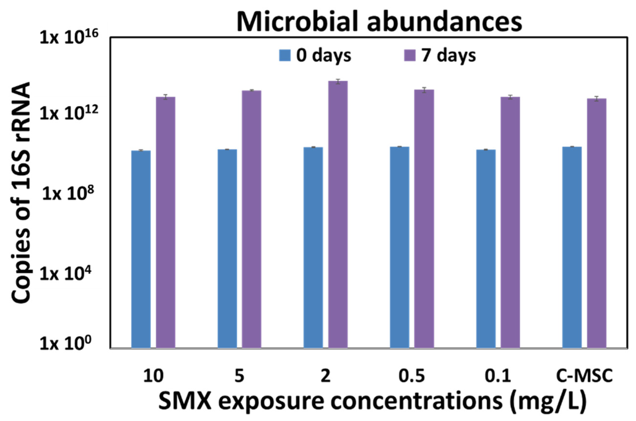
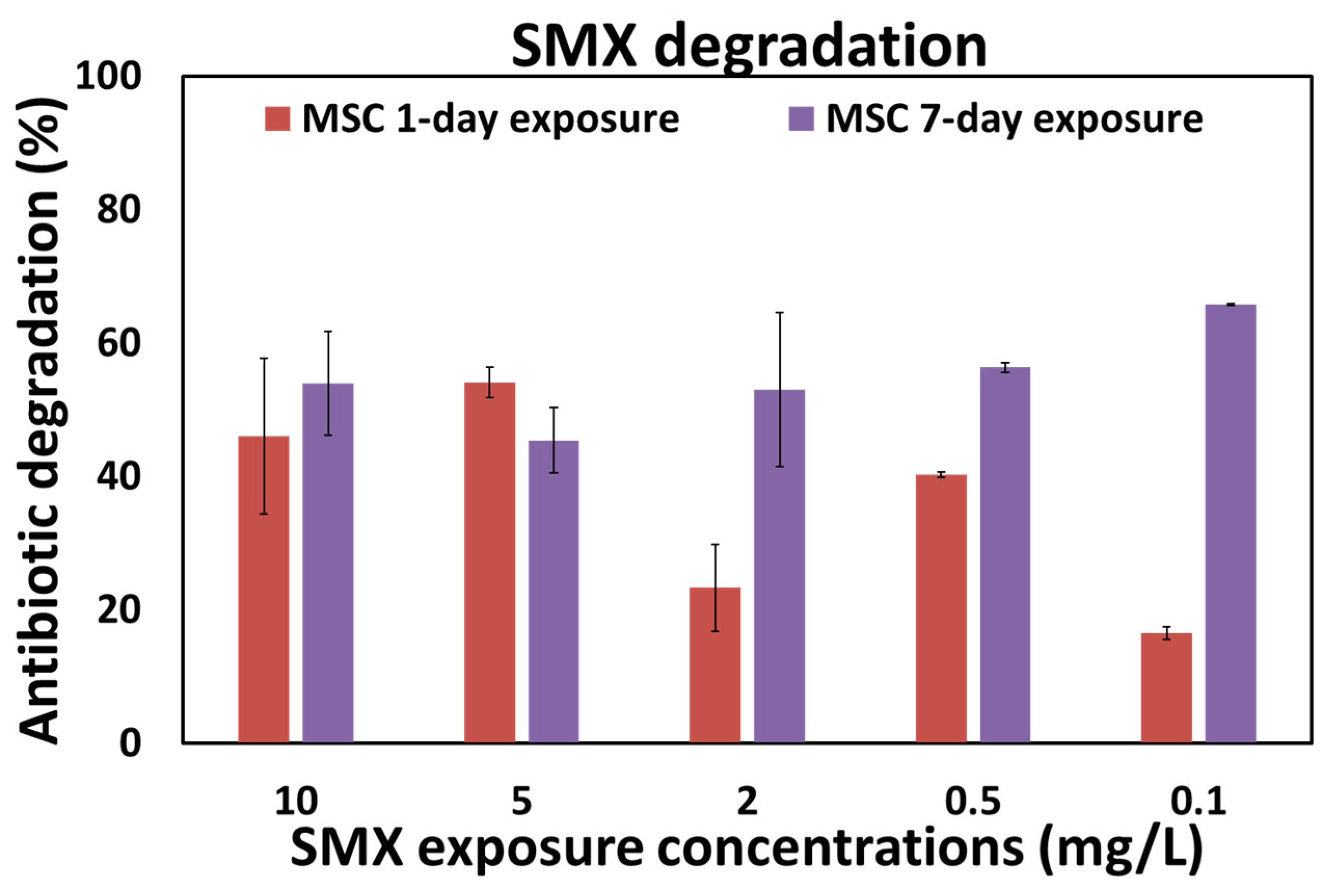
3.2. “Concentration-Dependent” Experiments

In the present study, the growth curves of the digestate mixed microbial communities exposed to various SMX concentrations (from 0.07 to 76 mg/L) were evaluated over 24 h (acute exposure). Antibiotic concentrations from 0.07 mg/L to 4.75 mg/L (4.75, 2.38, 1.19, 0.59, 0.30, 0.15, 0.07 mg/L) did not affect the microbial community (no significant differences compared with the control free of antibiotics). However, SMX concentrations higher than 4.75 mg/L (9.5, 19, 38, and 76 mg/L) significantly inhibited (p < 0.01) microbial growth (data not shown). In line with our results, a recent study showed that 5 mg/L of SMX had an acute detrimental effect on the abundance of a digestate microbial community [31].
The effect of SMX on microbial abundance was evaluated in terms of 16S rRNA copy number at the start (zero days) and end (seven days) of the antibiotic chronic exposure (MSC-experiment). As it is possible to see in Figure 1, all the concentrations tested (0.1, 0.5, 2, 5, and 10 mg/L) were not able to significantly affect microbial abundance.
In the present work, the SMX concentrations tested did not select for an increase in ARG spread among the microbial community (MSC-experiment). All genes decreased significantly at day seven (Table 2) and gene abundance differences due to exposure time (zero, seven days) or specific gene (sul1, sul2, intI1) were both affected by the SMX concentration (Table 3, Supplementary Table S3). The overall results confirm that natural microbial communities can react differently to single species to antibiotic exposure as reported by other authors [19].
Other seven-day chronic exposure works evaluated MSCs for other antibiotics at concentrations much lower than those of this work [10,12,24]. Stanton et al. [36] reported MSCs for some macrolides in a wastewater microbial community at concentrations considerably higher than the environmental ones.
The fact that SMX addition did not select for ARGs and intI1 gene maintenance or spread in the digestate microbial community at the relatively high concentrations tested can be ascribed to a continuous presence of antibiotic residues in inputs of the anaerobic digestion plant. Indeed, SMX is currently used in livestock raising and its residues are generally detected in manure [37,38].
Interestingly, prompt biodegradation (from 23% to 76% at 24 h) of all initial spiked concentrations (10, 5, 2, 0.5, 0.1 mg/L) was observed for both one-day acute exposure and seven-day chronic exposure in the MSC experiments (Figure 2). The fact that the different removal percentages were not concentration-dependent was in accordance with recent findings [39]. SMX degradation can occur through both metabolic and co-metabolic pathways [40] and the different degradation rates might be due to these different processes; however, this work did not aim to assess these aspects.
The percentages of antibiotic degradation were higher than those reported in other studies for both a bacterial consortium (i.e., 5% SMX removal after 300 h) and a single species assay (Rhodococcus equi: 5% SMX removal at 24 h) [41]. A more prolonged (seven days) SMX exposure significantly (p < 0.05) favored the degradation if compared to the one-day exposure, for the 2, 0.5, and 0.1 mg/L antibiotic concentrations. The latter result can be ascribed to a “selective effect” due to the daily transfer of the microbial community into a new fresh antibiotic solution. In other studies, microbial pre-exposure to an antibiotic, in a rich medium, favored its biodegradation by co-metabolism [42,43].
The antibiotic degradation in a few hours observed in the present work can explain the decrease in ARG abundance.
Indeed, complex natural microbial communities can show homeostatic capacities of resistance and resilience versus antibiotics; they can both resist antibiotics exposure through an increase in the ARG relative abundances among the community, and/or biodegrade them. Indeed, antibiotics can be an energy source for microbial growth [9,44,45]. ARGs have a fitness cost for bacteria and their selection depends on the specific microbial community and environmental conditions, but first of all, antibiotic presence [46]. In accordance with the results reported in the present study, in a digestate-amended soil microcosm experiment, a positive correlation between SMX degradation and an intI1 gene decrease was found [45]. A relationship between the persistence of this antibiotic and intI1 and sul1 genes was also observed in river water microcosm experiments [47,48].

4. Conclusions

The effects of SMX on microbial growth and antibiotic resistance in the digestate microbial community were evaluated with concentration-dependent experiments. The SMX concentrations which affected the microbial growth were higher (>4.75 mg/L) than those usually found in natural environments. The SMX concentrations tested did not exert a positive selection for the maintenance or spread of ARGs among the digestate microbial populations. This phenomenon was presumably due to a previous adaptation to the antibiotic. In fact, the microbial community was not only not inhibited, but was also able to promptly degrade (24 h) SMX in liquid cultures.
The overall data obtained in the present study show the complexity of this issue and how the effects of antibiotics on natural microbial communities can vary with different combinations of biotic and abiotic factors.

Supplementary Materials

The following are available online at https://www.mdpi.com/article/10.3390/w13091262/s1, Table S1: List of primers used for ARGs quantification, Table S2: MS/MS parameters used for the determination of SMX (sulfamethoxazole), Table S3: Tukey HSD (Tukey Honest Significant Differences) obtained from multiple pairwise-comparison between the means of groups.

Author Contributions

Conceptualization, J.R., L.P., A.B.C., A.V., P.G. and F.S.; methodology, J.R., AV., T.P., N.A. and F.S.; formal analysis, J.R., A.V., T.P., N.A. and F.S.; investigation, J.R., A.V., T.P., N.A. and F.S.; resources, L.P. and A.B.C.; data curation, J.R., A.V. and F.S.; writing—original draft preparation, J.R.; A.V. and F.S.; writing—review and editing, J.R., L.P. and A.B.C.; All authors have read and agreed to the published version of the manuscript.

Funding

This research was funded by Regione Lazio-Lazio Innova L.R. 13/08 (Progetti di Gruppi di Ricerca-Programma strategico Regionale per la ricerca, innovazione ed il trasferimento tecnologico 2017–2020-Italy) Azero Antibiotic Project N. 85-2017-15065.

Institutional Review Board Statement

Not applicable.

Informed Consent Statement

Not applicable.

Data Availability Statement

Not applicable.

Conflicts of Interest

The authors declare no conflict of interest. The funders had no role in the design of the study; in the collection, analyses, or interpretation of data; in the writing of the manuscript, or in the decision to publish the results.

References

  1. Hawkey, P.M. The origins and molecular basis of antibiotic resistance. Br. Med. J. 1998, 317, 657–660. [Google Scholar] [CrossRef] [PubMed] [Green Version]
  2. Berti, A.D.; Hirsch, E.B. Tolerance to antibiotics affects response. Science 2020, 367, 2019–2021. [Google Scholar] [CrossRef] [PubMed]
  3. García, J.; García-Galán, M.J.; Day, J.W.; Boopathy, R.; White, J.R.; Wallace, S.; Hunter, R.G. A review of emerging organic contaminants (EOCs), antibiotic resistant bacteria (ARB), and antibiotic resistance genes (ARGs) in the environment: Increasing removal with wetlands and reducing environmental impacts. Bioresour. Technol. 2020, 307, 123228. [Google Scholar] [CrossRef] [PubMed]
  4. Nadimpalli, M.L.; Marks, S.J.; Montealegre, M.C.; Gilman, R.H.; Pajuelo, M.J.; Saito, M.; Tsukayama, P.; Njenga, S.M.; Kiiru, J.; Swarthout, J.; et al. Urban informal settlements as hotspots of antimicrobial resistance and the need to curb environmental transmission. Nat. Microbiol. 2020, 5, 787–795. [Google Scholar] [CrossRef]
  5. Bengtsson-Palme, J.; Kristiansson, E.; Larsson, D.G.J. Environmental factors influencing the development and spread of antibiotic resistance. FEMS Microbiol. Rev. 2018, 42. [Google Scholar] [CrossRef]
  6. Lee, K.; Kim, D.W.; Lee, D.H.; Kim, Y.S.; Bu, J.H.; Cha, J.H.; Thawng, C.N.; Hwang, E.M.; Seong, H.J.; Sul, W.J.; et al. Mobile resistome of human gut and pathogen drives anthropogenic bloom of antibiotic resistance. Microbiome 2020, 8, 1–14. [Google Scholar] [CrossRef] [Green Version]
  7. Collignon, P. Antibiotic resistance: Are we all doomed? Intern. Med. J. 2015, 45, 1109–1115. [Google Scholar] [CrossRef]
  8. Finley, R.L.; Collignon, P.; Larsson, D.G.J.; Mcewen, S.A.; Li, X.Z.; Gaze, W.H.; Reid-Smith, R.; Timinouni, M.; Graham, D.W.; Topp, E. The scourge of antibiotic resistance: The important role of the environment. Clin. Infect. Dis. 2013, 57, 704–710. [Google Scholar] [CrossRef] [Green Version]
  9. Grenni, P.; Ancona, V.; Barra Caracciolo, A. Ecological effects of antibiotics on natural ecosystems: A review. Microchem. J. 2018, 36, 25–39. [Google Scholar] [CrossRef]
  10. Le Page, G.; Gunnarsson, L.; Snape, J.; Tyler, C.R. Integrating human and environmental health in antibiotic risk assessment: A critical analysis of protection goals, species sensitivity and antimicrobial resistance. Environ. Int. 2017, 109, 155–169. [Google Scholar] [CrossRef]
  11. Chow, L.K.M.; Ghaly, T.M.; Gillings, M.R. A survey of sub-inhibitory concentrations of antibiotics in the environment. J. Environ. Sci. 2021, 99, 21–27. [Google Scholar] [CrossRef]
  12. Gullberg, E.; Albrecht, L.M.; Karlsson, C.; Sandegren, L.; Andersson, D.I. Selection of a multidrug resistance plasmid by sublethal levels of antibiotics and heavy metals. MBio 2014, 5, e01918-14. [Google Scholar] [CrossRef] [Green Version]
  13. Gullberg, E.; Cao, S.; Berg, O.G.; Ilbäck, C.; Sandegren, L.; Hughes, D.; Andersson, D.I. Selection of resistant bacteria at very low antibiotic concentrations. PLoS Pathog. 2011, 7, e1002158. [Google Scholar] [CrossRef] [PubMed] [Green Version]
  14. Zarei-Baygi, A.; Harb, M.; Wang, P.; Stadler, L.B.; Smith, A.L. Microbial community and antibiotic resistance profiles of biomass and effluent are distinctly affected by antibiotic addition to an anaerobic membrane bioreactor. Environ. Sci. Water Res. Technol. 2020, 6, 724–736. [Google Scholar] [CrossRef]
  15. Yan, M.; Xu, C.; Huang, Y.; Nie, H.; Wang, J. Tetracyclines, sulfonamides and quinolones and their corresponding resistance genes in the Three Gorges Reservoir, China. Sci. Total Environ. 2018, 631–632, 840–848. [Google Scholar] [CrossRef]
  16. Ma, J.; Cui, Y.; Li, A.; Zhang, W.; Liang, J.; Wang, S.; Zhang, L. Evaluation of the fate of nutrients, antibiotics, and antibiotic resistance genes in sludge treatment wetlands. Sci. Total Environ. 2020, 712, 136370. [Google Scholar] [CrossRef]
  17. Gillings, M.R.; Gaze, W.H.; Pruden, A.; Smalla, K.; Tiedje, J.M.; Zhu, Y.G. Using the class 1 integron-integrase gene as a proxy for anthropogenic pollution. ISME J. 2015, 9, 1269–1279. [Google Scholar] [CrossRef]
  18. Hernando-Amado, S.; Sanz-García, F.; Blanco, P.; Martínez, J.L. Fitness costs associated with the acquisition of antibiotic resistance. Essays Biochem. 2017, 61, 37–48. [Google Scholar] [PubMed]
  19. Klümper, U.; Recker, M.; Zhang, L.; Yin, X.; Zhang, T.; Buckling, A.; Gaze, W.H. Selection for antimicrobial resistance is reduced when embedded in a natural microbial community. ISME J. 2019, 13, 2927–2937. [Google Scholar] [CrossRef] [PubMed]
  20. Du, B.; Yang, Q.; Wang, R.; Wang, R.; Wang, Q.; Xin, Y. Evolution of antibiotic resistance and the relationship between the antibiotic resistance genes and microbial compositions under long-term exposure to tetracycline and sulfamethoxazole. Int. J. Environ. Res. Public Health 2019, 16, 4681. [Google Scholar] [CrossRef] [PubMed] [Green Version]
  21. Murray, A.K.; Zhang, L.; Snape, J.; Gaze, W.H. Comparing the selective and co-selective effects of different antimicrobials in bacterial communities. Int. J. Antimicrob. Agents 2019, 53, 1–7. [Google Scholar] [CrossRef]
  22. Di Lenola, M.; Barra Caracciolo, A.; Grenni, P.; Ancona, V.; Rauseo, J.; Laudicina, V.A.; Uricchio, V.F.; Massacci, A. Effects of Apirolio Addition and Alfalfa and Compost Treatments on the Natural Microbial Community of a Historically PCB-Contaminated Soil. Water Air Soil Pollut. 2018, 229, 143. [Google Scholar] [CrossRef]
  23. Barra Caracciolo, A.; Bottoni, P.; Crobe, A.; Fava, L.; Funari, E.; Giuliano, G.; Silvestri, C. Microbial degradation of two carbamate insecticides and their main metabolites in soil. Chem. Ecol. 2002, 18, 245–255. [Google Scholar] [CrossRef]
  24. Murray, A.K.; Zhang, L.; Yin, X.; Zhang, T.; Buckling, A.; Snape, J.; Gaze, W.H. Novel insights into selection for antibiotic resistance in complex microbial communities. MBio 2018, 9, e00969-18. [Google Scholar] [CrossRef] [PubMed] [Green Version]
  25. EUCAST (The European Committee on Antimicrobial Susceptibility Testing). Breakpoint Tables for Interpretation of MICs and Zone Diameters; Version 10.0; EUCAST: Växjö, Sweden, 2020. [Google Scholar]
  26. Göbel, A.; McArdell, C.S.; Suter, M.J.F.; Giger, W. Trace determination of macrolide and sulfonamide antimicrobials, a human sulfonamide metabolite, and trimethoprim in wastewater using liquid chromatography coupled to electrospray tandem mass spectrometry. Anal. Chem. 2004, 76, 4756–4764. [Google Scholar] [CrossRef] [PubMed]
  27. Göbel, A.; Thomsen, A.; McArdell, C.S.; Alder, A.C.; Giger, W.; Theiß, N.; Löffler, D.; Ternes, T.A. Extraction and determination of sulfonamides, macrolides, and trimethoprim in sewage sludge. J. Chromatogr. A 2005, 1085, 179–189. [Google Scholar] [CrossRef] [PubMed]
  28. Spataro, F.; Ademollo, N.; Pescatore, T.; Rauseo, J.; Patrolecco, L. Antibiotic residues and endocrine disrupting compounds in municipal wastewater treatment plants in Rome, Italy. Microchem. J. 2019, 148, 634–642. [Google Scholar] [CrossRef]
  29. Thompson, M.; Ellison, S.L.R.; Wood, R. Harmonized guidelines for single-laboratory validation of methods of analysis (IUPAC Technical Report). Pure Appl. Chem. 2002, 74, 835–855. [Google Scholar] [CrossRef]
  30. Gurmessa, B.; Pedretti, E.F.; Cocco, S.; Cardelli, V.; Corti, G. Manure anaerobic digestion effects and the role of pre- and post-treatments on veterinary antibiotics and antibiotic resistance genes removal efficiency. Sci. Total Environ. 2020, 721, 137532. [Google Scholar] [CrossRef]
  31. Mazzurco Miritana, V.; Massini, G.; Visca, A.; Grenni, P.; Patrolecco, L.; Spataro, F.; Rauseo, J.; Garbini, G.L.; Signorini, A.; Rosa, S.; et al. Effects of Sulfamethoxazole on the Microbial Community Dynamics During the Anaerobic Digestion Process. Front. Microbiol. 2020, 11, 1–12. [Google Scholar] [CrossRef]
  32. Mohring, S.A.I.; Strzysch, I.; Fernandes, M.R.; Kiffmeyer, T.K.; Tuerk, J.; Hamscher, G. Degradation and elimination of various sulfonamides during anaerobic fermentation: A promising step on the way to sustainable pharmacy? Environ. Sci. Technol. 2009, 43, 2569–2574. [Google Scholar] [CrossRef]
  33. Gillings, M.; Boucher, Y.; Labbate, M.; Holmes, A.; Krishnan, S.; Holley, M.; Stokes, H.W. The evolution of class 1 integrons and the rise of antibiotic resistance. J. Bacteriol. 2008, 190, 5095–5100. [Google Scholar] [CrossRef] [PubMed] [Green Version]
  34. Adelowo, O.O.; Helbig, T.; Knecht, C.; Reincke, F.; Mäusezahl, I.; Müller, J.A. High abundances of class 1 integrase and sulfonamide resistance genes, and characterisation of class 1 integron gene cassettes in four urban wetlands in Nigeria. PLoS ONE 2018, 13, e0208269. [Google Scholar] [CrossRef] [PubMed] [Green Version]
  35. Luo, Y.; Mao, D.; Rysz, M.; Zhou, Q.; Zhang, H.; Xu, L.; Alvarez, P.J.J. Trends in antibiotic resistance genes occurrence in the Haihe River, China. Environ. Sci. Technol. 2010, 44, 7220–7225. [Google Scholar] [CrossRef]
  36. Stanton, I.C.; Murray, A.K.; Zhang, L.; Snape, J.; Gaze, W.H. Evolution of antibiotic resistance at low antibiotic concentrations including selection below the minimal selective concentration. Commun. Biol. 2020, 3, 1–11. [Google Scholar] [CrossRef]
  37. Karci, A.; Balcioǧlu, I.A. Investigation of the tetracycline, sulfonamide, and fluoroquinolone antimicrobial compounds in animal manure and agricultural soils in Turkey. Sci. Total Environ. 2009, 407, 4652–4664. [Google Scholar] [CrossRef] [PubMed]
  38. Hu, X.; Zhou, Q.; Luo, Y. Occurrence and source analysis of typical veterinary antibiotics in manure, soil, vegetables and groundwater from organic vegetable bases, northern China. Environ. Pollut. 2010, 158, 2992–2998. [Google Scholar] [CrossRef]
  39. Visca, A.; Barra Caracciolo, A.; Grenni, P.; Patrolecco, L.; Rauseo, J.; Massini, G.; Miritana, V.M.; Spataro, F. Anaerobic Digestion and Removal of Sulfamethoxazole, Enrofloxacin, Ciprofloxacin and Their Antibiotic Resistance Genes in a Full-Scale Biogas Plant. Antibiotic 2021, 10, 502. [Google Scholar] [CrossRef]
  40. Radke, M.; Lauwigi, C.; Heinkele, G.; Múrdter, T.E.; Letzel, M. Fate of the antibiotic sulfamethoxazole and its two major human metabolites in a water sediment test. Environ. Sci. Technol. 2009, 43, 3135–3141. [Google Scholar] [CrossRef]
  41. Larcher, S.; Yargeau, V. Biodegradation of sulfamethoxazole by individual and mixed bacteria. Appl. Microbiol. Biotechnol. 2011, 91, 211–218. [Google Scholar] [CrossRef]
  42. Larcher, S.; Yargeau, V. Biodegradation of sulfamethoxazole: Current knowledge and perspectives. Appl. Microbiol. Biotechnol. 2012, 96, 309–318. [Google Scholar] [CrossRef] [Green Version]
  43. Cycoń, M.; Mrozik, A.; Piotrowska-Seget, Z. Antibiotics in the soil environment—Degradation and their impact on microbial activity and diversity. Front. Microbiol. 2019, 10, 338. [Google Scholar] [CrossRef]
  44. Topp, E.; Chapman, R.; Devers-Lamrani, M.; Hartmann, A.; Marti, R.; Martin-Laurent, F.; Sabourin, L.; Scott, A.; Sumarah, M. Accelerated Biodegradation of Veterinary Antibiotics in Agricultural Soil following Long-Term Exposure, and Isolation of a Sulfamethazine-degrading Microbacterium sp. J. Environ. Qual. 2013, 42, 173–178. [Google Scholar] [CrossRef] [PubMed]
  45. Rauseo, J.; Barra Caracciolo, A.; Ademollo, N.; Cardoni, M.; Di Lenola, M.; Gaze, W.H.; Stanton, I.C.; Grenni, P.; Pescatore, T.; Spataro, F.; et al. Dissipation of the antibiotic sulfamethoxazole in a soil amended with anaerobically digested cattle manure. J. Hazard. Mater. 2019, 378, 120769. [Google Scholar] [CrossRef]
  46. Norman, A.; Hansen, L.H.; Sørensen, S.J. Conjugative plasmids: Vessels of the communal gene pool. Philos. Trans. R. Soc. B Biol. Sci. 2009, 364, 2275–2289. [Google Scholar] [CrossRef] [Green Version]
  47. Grenni, P.; Patrolecco, L.; Rauseo, J.; Spataro, F.; Di Lenola, M.; Aimola, G.; Zacchini, M.; Pietrini, F.; Di Baccio, D.; Stanton, I.C.; et al. Sulfamethoxazole persistence in a river water ecosystem and its effects on the natural microbial community and Lemna minor plant. Microchem. J. 2019, 149, 103999. [Google Scholar] [CrossRef]
  48. Patrolecco, L.; Rauseo, J.; Ademollo, N.; Grenni, P.; Cardoni, M.; Levantesi, C.; Luprano, M.L.; Barra Caracciolo, A. Persistence of the antibiotic sulfamethoxazole in river water alone or in the co-presence of ciprofloxacin. Sci. Total Environ. 2018, 640–641, 1438–1446. [Google Scholar] [CrossRef] [PubMed]
Figure 1. Microbial abundances expressed as 16S rRNA copies under the various SMX concentrations at zero and seven days. C-MSC: free-antibiotic control. The vertical bars represent standard errors.
Water 13 01262 g001
Figure 2. SMX degradation (%) in MSC experiments at one-day (acute experiment) and seven-day exposure (chronic exposure). n.d.: not detected. The vertical bars represent the standard errors.
Water 13 01262 g002
Table 1. Average SMX concentration (µg/kg), SMX-ARGs (sul1 and sul2), and intI1 gene relative abundances measured in the digestate. s.e.: standard error.
SMXintI1 Genesul1 Genesul2 Gene
Average values2.403.03 ×10−42.71 ×10−48.06 ×10−2
s.e.0.501.20 ×10−32.02 ×10−30.03
Table 2. SMX-ARGs (sul1 and sul2) and intI1 gene relative abundances at days zero and seven for each SMX concentration in MSC-experiment and control (C-MSC).
sul1 Genesul2 GeneintI1 Gene
0 Days7 Days0 Days7 Days0 Days7 Days
C-MSC2.6 × 10−48.3 × 10−88.1 × 10−22.0 × 10−43.2 × 10−42.8 × 10⁻⁶
10 mg/L2.7 × 10−43.9 × 10−88.1 × 10−21.9 × 10−42.32 × 10−83.4 × 10⁻⁷
5 mg/L2.7 × 10−40 8.0 × 10−202.32 × 10−82.3 × 10−6
2 mg/L2.8 × 10−43.1 × 10⁻¹⁰ 8.3 × 10−23.1 × 10−72.32 × 10−81.5 × 10−4
0.5 mg/L2.5 × 10−47.3 × 10−108.1 × 10−24.7 × 10−52.32 × 10−81.2 × 10−5
0.1 mg/L2.3 × 10−41.1 × 10⁻⁶ 8.0 × 10−23.9 × 10−42.32 × 10−87.8 × 10−7
Table 3. Effect of SMX concentrations (10, 5, 2, 0.5, 0.1 mg/L and C-MSC), time (zero or seven days), specific gene (sul1, sul2 or intI1) on gene abundances.
Factorsp-Value
(A) SMX concentrations 0.00376 **
(B) time0.01253 *
(C) specific gene1.25 × 10−7 ***
A × B Interaction0.01084 *
A × C Interaction3.72 × 10−6 ***
B × C Interaction0.10162
Two-way ANOVA results: p-values (signif. codes: 0: ***; 0.001: **; 0.01: *) for the main factors and interactions.
Publisher’s Note: MDPI stays neutral with regard to jurisdictional claims in published maps and institutional affiliations.

