
Investigating Landfill Leachate Influence on Soil Microbial Biodiversity and Its Cytotoxicity
by
 , , and
Urszula Wydro
*,
Elżbieta Wołejko
,
Gabriela Sokołowska
,
Jacek Leszczyński
and
Agata Jabłońska-Trypuć
* Faculty of Civil Engineering and Environmental Sciences, Bialystok University of Technology, Wiejska Str. 45E, 15-351 Białystok, Poland
*
Authors to whom correspondence should be addressed.
Water 2022, 14(22), 3634; https://doi.org/10.3390/w14223634
Submission received: 27 October 2022
/
Revised: 8 November 2022
/
Accepted: 9 November 2022
/
Published: 11 November 2022
(This article belongs to the Special Issue Presence of Microorganisms in Soil and Water)
Abstract
:The most important problem related to the collection of waste in landfills is the presence of harmful substances contained in leachate resulting from the flow of atmospheric precipitation through the deposited waste, which may end up in the soil. As a result, the soil’s chemical degradation occurs, which may result in a change in the structure and activity of microorganisms. In addition, leachate can end up in groundwater that may be hazardous to human health. The aim of the study was to determine the effect of landfill leachate on selected physicochemical, biological and biochemical properties of soils. The cytotoxicity of soil filtrates (SF) in human cell lines and their effect on apoptosis were also analyzed. In the pot experiment, leachate was added to the soil in two doses: 50LL (50% of the N dose) and 100LL (100% of the N dose). Selected physicochemical properties, activity of enzymes, diversity of bacteria and fungi by the T-RFLP approach were determined in soil samples. As a result of the analyses, it was found that LL causes an increase in the activity of dehydrogenases and a decrease in β-glucosidase. In the soils treated with LL, bacterial T-RF (144) bp, (179) bp and (413) bp were specific, while fungal were T-RF (390) bp, (441) bp and (595) bp. Potentially cytotoxic effects of SF and reduction in apoptosis in neoplastic cells were found in in vitro tests. LL and SF are complex matrices with a variable composition, which means that their impact on the soil environment must be constantly monitored.
1. Introduction
Taking into account an increase in the size of the human population, the progressive urbanization and the resulting increase in the production of waste per capita, the utilization and neutralization of waste becomes a significant problem for society. Proper waste management is a key aspect in both developed and developing countries. Disposal of waste in landfills is one of the oldest waste management methods in use today, but still very common all over the world. During storage, waste undergoes many complex biological and physicochemical processes, resulting in a complex product—the so-called landfill leachate (LL) [1]. The composition of such a product almost always identifies the basic chemical categories of compounds from the groups of important environmental pollutants, such as biodegradable and non-biodegradable organic compounds, heavy metals and some inorganic salts [2].
The chemical composition of the leachate depends mainly on the type of waste stored in the landfill, but also on the degree of water infiltration, the degree of irrigation, the technology used in the landfill and the degree of waste degradation. The negative effect of the LL reflects the amount of pollutants contained in it [3,4,5,6].
Soils, which are subject to chemical degradation, are particularly exposed to the negative effects of LL, which in turn may disturb the ecological balance, which is visible, among others, by changes in the species composition of microorganisms [7]. Hence, leachate can leak into groundwater and migrate for a long distance, posing a threat to other components of the ecosystem and to public health. As a result, the leachate chemicals can accumulate in different links in the food chain [8,9].
In order to determine the toxicity of leachate in various elements of the environment, including soil, the most common methods are traditional applications involving chemical analyses, mainly limited to the determination of heavy metals and organic compounds with carcinogenic, estrogenic and toxic properties. They allow for an initial estimation of the hazard and risk that leachates pose to the environment and human [10]. More sensitive to changes in the properties of the soil environment are biological parameters, which include, e.g., the activity of selected enzymes such as dehydrogenases (Dha) and β-glucosidases (β-Glu) as well as indicators determining the number, genetic diversity and community structure of microorganisms [7,11].
Extracellular enzymes, as indicators of soil quality, are very sensitive to environmental stress and react quickly to changes caused by human activity, including anthropogenic pollution. Dehydrogenases are oxidoreductases that catalyze the reversible oxidation and reduction reactions, i.e., transformations related to the transfer of protons and electrons. In turn, β-glucosidases are hydrolytic enzymes involved in the mineralization of organic matter and soil carbon [11]. Methods based on DNA extraction are used in the study of soil biodiversity. Despite the very popular NGS techniques, indirect methods for determining the diversity of a community of microorganisms are still often used, including fingerprinting methods such as T-RFLP (terminal restriction fragments length polymorphism). This approach allows for a relatively quick and accurate determination of changes in the structure of the soil microflora as a result of changes in the soil environment due to pollution [12].
LL flowing through the soil may end up in groundwater, which is a common source of drinking water. It is therefore important to determine the human toxicity of such filtrates. Baderna et al. [13] in their work focused on in vitro human cell cultures as the main biological model used in order to estimate the potential risk of LL to human health. One of the main reasons for an increasing use of biological tests based on in vitro human cell cultures is their very high sensitivity to mutagenic and carcinogenic toxins present in environmental matrices. The metabolic competence of human cell lines is an important property, because many components of environmental matrices, such as LL, show their genotoxic and/or carcinogenic potential only after metabolic activation [14]. The most commonly used cell line in toxicological studies is Caco-2 cell line. These are cells derived from colorectal adenocarcinoma of a 72-year-old man, which are available in the collections of ATCC (USA). They are characterized by adherent growth and, above all, they have the ability to create a brush border, i.e., a system of microvilli on the cell surface. This feature distinguishes Caco-2 cells from other lines. In addition, as in the case of enterocytes, these cells create tight connections with each other and systems for transporting substances. Despite many limitations, this cell line has been accepted by regulatory institutions (e.g., the Food and Drug Administration) as a reliable model of biological barriers occurring in the gastrointestinal tract. The simplicity of the model as well as the morphological and functional similarity of Caco-2 cells make them an appropriate model for toxicological studies [15,16].
The premise for undertaking the research topic is the lack of the literature data focusing on the impact of LL on the biological parameters of soils located in their vicinity. Moreover, no data on the toxic effect of SF on the human organism were reported. Therefore, a research hypothesis was formulated, in which it was assumed that LL causes changes in the activity, number and structure of soil microorganisms. Moreover, LL flowing through soil exhibits a cytotoxic effect on the Caco-2 cell line. In order to prove the hypothesis, soil tests were performed, including the determination of the activity of selected soil enzymes (Dha and β-Glu), the total number of bacteria, fungi and fluorescent Pseudomonas sp. and the genetic diversity of bacteria and fungi in the soil in the pot experiment. In addition, the cytotoxicity and the level of apoptosis in the Caco-2 cell line treated with SF was determined.
2. Materials and Methods
2.1. Landfill Leachate and Soil for Pot Experiment Characteristic
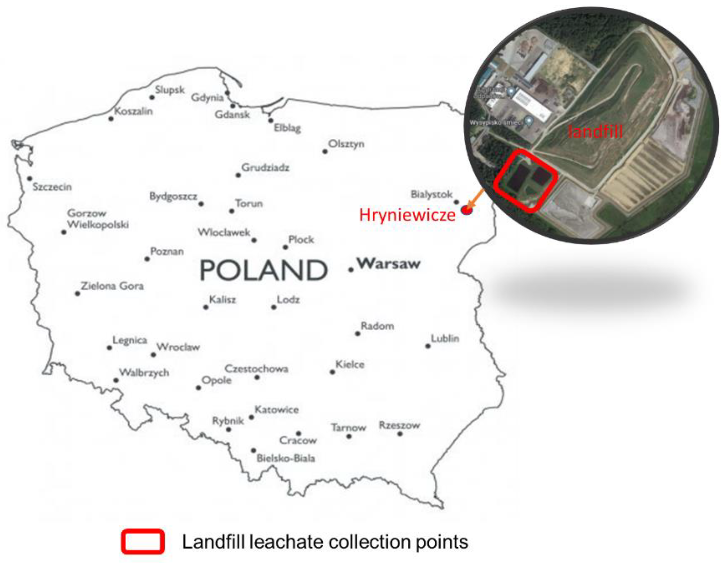
The LL for research came from a landfill, which is located in the Podlaskie voivodship in Hryniewicze (Poland), receiving waste from Białystok and the surrounding towns and communes (Figure 1). The landfill accepts municipal solid waste and industrial solid waste—non-toxic—which do not require the development of a special storage technology. Leachate separated during waste storage is collected in two ground-structure retention tanks, sealed with a geomembrane. A total of approx. 40 dm3 of LL was collected for the tests at various points from the retention reservoirs. The collected LL in the spring was mixed, thus obtaining a uniform sample intended for analytical and technological research. Properties of the analyzed LL were characterized in manuscript by Jabłońska-Trypuć et al. [17].
The soil intended for the pot experiment was typical brown soil from farmland collected in the vicinity of Białystok from a depth of 0–20 cm from several points. The collected soil was transported to the laboratory of the Department of Chemistry, Biology and Biotechnology of the Bialystok University of Technology; the roots and stones were removed, then mixed and sieved through a sieve with a mesh size of 1.0 cm. The main soil characteristics are as follows: pH 6.82, TOC—20.18%; NT.—0.28%; heavy metals (mg/kg): Cr—13.22, Zn—37.98; Cd—0.13; Cu—15.65; Ni—4.73; Pb—12.97; Hg—0.13.
2.2. Pot Experiment and Sampling Preparation
In order to avoid the influence of uncontrolled factors on the effect of the experiment, it was conducted in ex situ conditions. The experiment was conducted in pots with dimensions: 20 × 24 cm. Each pot with soil was successively applied with a single dose of 85 kg/ha (50% of the N dose, 50LL) and 170 kg/ha (100% of the N dose, 100LL) in three repetitions. The LL dose was determined on the basis of the nitrogen content, the annual amount of which for agricultural purposes may not exceed 170 kg N/ha, and on the basis of Panchoni et al. [18]. Pots were also prepared with no LL added, as the control (C). Mustard (Sinapis alba) seeds were sown before the LL was added. During the experiment, the pots were watered regularly, keeping the humidity at the level of 60–70% of the maximum water-holding capacity using deionized water.
The experiment was conducted for 6 weeks. Soil samples were collected three times with a two-week interval (T1, T2, T3). From each pot, 3 soil samples were taken from a depth of 0–10 cm. A sample of each variant was 3 sub-samples taken from each pot. The soil was dried to obtain air dry mass. The samples prepared in this way were stored in closed containers for physicochemical and biochemical analyses. Fresh soil samples intended for genetic and microbiological tests were collected in sterile containers and stored in a freezer (−70 °C) for further analysis.
At the end of the experiment, SF was prepared according to Ming et al. [19] with modifications. Extraction of soil with deionized water 1: 2.5 (w/v) was performed by shaking at 180 rpm for 2 h at 25 °C. The mixture was then centrifuged at 3800 rpm for 10 min, then the supernatant was immediately passed through a 0.45 µm membrane filter. The obtained SF was used for physicochemical analyses and determination of its cytotoxicity.
2.3. Physicochemical Analyses of Soil and SF Samples
The soil pH was measured in distilled water soil extract (1:2.5, w/v) using a pH-meter (Mettler-Toledo, Polaris Parkway Columbus, OH, USA). Total organic carbon content (TOC) in the FS and in the soil was estimated by using Multi N/C 3100 (Analytik Jena, Tewksbury, MA, US) apparatus. The content of selected ions (F−, Cl−, NO2−, NO3−, SO42−, Na+, K+, Mg2+, Ca2+, NH4+) was determined on an ion chromatograph Dionex ICS 5000+ (Thermo Scientific, Waltham, MA, USA). The content of Na, K, Ca and Mg was determined on a flame atomic absorption spectrometer iCE3300 (Thermo Scientific, Waltham, MA, USA). Selected metals (Al, Fe), including heavy metals (Ni, Cu, Zn, Cd, Pb, Cr) and other elements (S, P) were determined on the X-ray fluorescence spectrometer S2 PICOFOX (Bruker, Poznań, Poland). Soil samples were mineralized with aqua regia (HCl and HNO3 in a ratio of 3:1, 9 mL:3 mL) using Advanced Microwave Digestion System Ethos Easy (Milestone, Sorisole (BG), Italy) before the analyses. The content of 16 PAHs (naphthalene—NA; acenaphthylene—Ace; acenaphthene—Acy; fluorene—FL; phenanthrene—Phe; anthracene—AN; fluoranthene—Flu; pyrene—PY; benzo (a) anthracene—BaA; chrysene—Chry; benzo (b) fluoranthene—BbF; benzo (k) fluoranthene—BkF; benzo (a) pyrene—BaP; indeno (1, 2,3-cd) pyrene—IP; dibenzo (a, h) anthracene—DBA; benzo (g, h, i) perylene)—BP was determined using the system GC-MS Triple Quad 7890B (Agilent Technologies, Inc., Santa Clara, CA, USA). The analyses were carried out at the Faculty Chemical Laboratory of the Białystok University of Technology.
2.4. Bacteria Abundance and Soil Enzymatic Activity
The total bacteria number was determined by using plate-count techniques with 10% of trypticase soy agar [20] and King B agar for isolation of fluorescent Pseudomonas sp. [21]. The number of fungi was determined using the plate-count techniques on a medium containing Bengal rose [22]. The abundance of studied microorganisms was expressed as cfu (colony forming unit) per g of soil DW (cfu/g DW).
2.5. DNA Extraction and Microbial Communities Fingerprinting by T-RFLP
Genomic DNA from the soil was extracted using the GeneMATRIX Soil DNA Purification Kit (EURx, Gdańsk, Poland) according to the manufacturer’s protocol. The amount of DNA obtained was determined using the QuantiFluor® dsDNA Dye (Promega, WI, USA) on GloMax®-Multi Microplate Multimode Reader (Promega Corporation, Madison, WI, USA). The quality of the soil DNA was assessed using Lambda Bio+ spectrophotometer (PerkinElmer, Boston, MA, USA) with ultra-micro sample-volume spectrometer cell TrayCell (Hellma GmbH, Müllheim/Baden, Germany).
Bacterial (16S rDNA) and fungal (ITS) gene amplification was performed using the primers and conditions reported by Mattana et al. [25] with the use of Labcycler SensoQuest termocycler (SensoQuest GmbH, Germany). The 25 µL reaction mixture consisted of: 15 µL GoTaq® G2 Colorless Master Mix (Promega Corporation, Madison, WI, USA), 0.5 µM of each primer and 2 µL (5 ng/µL) of soil DNA. The purity of the PCR product was determined by agarose gel electrophoresis (1.2%, w/v).
The amplification products were enzymatically purified using Exo-BAP Mix (EURx, Gdańsk, Poland) according to the manufacturer’s assay, and then 7 µL of the purified product was digested with 6U of the restriction enzyme HaeIII (10 U/µL) (Promega Corporation, Madison, WI, USA) in a restriction buffer in a final volume of 10 µL for 2 h at 37 °C. The resulting product was desalted using the PCR/DNA Clean-Up Purification Kit (Promega Corporation, Madison, WI, USA). Further procedures, including the T-RFLP protocol, are described in Wydro et al. [26].
The relative abundance of restriction fragments (T-RFs) was determined by the quotient of the area of each peak and the sum of the area of all peaks in one sample. The analysis included peaks in the 50–600 bp range with a relative abundance greater than 1%.
2.6. Analysis of FL Cytotoxicity in Eukaryotic Cells
The potential toxicity of the SF was studied using human cell line—Caco-2—which was obtained from the American Type Culture Collection (ATCC Collection). This cell line is an appropriate model approved by the FDA and used in toxicology research. Cells were cultured in EMEM medium containing 4.5 mg/mL glucose supplemented with 10% FBS (fetal bovine serum), penicillin (100 U/mL) and streptomycin (100 μg/mL) at 37 °C in a 5% CO2 atmosphere. Cell viability was tested at the following concentrations of SF samples: 0.1%, 0.5%, 1%, 1.5%, 2%, 2.5%, 5%, 10%, 15%, 20%, and 30%. The incubation time was 24 h.
The tested SF samples were stored in a refrigerator at 4 °C. They were added to the culture medium in an amount allowing to achieve final concentrations in the range: 0.1%, 0.5%, 1%, 1.5%, 2%, 2.5%, 5%, 10%, 15%, 20% and 30%. Control cells were incubated without test compounds in the culture medium.
Cytotoxicity was measured using an MTT assay based on the reaction with 3-(4,5-dimethylthiazol-2-yl)-2,5-diphenyltetrazolium bromide [17]. Caco-2 cells were seeded in 96-well plates at a density of 2 × 10⁴ cells/well. Cells were then cultivated for 24 h, after which the medium was changed to contain the test compounds at the described concentrations. After the 24-h incubation time, cells were washed three times with PBS and incubated with a MTT solution (5 mg/mL in PBS) for 2 h at 37 °C in 5% CO2 in an incubator. Then, 100 µL of DMSO was added and the culture was incubated in the dark for another 2 h. After this time, the absorbance at 570 nm was measured in a GloMax®-Multi Microplate Multimode Reader (Promega Corporation, Madison, WI, USA). Cell viability of both lines was calculated as the percentage of control cells incubated without test compounds. All experiments were performed in triplicate.
2.7. Study of the Level of Apoptosis
Apoptosis was tested using a Caspase-Glo® 3/7 assay bioluminescent assay based on Caspase 3/7 activity measurements. Caco-2 cells were seeded in white 96-well plates and grown for 24 h. After this time, they were subjected to the test SF in the concentrations described above. The incubation time was 24 h. After the specified incubation time, the culture medium was removed and the test was performed according to the manufacturer’s instructions. Caspases 3/7 activity was measured with the use of luminescent assay. An utilized assay is based on the presence of substrate which contains the tetrapeptide sequence DEVD in a reagent optimized for caspase activity, luciferase activity and cell lysis. Cell lysis is followed by the caspase cleavage of the substrate and generation of the luminescent signal. The amount of caspase activity present in the sample is proportional to the luminescence level. The assay is based on the thermostable luciferase activity, which generates a stable, “glow-type” luminescent signal [17]. A GloMax®-Multi Microplate Multimode Reader plate reader (Promega Corporation, Madison, WI, USA) was used to perform the measurements. The test was carried out in triplicate to ensure the reproducibility of the results and to perform the statistical analysis.
2.8. Statistical Analysis
The differences between the variants of the experiment for the tested soil properties were determined using the ANOVA analysis of variance. If there were significant differences between the experimental factors, the means were compared using the HSD post-hoc Tukey test for p < 0.05. Dunnett’s test at significance levels p < 0.05 and p < 0.01 was used to compare means for controls. In order to determine the relationships between studied variables, the Pearson correlation analysis was performed by significance level at p < 0.05.
Genetic diversity of bacteria/fungi was assessed by calculating the Shannon (H’) and Evenness (E) indices [27]. The similarity between the defined T-RFs for bacteria and fungi in the test variants is presented in Venn diagrams (https://bioinformatics.psb.ugent.be/webtools/Venn/ (accessed on 10 October 2022)). The analyses and calculations were performed using the Statistica 13.3 package and PAST 4.03 (https://www.nhm.uio.no/english/research/resources/past/ (accessed on 10 October 2022)).
3. Results
3.1. Physicochemical Properties of Soil and SF Samples
The main physicochemical properties of the analyzed soil and SF samples are presented in Table 1. In soil and in SF, the pH was similar, ranging from 6.8 for the control pots to 7.6 after 100LL application. Among all the analyzed metals in the soil, the highest values were noted for Al, Pb and Fe. In turn, in the SF after the application of the LL, the highest content were observed for Cl, Na, Ca and K. Moreover, among the tested ions, the highest values were obtained for Cl−, K+, NO3−, Na+, Ca2+ and NH4+. Among the PAHs group of compounds present in soil, after the LL application, the highest values were for BbF, Flu, Py, Chry and BaA. On the other hand, in SF, the highest PAH values were obtained for NA and Flu. On average, the sum of 16 PAHs in soil was at the level of about 141.26 µg/kg, while in SF it was 14.29 ng/L.
3.2. Bacteria Abundance and Soil Enzymatic Activity
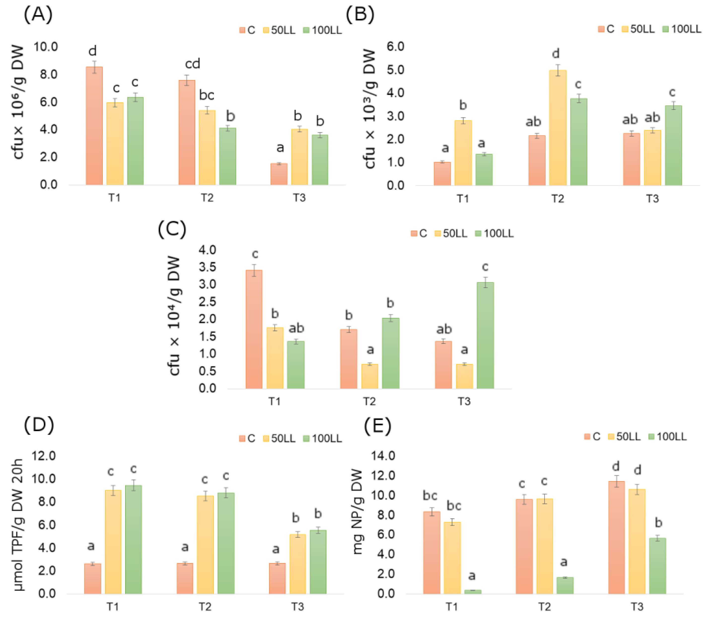
Based on the obtained results, a positive effect of LL on the total number of bacteria was found (Figure 2A). The average total number of bacteria in the soils after LL application varied depending on the date and dose of the LL. The highest total number of bacteria was observed in pots after 50LL application (1.05 × 10⁷ cfu/g DM, T1), while the lowest was in the C pots (1.43 × 10⁶ cfu/g DM, T3). On average, for all fertilization variants used, the highest total number of bacteria was observed in T1 and the lowest in T3.
On average, the highest total number of fungi was recorded in pots after 50LL application (approx. 3.40 × 10³ cfu/g DM) and the lowest in C pots (1.81 × 10³ cfu/g DM). Regarding the sampling date, an increase in the total number of fungi in T2 was obtained and it was about 53% higher as compared to T1 and about 25% higher as compared to T3 (Figure 2B). In turn, on average, the highest number of fluorescent Pseudomonas sp. was observed in samples taken from pots with 100LL (approx. 3.37 × 10⁴ cfu/g DM) and the lowest in pots after 50LL (1.73 × 10⁴ cfu/g DM) (Figure 2C).
The LL doses used in the study influenced the activity of Dha and β-Glu (Figure 2D,E). Dha activity was the highest after application of 100LL (7.78 µmol TPF g/DM × 20 h) and the lowest in C (2.40 µmol TPF g/DM × 20 h). Generally, a higher mean Dha activity was obtained in the samples collected in T1, while the lowest was in the last sampling date (T3). On the other hand, the highest β-Glu activity was found in the C pots and amounted to 9.69 µg NP/g DM, while the lowest in pots after 100LL application (2.40 µg NP/g DM). In subsequent measurements, the activity of β-Glu increased and in the last one the average for all variants was 9.09 µg NP/g DM.
3.3. Microbial Community Structure Shifts
The results showed that the genetic patterns characterizing the community of bacteria and fungi in the soil, determined by the T-RFLP method, differed depending on the research variant and the sampling date. This dissimilarity was demonstrated in the form of the relative abundance of defined T-RFs and by calculating the H ‘and E indices and in Venn diagrams.
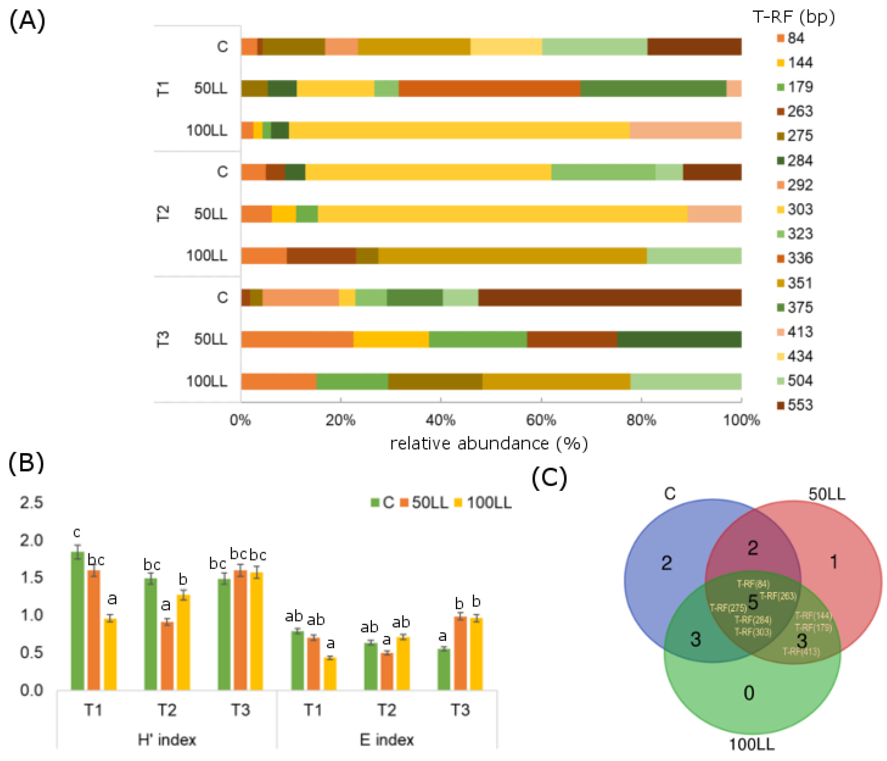
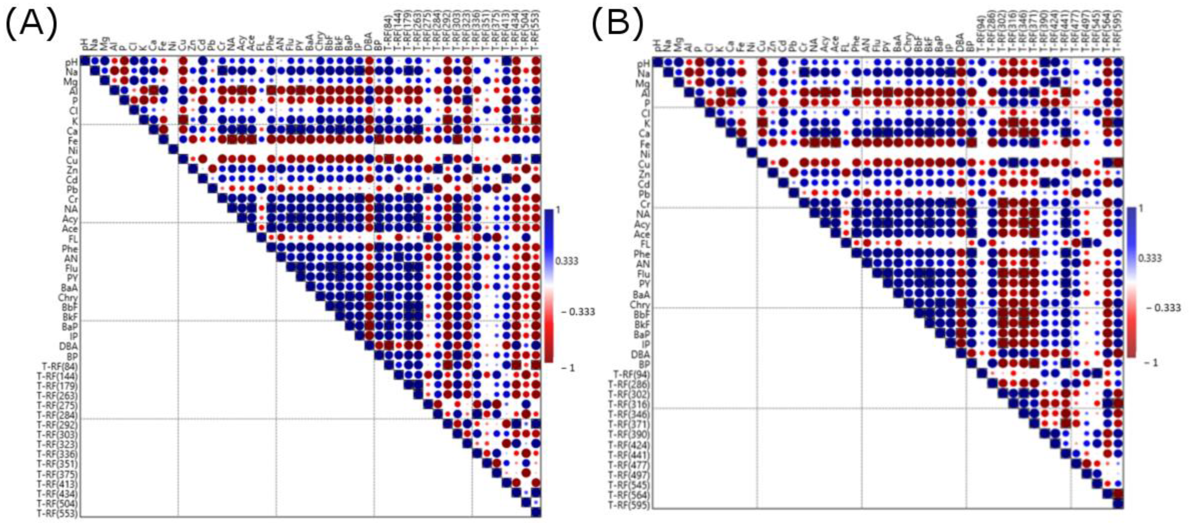
By analyzing the T-RFs patterns defined for the bacteria, it was found that their length ranged from 84 to 553 bp (Figure 3). After digestion with HaeIII, 16 T-RFs were obtained. It was observed that for all tested variants throughout the research period, the specific patterns were T-RF (84) bp, (263) bp, (275) bp, (284) bp and (303) bp. Among them, the highest relative abundance was found in T-RF (303) bp, ranging from 3% to 74%. In the soils treated with LL, T-RF (144) bp, (179) bp and (413) bp were specific, and their relative abundance did not exceed 22%. The data regarding H’ index showed that they have a low level, with values ranging from 0.9 (50LL, T2) to 1.8 (C, T1) and higher values in T1 and T2 were recorded than in C in variants with LL. Comparing the determined E coefficients for bacterial T-RFs, it was observed that in T1 a significantly higher level of the E coefficient was in C, while in T3 higher values were noted for variants with LL.
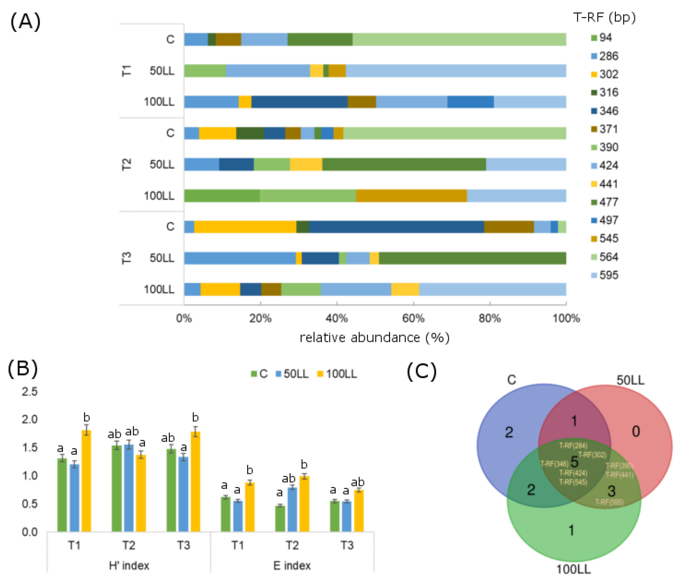
As shown by the data presented in Figure 4, 14 fungal T-RFs ranging in length from 94 to 595 bp were obtained after digestion with HaeIII. T-RF (284) bp, (302) bp, (346) bp, (424) bp and (545) bp were found to be characteristic of all test variants. Among them, the highest relative abundance was observed for T-RF (346) bp and its value ranged from 9% to 46%. T-RF (390) bp, (441) bp and (595) bp were characteristic for the LL variants, however the highest realty abundance was noted for T-RF (595) bp, ranging from 10% to 58%. Similar for bacteria, the H’ index calculated for fungal T-RFs was not high and ranged from 1.2 (50LL, T1) to 1.8 (100LL, T1, T3). The designated E index for all research variants did not exceed 1.0. and was the highest at 100LL at each of the three sampling dates.
As presented in Figure 5, there were both positive and negative significant correlations between the tested soil properties and the relative abundance of the defined TRFs. As a result of the statistical analysis, the most significant correlations were found between bacteria and fungi abundance and heavy metals and PAHs content.
3.4. Effect of FL on Cytotoxicity and Apoptosis Level
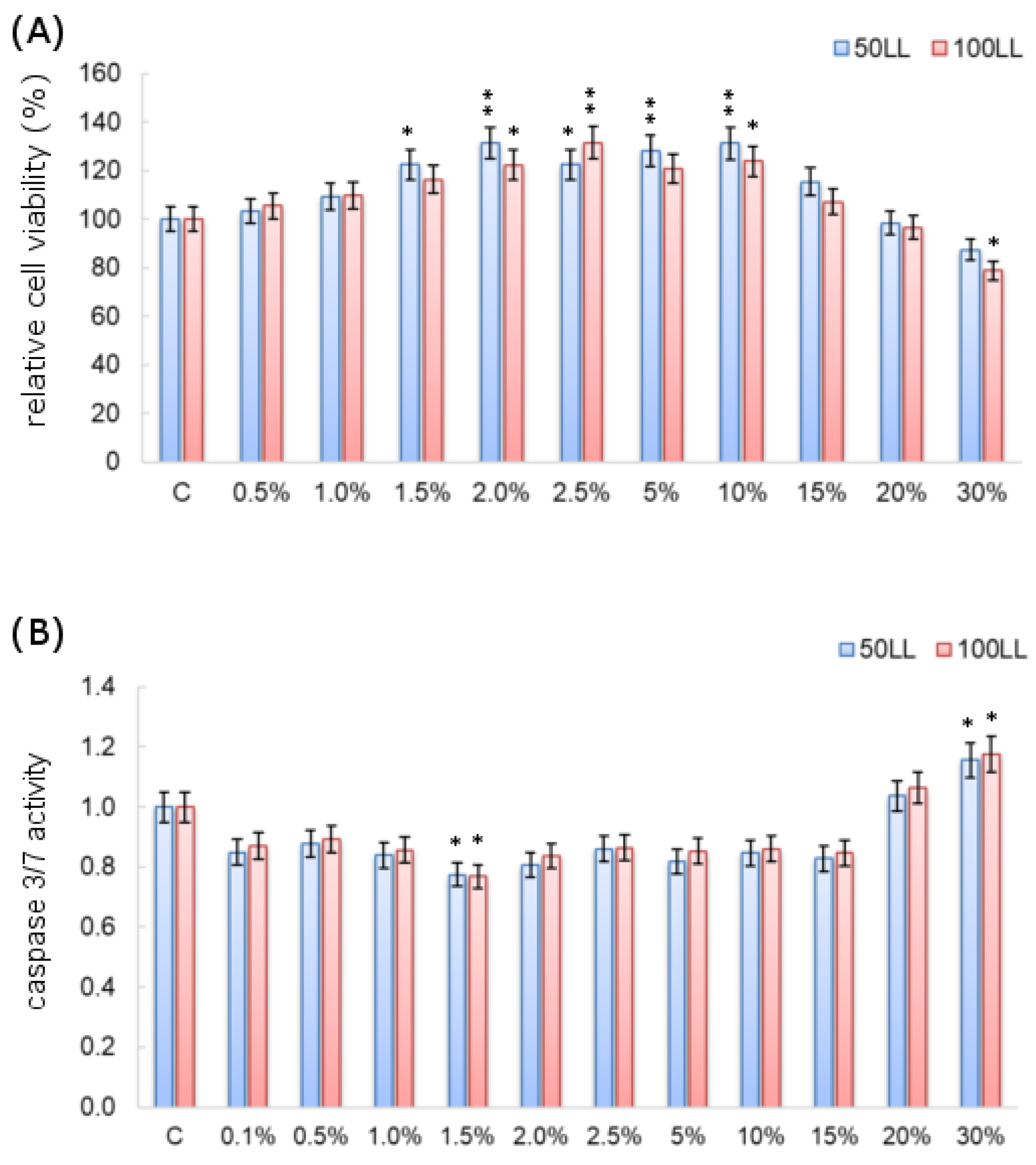
In the Caco-2 cell line, no significant differences were observed between the results obtained for both the tested SF doses and the cytotoxicity/apoptosis tests (Figure 6). The results of the MTT test showed that the viability of the Caco-2 cells significantly increased under the influence of the analyzed SF, with the highest increases in the tested parameter observed at the concentrations of 2%, 2.5%, 5% and 10%, where they reached about 130% compared to 100% relative lifetime control sample. On the other hand, as could be expected, a significant decrease in cell proliferation was observed under the influence of the highest applied SF concentrations. As the SF concentration increased, the cell viability decreased.
The results of the analysis of the level of apoptosis also showed a large decrease in the activity of caspases 3/7 in the case of Caco-2 cells compared to the control cells at the concentrations that significantly stimulated the viability of the cells. This is in agreement with the results of the cell viability assay. The tested effluents did not cause apoptosis in neoplastic cells, and even inhibited it at low concentrations. On the other hand, at the highest concentrations used, the SF caused a significant increase in the caspase 3/7 activity level, even by about 17% at the highest concentration analyzed.
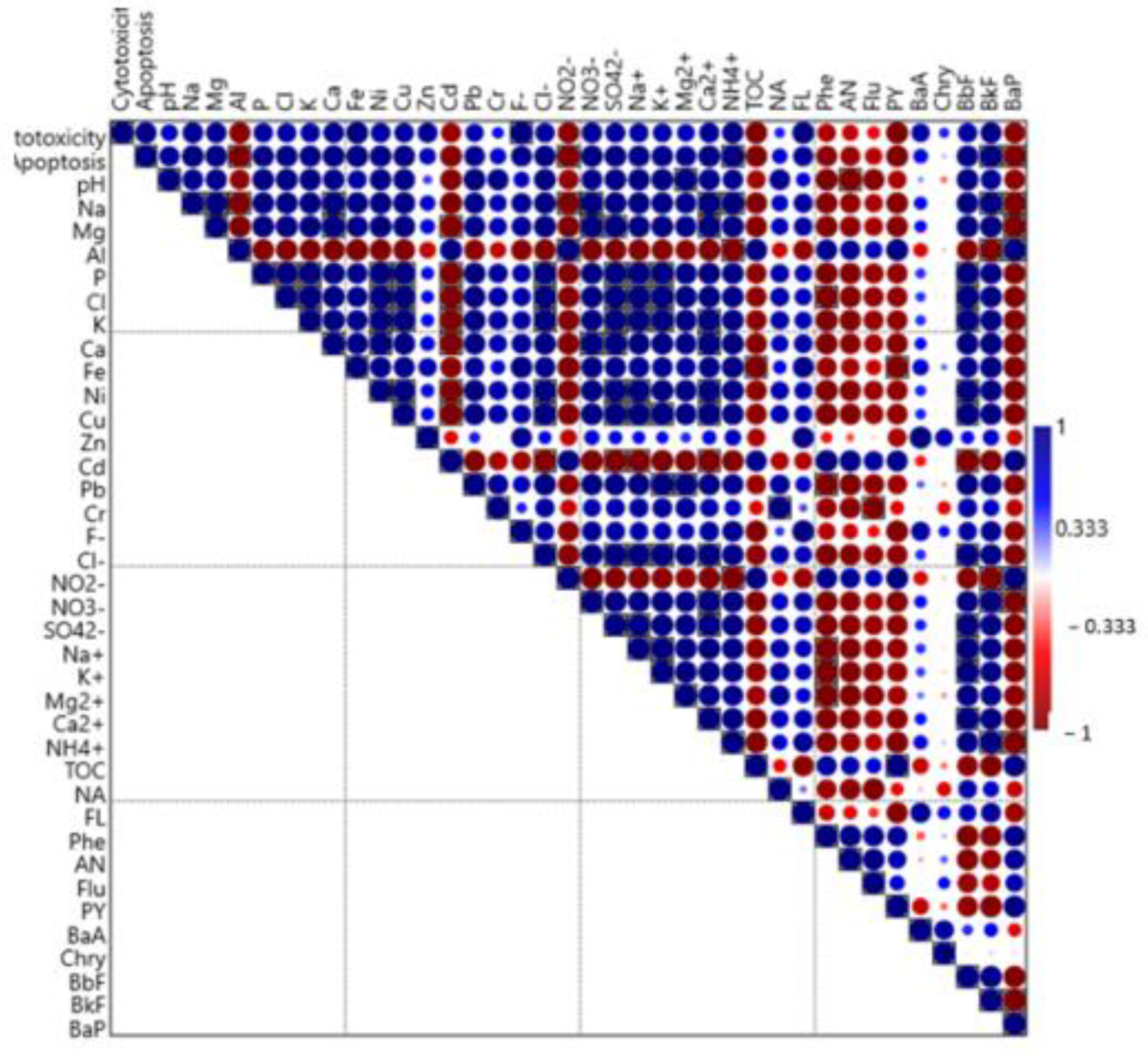
The results of the statistical analysis presented in Figure 7 clearly indicate a significant correlation between the cytotoxicity of tested SF towards human cells and the presence of heavy metals, PAHs and ammonia in the studied SF. An apoptosis level is also positively related with the analyzed physicochemical properties of analyzed complex matrix.
4. Discussion
4.1. Effect of LL on Microbial Activity and Community Structure
Leachate from landfills are potentially hazardous by-products resulting from the storage of various types of waste. Leachate entering the groundwater circulation becomes a threat not only to the environment but also to human health [17]. One of the elements of the environment that may be directly affected by LL is the soil as well as the microorganisms that inhabit it [2,7]. Therefore, biological parameters such as the activity of soil enzymes, the number and structure of microorganisms are valuable tools used to assess changes in the soil environment under the influence of heavy metals and organic pollutants [28,29].
According to Panchoni et al. [18], an important bioindicator of soil quality is its enzymatic activity. It is a sensitive parameter and even the smallest changes in soil environment affect its shaping. Moreover, the enzymatic activity of soils is one of the main measurements of the metabolic potential as well as its fertility and quality. To assess biochemical activity, enzymes such as Dha and β-Glu are often taken into account, as they play a key role in the decomposition of organic matter and in the carbon cycle [11,18].
Our own studies showed an increase in Dha activity in the soil samples treated with LL compared to the control. This was not confirmed by the studies conducted by Panchoni et al. [18]. In turn, the activity of β-Glu was lower in variants with LL than in C, but its activity increased in subsequent pieces of research. The studies conducted by Kandzior-Ciupa et al. [28] and Baran et al. [30] indicated that the activity of soil enzymes was related to the content of heavy metals and organic pollutants (including PAHs) in soils, the source of which are organic mixtures. According to Zhu et al. [31], LL biotoxicity was mainly correlated with the content of ammonia and heavy metals in them. Some metals, present in low concentrations in soil, can stimulate the action of some enzymes, while others may be inhibitors of their activity This is determined by the composition and properties of soil [32].
As indicated by the data obtained, the number of the soil microorganisms under the influence of LL changed, i.e., the total number of bacteria, fungi and fluorescent Pseudomonas sp. It was also observed that the trend of changes depended on the sampling date. According to Teng et al. [2], LL is a mixture of soluble organic matter, heavy metals, PAHs and others, which are toxic and, after introduction into the soil, may affect its activity and the number of microorganisms. The presence of substances with toxic properties may interfere with the adaptability of some microorganisms, which results, e.g., in reducing their number [33]. This may explain the decreasing tendency of the total number of bacteria and fluorescent Pseudomonas sp. observed in soil samples with LL collected at the first date. In the case of fungi, no such relationship was found after the application of 50LL. The ability to adapt microorganisms to difficult environmental conditions is a feature that depends on the type and structure of the microorganism. In addition, the mechanisms that protect the cells against the toxic effects of pollutants often result from the inherent physiological properties of individual microorganisms. According to the literature’s data, the phenomenon of reducing the population of soil bacteria under the influence of organic treatment as opposed to the population of fungi was observed [34], which is in accordance with the obtained results. In addition, as Macdonald et al. [35] and Salehi et al. [1] state, it should also be emphasized that many of the components contained in the LL are a medium for microorganisms that provide many biodegradable organic compounds, ions, micro- and macronutrients (especially nitrogen) necessary for growth and development. In addition, LL introduced into the soil changes the pH (often making it higher), which may improve the bioavailability of its components. This may explain an increase in the number of tested microorganisms in the final phase of the experiment (T3) in the variants with LL. It is also worth mentioning that the experiment used plants that could contribute to the reduction in some pollutants through phytoextraction and phytodegradation.
In our research, the T-RFLP approach was used to assess the community structure of microorganisms, which is an indirect technique from the genetic fingerprinting group of methods. It allows for the differentiation of microorganisms and the assessment of the dynamics of changes in their structure depending on soil conditions [36].
The presented results indicate that the application of LL to the soil changes the structure of the microbial community, their richness and relative abundance. The direction of these changes varies and depends on the type of pollutants and their concentration. Heavy metals play an important role in shaping the structure of soil microflora, as reported by Li et al. [33]. Moreover, the addition of LL containing mainly organic matter to the soil causes a modification in the environment as a result of changes taking place in it, which results in shifts in the structure of the microbial community. Differentiation in the structure of soil microorganisms under the influence of LL in comparison to the control were also noted by Gu et al. [7].
The presence of important nutrients in LL ensures that some consortia of microorganisms have the ability to acclimatize in difficult conditions and at the same time are capable of decomposing pollutants. This may explain the existence of specific T-RFs common to both C and LL variants. Research results also indicate that LL contributes to the development of other microorganisms resistant to potential toxic components in LL. This is evidenced by the specific T-RFs found only in soils with LL obtained in our study. In an investigation conducted by Gu et al. [7], Bacilli, Clostridia, Thermacetogeniaza and Firmicutes phyla were key taxa in soils exposed to LL.
It is important to maintain an appropriate structure of bacteria and fungi in soil in order to ensure high soil fertility and the proper course of processes involving the circulation of nutrients in the ecosystem [37]. In the presented study, the fungi community was generally characterized by higher diversity (expressed by H’ and E indices) in the soils treated with LL. As indicated by Ondreičková et al. [27], fungi communities are able to accept even higher concentrations of pollutants and the changes in them are smaller as compared to bacterial communities. In addition, fungi play an important role in the soil/plant system, as they are responsible for the bioavailability of some nutrients (e.g., N, P, K, Fe), they are involved in the production of enzymes and they are also responsible for protecting the functionality of the agroecosystem [38,39,40].
4.2. Effect of SF on Cytotoxicity and Apoptosis Level
Human cell lines are an increasingly widely used model in the analysis of the impact of environmental pollutants on the human body. They allow for quick, precise and relatively less expensive obtaining of a large number of cytotoxicity results—the so-called screening, in no time. Taking into account bioethical considerations, they are also an excellent alternative to animal testing. The most frequently used lines in cytotoxicity studies include lines such as Caco-2, HepG2 or lymphocytes and other cell lines.
According to Gosh et al. [41], the HepG2 cell line was exposed to LL in the concentration range of 0 to 20% in the MTT test and they observed a significant cytotoxic effect. The obtained results indicate changes in the relative viability of cells under the influence of SF. Baderna et al. [13] also investigated the cytotoxicity of the LL in a similar concentration range and found them to be cytotoxic even at low concentrations, but also after longer incubation times. The presented results indicating the cytotoxic and carcinogenic potential of the studied SF are consistent with the literature data. The obtained results may be related to the physicochemical properties of SF. The SF contain cytotoxic and carcinogenic compounds, as indicated by the results of the chemical analyses. Heavy metals, PAHs and ammonia present in the samples can cause oxidative stress and thus induce apoptosis. This has also been demonstrated by Eckers et al. [42] in their research on the HepG2 line. The obtained data support the hypothesis that the apoptotic pathway may be one of the mechanisms of cytotoxicity of the studied SF.
5. Conclusions
LL as a by-product resulting from waste disposal is a mixture of various compounds, including those potentially hazardous to the environment and human health. The presented study focused on two aspects: (a) the influence of LL on selected microbiological properties of soil; and (b) the cytoxicity of SF (constituting a liquid mixture flowing through the soil) in the Caco-2 cell line. It was found that under the influence of LL, the activity of Dha in the soil increased as compared to the control and decreased during the experiment. On the other hand, the activity of β-Glu in the soil was significantly lower than in the control after the application of 100LL throughout the research period, although its level was higher at the end of the experiment. It was also shown that the abundance and structure of the microbial community in the soil treated with LL were significantly different and changed over time. It transpired that at the beginning of the experiment the diversity of bacteria in the soils with LL was lower than in the control and increased during the experiment. Taking into account the fungal community, the significantly highest diversity and evenness was found in soils with 100LL out of all the study data. The obtained dependencies were influenced by the physicochemical properties of soil such as the content of heavy metals and micro- and macronutrients constituting a potential reservoir of nutrients for microorganisms.
Considering the effect of SF on the Caco-2 cell line, no significant differences were observed between the results obtained with the tested doses of SF for both the cytotoxicity and apoptosis tests. Due to the observed proliferation of neoplastic cells, it can be concluded that the tested matrices are potentially toxic material, the introduction of which to the environment should be conducted with caution.
In summary, the obtained results allowed the confirmation of the research hypothesis and at the same time provided an initial insight into the biological characteristics of the soil exposed to direct contact with LL from the landfill and provided valuable information from the point of view of soil microbiology and human toxicology. For future research on the effects of LL and SF on the soil and human, the focus should be on understanding soil/plant/human interactions and identifying LL exposure-resistant strains that may be important in bioaugmentation techniques.
Author Contributions
Conceptualization, U.W. and A.J.-T.; methodology, U.W., E.W. and A.J.-T.; investigation, U.W., J.L., E.W. and G.S.; writing—original draft preparation, U.W. and E.W.; writing—review and editing, A.J.-T.; visualization, U.W. and E.W. All authors have read and agreed to the published version of the manuscript.
Funding
This work was financially supported by the Ministry of Education and Science, Poland, under the research project number WZ/WB-IIŚ/6/2022.
Data Availability Statement
Not applicable.
Conflicts of Interest
The authors declare no conflict of interest.
References
- Salehi, N.; Azhdarpoor, A.; Shirdarreh, M. The Effect of Different Levels of Leachate on Phytoremediation of Pyrene-Contaminated Soil and Simultaneous Extraction of Lead and Cadmium. Chemosphere 2020, 246, 125845. [Google Scholar] [CrossRef] [PubMed]
- Teng, C.; Zhou, K.; Peng, C.; Chen, W. Characterization and Treatment of Landfill Leachate: A Review. Water Res. 2021, 203, 117525. [Google Scholar] [CrossRef] [PubMed]
- Omar, H.; Rohani, S. Treatment of Landfill Waste, Leachate and Landfill Gas: A Review. Front. Chem. Sci. Eng. 2015, 9, 15–32. [Google Scholar] [CrossRef]
- Torretta, V.; Ferronato, N.; Katsoyiannis, I.A.; Tolkou, A.K.; Airoldi, M. Novel and Conventional Technologies for Landfill Leachates Treatment: A Review. Sustainability 2017, 9, 9. [Google Scholar] [CrossRef] [Green Version]
- Peng, Y. Perspectives on Technology for Landfill Leachate Treatment. Arab. J. Chem. 2017, 10, S2567–S2574. [Google Scholar] [CrossRef] [Green Version]
- Christensen, T.H.; Kjeldsen, P.; Bjerg, P.L.; Jensen, D.L.; Christensen, J.B.; Baun, A.; Albrechtsen, H.-J.; Heron, G. Biogeochemistry of Landfill Leachate Plumes. Appl. Geochem. 2001, 16, 659–718. [Google Scholar] [CrossRef]
- Gu, Z.; Feng, K.; Li, Y.; Li, Q. Microbial Characteristics of the Leachate Contaminated Soil of an Informal Landfill Site. Chemosphere 2022, 287 Pt 2, 132155. [Google Scholar] [CrossRef]
- Khalil, C.; Al Hageh, C.; Korfali, S.; Khnayzer, R.S. Municipal Leachates Health Risks: Chemical and Cytotoxicity Assessment from Regulated and Unregulated Municipal Dumpsites in Lebanon. Chemosphere 2018, 208, 1–13. [Google Scholar] [CrossRef]
- Sang, N.; Li, G. Genotoxicity of Municipal Landfill Leachate on Root Tips of Vicia Faba. Mutat. Res. 2004, 560, 159–165. [Google Scholar] [CrossRef]
- Clarke, B.O.; Anumol, T.; Barlaz, M.; Snyder, S.A. Investigating Landfill Leachate as a Source of Trace Organic Pollutants. Chemosphere 2015, 127, 269–275. [Google Scholar] [CrossRef]
- Tiwari, R.; Dwivedi, B.S.; Sharma, Y.M.; Sharma, A.; Dwivedi, A.K. Activities of β-Glucosidase, Phosphatase and Dehydrogenase as Soil Quality Indicators: A Review. Int. J. Curr. Microbiol. Appl. Sci. 2019, 8, 834–846. [Google Scholar] [CrossRef]
- De Vrieze, J.; Ijaz, U.Z.; Saunders, A.M.; Theuerl, S. Terminal Restriction Fragment Length Polymorphism Is an “Old School” Reliable Technique for Swift Microbial Community Screening in Anaerobic Digestion. Sci. Rep. 2018, 8, 16818. [Google Scholar] [CrossRef] [PubMed] [Green Version]
- Baderna, D.; Caloni, F.; Benfenati, E. Investigating Landfill Leachate Toxicity in Vitro: A Review of Cell Models and Endpoints. Environ. Int. 2019, 122, 21–30. [Google Scholar] [CrossRef] [PubMed]
- Glatt, H.; Gemperlein, I.; Setiabudi, F.; Platt, K.L.; Oesch, F. Expression of Xenobiotic-Metabolizing Enzymes in Propagatable Cell Cultures and Induction of Micronuclei by 13 Compounds. Mutagenesis 1990, 5, 241–249. [Google Scholar] [CrossRef] [PubMed]
- Sun, H.; Chow, E.C.; Liu, S.; Du, Y.; Pang, K.S. The Caco-2 Cell Monolayer: Usefulness and Limitations. Expert Opin. Drug Metab. Toxicol. 2008, 4, 395–411. [Google Scholar] [CrossRef]
- Sambuy, Y.; De Angelis, I.; Ranaldi, G.; Scarino, M.L.; Stammati, A.; Zucco, F. The Caco-2 Cell Line as a Model of the Intestinal Barrier: Influence of Cell and Culture-Related Factors on Caco-2 Cell Functional Characteristics. Cell Biol. Toxicol. 2005, 21, 1–26. [Google Scholar] [CrossRef]
- Jabłońska-Trypuć, A.; Wydro, U.; Wołejko, E.; Pietryczuk, A.; Cudowski, A.; Leszczyński, J.; Rodziewicz, J.; Janczukowicz, W.; Butarewicz, A. Potential Toxicity of Leachate from the Municipal Landfill in View of the Possibility of Their Migration to the Environment through Infiltration into Groundwater. Environ. Geochem. Health 2021, 43, 3683–3698. [Google Scholar] [CrossRef]
- Panchoni, L.C.; Santos, C.A.; Kuwano, B.H.; Carmo, K.B.; Cely, M.V.T.; Oliveira-Júnior, A.G.; Fagotti, D.S.L.; Cervantes, V.N.M.; Zangaro, W.; Andrade, D.S.; et al. Effect of Landfill Leachate on Cereal Nutrition and Productivity and on Soil Properties. J. Environ. Qual. 2016, 45, 1080–1086. [Google Scholar] [CrossRef]
- Ming, L.; Li, L.; Zhang, Y.; Lin, W.; Wang, G.; Zhang, S.; Guo, P. Effects of Dissolved Organic Matter on the Desorption of Cd in Freeze–Thaw Treated Cd-Contaminated Soils. Chem. Ecol. 2014, 30, 76–86. [Google Scholar] [CrossRef]
- Kaczynski, P.; Lozowicka, B.; Wolejko, E.; Iwaniuk, P.; Konecki, R.; Dragowski, W.; Lozowicki, J.; Amanbek, N.; Rusilowska, J.; Pietraszko, A. Complex Study of Glyphosate and Metabolites Influence on Enzymatic Activity and Microorganisms Association in Soil Enriched with Pseudomonas Fluorescens and Sewage Sludge. J. Hazard. Mater. 2020, 393, 122443. [Google Scholar] [CrossRef]
- Wołejko, E.; Wydro, U.; Jabłońska-Trypuć, A.; Butarewicz, A.; Łoboda, T. Pseudomonas fluoresces occurrence in soil after fertilization with sewage sludge. Econ. Environ. 2018, 65, 10. [Google Scholar]
- Kramer, C.L.; Pady, S.M. Inhibition of Growth of Fungi on Rose Bengal Media by Light. Trans. Kans. Acad. Sci. 1961, 64, 110–116. [Google Scholar] [CrossRef] [PubMed]
- Tabatabai, M.A. Soil enzymes. In Methods of Soil Analysis: Microbiological and Biochemical Properties; Weaver, R.W., Angle, J.S., Botttomley, P.S., Eds.; Soil Science Society of America: Madison, WI, USA, 1994. [Google Scholar]
- Alef, K.; Nannipieri, P. Β-Glucosidase Activity. In Methods in Applied Soil Microbiology and Biochemistry; Academic Press: Cambridge, MA, USA; Harcourt Brace & Coy Publishers: London, UK, 1995; pp. 350–352. [Google Scholar]
- Mattana, S.; Chelinho, S.; Sousa, J.P.; Alcañiz, J.M.; Domene, X. Nonylphenol Causes Shifts in Microbial Communities and Nitrogen Mineralization in Soil Microcosms. Ecotoxicol. Environ. Saf. 2019, 181, 395–403. [Google Scholar] [CrossRef]
- Wydro, U.; Wołejko, E.; Łozowicka, B.; Jabłońska-Trypuć, A. Microbial Diversity and P Content Changes after the Application of Sewage Sludge and Glyphosate to Soil. Minerals 2021, 11, 1423. [Google Scholar] [CrossRef]
- Ondreičková, K.; Gubišová, M.; Piliarová, M.; Horník, M.; Matušinský, P.; Gubiš, J.; Klčová, L.; Hudcovicová, M.; Kraic, J. Responses of Rhizosphere Fungal Communities to the Sewage Sludge Application into the Soil. Microorganisms 2019, 7, 505. [Google Scholar] [CrossRef] [Green Version]
- Kandziora-Ciupa, M.; Nadgórska-Socha, A.; Barczyk, G. The Influence of Heavy Metals on Biological Soil Quality Assessments in the Vaccinium Myrtillus, L. Rhizosphere under Different Field Conditions. Ecotoxicology 2021, 30, 292–310. [Google Scholar] [CrossRef]
- Chakravarty, P.; Chowdhury, D.; Deka, H. Ecological Risk Assessment of Priority PAHs Pollutants in Crude Oil Contaminated Soil and Its Impacts on Soil Biological Properties. J. Hazard. Mater. 2022, 437, 129325. [Google Scholar] [CrossRef] [PubMed]
- Baran, S.; Bielińska, J.E.; Oleszczuk, P. Enzymatic Activity in an Airfield Soil Polluted with Polycyclic Aromatic Hydrocarbons. Geoderma 2004, 118, 221–232. [Google Scholar] [CrossRef]
- Zhu, N.; Ku, T.; Li, G.; Sang, N. Evaluating Biotoxicity Variations of Landfill Leachate as Penetrating through the Soil Column. Waste Manag. 2013, 33, 1750–1757. [Google Scholar] [CrossRef]
- Yeboah, J.O.; Shi, G.; Shi, W. Effect of Heavy Metal Contamination on Soil Enzymes Activities. J. Geosci. Environ. Prot. 2021, 09, 135–154. [Google Scholar] [CrossRef]
- Li, X.; Meng, D.; Li, J.; Yin, H.; Liu, H.; Liu, X.; Cheng, C.; Xiao, Y.; Liu, Z.; Yan, M. Response of Soil Microbial Communities and Microbial Interactions to Long-Term Heavy Metal Contamination. Environ. Pollut. 2017, 231, 908–917. [Google Scholar] [CrossRef] [PubMed]
- Suhadolc, M.; Schroll, R.; Hagn, A.; Dörfler, U.; Schloter, M.; Lobnik, F. Single Application of Sewage Sludge–Impact on the Quality of an Alluvial Agricultural Soil. Chemosphere 2010, 81, 1536–1543. [Google Scholar] [CrossRef] [PubMed]
- Macdonald, N.W.; Rediske, R.R.; Scull, B.T.; Wierzbicki, D. Landfill Cover Soil, Soil Solution, and Vegetation Responses to Municipal Landfill Leachate Applications. J. Environ. Qual. 2008, 37, 1974–1985. [Google Scholar] [CrossRef] [PubMed]
- Gryta, A.; Frąc, M.; Oszust, K. Community Shift in Structure and Functions across Soil Profile in Response to Organic Waste and Mineral Fertilization Strategies. Appl. Soil Ecol. 2019, 143, 55–60. [Google Scholar] [CrossRef]
- Frąc, M.; Hannula, S.E.; Bełka, M.; Jędryczka, M. Fungal Biodiversity and Their Role in Soil Health. Front. Microbiol. 2018, 9, 707. [Google Scholar] [CrossRef] [PubMed] [Green Version]
- Rashid, M.I.; Mujawar, L.H.; Shahzad, T.; Almeelbi, T.; Ismail, I.M.I.; Oves, M. Bacteria and Fungi Can Contribute to Nutrients Bioavailability and Aggregate Formation in Degraded Soils. Microbiol. Res. 2016, 183, 26–41. [Google Scholar] [CrossRef] [PubMed]
- Hashem, A.; Abd Allah, E.F.; Alqarawi, A.A.; Egamberdieva, D. Arbuscular Mycorrhizal Fungi and Plant Stress Tolerance. In Plant Microbiome: Stress Response; Springer: Singapore, 2018; pp. 81–103. [Google Scholar] [CrossRef]
- Talbot, J.M.; Martin, F.; Kohler, A.; Henrissat, B.; Peay, K.G. Functional Guild Classification Predicts the Enzymatic Role of Fungi in Litter and Soil Biogeochemistry. Soil Biol. Biochem. 2015, 88, 441–456. [Google Scholar] [CrossRef] [Green Version]
- Ghosh, P.; Gupta, A.; Thakur, I.S. Combined Chemical and Toxicological Evaluation of Leachate from Municipal Solid Waste Landfill Sites of Delhi, India. Environ. Sci. Pollut. Res. Int. 2015, 22, 9148–9158. [Google Scholar] [CrossRef]
- Eckers, A.; Reimann, K.; Klotz, L.-O. Nickel and Copper Ion-Induced Stress Signaling in Human Hepatoma Cells: Analysis of Phosphoinositide 3′-Kinase/Akt Signaling. Biometals 2009, 22, 307–316. [Google Scholar] [CrossRef]
Figure 1.
Location of the landfill in Hryniewicze (53°4′48″ N, 23°8′7″ E) and sampling site of LL.

Figure 2.
Abundance of soil total bacteria (A), fungi (B), fluorescent bacteria Pseudomonas sp. (C), and enzymatic activity of Dha (D) and β-Glu (E) in samples from C (control), 50LL (50% of dose N), and 100LL (100% of dose N) collected at T1, T2 and T3; the same letters mean non-significant differences between treatments assessed by Tukey post-hoc test at p < 0.05.

Figure 3.
Relative abundance (%) of soil bacteria terminal restriction fragments (T-RFs, bp) after HaeIII digestion (A); Shannon—H’ and Evenness—E indices (B); relationships between bacterial T-RFs presented as Venn diagram (C); The same letters presented on (B) describe non-significant differences between means assessed by Tukey HSD post-hoc test at p < 0.05.

Figure 4.
Relative abundance (%) of soil fungal terminal restriction fragments (T-RFs, bp) after HaeIII digestion (A); Shannon—H’ and Evenness—E indices (B); relationships between fungal T-RFs presented as Venn diagram (C); The same letters presented on (B) describe non-significant differences between means assessed by Tukey HSD post-hoc test at p < 0.05.

Figure 5.
Pearson’s correlation analysis between the main properties of soil and TRFs defined for bacteria (A) and fungi (B). The boxed points correspond with significance correlation at p < 0.05.
Figure 5.
Pearson’s correlation analysis between the main properties of soil and TRFs defined for bacteria (A) and fungi (B). The boxed points correspond with significance correlation at p < 0.05.

Figure 6.
Cytotoxicity (A) and apoptosis level (B) in Caco-2 cell line after application of soil filtrates (SF) in different concentrations for 24h incubation; each value presented as column with bars represent the mean (n = 3) with the standard deviation (±SD); *—p < 0.05 and **—p < 0.01 describe significant effects between treatments and control assessed by Dunnett’s post-hoc test.

Figure 7.
Pearson’s correlation analysis between the main properties of SF and cytotoxicity and apoptosis level. The boxed points correspond with significance correlation at p < 0.05.
Figure 7.
Pearson’s correlation analysis between the main properties of SF and cytotoxicity and apoptosis level. The boxed points correspond with significance correlation at p < 0.05.


Table 1.
Physicochemical properties of soil and SF.
| Chemical Properties | C | 50LL | 100LL | C | 50LL | 100LL |
|---|---|---|---|---|---|---|
| Soil (g/kg DW) | SF (mg/L) | |||||
| pH | 6.82 | 7.28 | 7.63 | 6.81 | 7.21 | 7.51 |
| Na | 0.075 | 0.557 | 0.431 | 3.047 | 827.58 | 903.99 |
| Mg | 1.536 | 1.668 | 1.860 | 14.26 | 73.02 | 81.75 |
| Al | 11.26 | 10.62 | 10.92 | 2.512 | nd | nd |
| p | 0.671 | 0.616 | 0.607 | 2.395 | 14.38 | 18.34 |
| Cl | 0.648 | 2.166 | 5.583 | 2.894 | 2628.80 | 3523.09 |
| K | 2.108 | 3.317 | 3.339 | 6.899 | 447.23 | 586.54 |
| Ca | 0.164 | 0.667 | 0.450 | 116.70 | 597.12 | 675.03 |
| Fe | 7.334 | 7.005 | 7.198 | 0.110 | 0.321 | 0.293 |
| Ni | 0.008 | 0.008 | 0.008 | 0.005 | 0.031 | 0.038 |
| Cu | 0.009 | 0.008 | 0.008 | 0.034 | 0.055 | 0.061 |
| Zn | 0.057 | 0.203 | 0.045 | 0.078 | 0.187 | 0.111 |
| Cd | 0.131 | 0.213 | 0.251 | 0.031 | 0.023 | 0.021 |
| Pb | 11.47 | 11.41 | 11.52 | 3.312 | 4.141 | 4.578 |
| Cr | 0.021 | 0.028 | 0.026 | <0.001 | 0.017 | 0.058 |
| F− | - | - | - | nd | 22.688 | 14.83 |
| Cl− | - | - | - | 58.37 | 2446.22 | 3178.97 |
| NO2− | - | - | - | 16.705 | nd | nd |
| NO3− | - | - | - | 270.90 | 948.50 | 1039.05 |
| SO42− | - | - | - | 4.432 | 6.481 | 7.001 |
| Na+ | - | - | - | 15.112 | 1036.26 | 1381.82 |
| K+ | - | - | - | 2.829 | 326.78 | 457.48 |
| Mg2+ | - | - | - | 20.427 | 39.061 | 50.35 |
| Ca2+ | - | - | - | 130.31 | 547.52 | 629.89 |
| NH4+ | - | - | - | 0.895 | 7.978 | 8.044 |
| TOC | 20.960 | 13.026 | 17.249 | 5.452 | 3.517 | 3.794 |
| PAHs in soil (µg/kg DW) | PAHs in SF (ng/L) | |||||
| NA | 1.989 | 7.237 | 14.218 | 4.726 | 6.151 | 10.852 |
| Acy | 0.0143 | 0.0286 | 0.0396 | nd | nd | nd |
| Ace | 0.0051 | 0.0119 | 0.0208 | nd | nd | nd |
| FL | 0.0182 | 0.0251 | 0.0120 | 1.4511 | 5.2751 | 3.5854 |
| Phe | 7.4624 | 9.234 | 11.123 | 0.4936 | 0.1603 | nd |
| AN | 3.8322 | 3.9724 | 5.1698 | 0.1960 | 0.0911 | nd |
| Flu | 15.035 | 23.263 | 28.333 | 1.2519 | 0.8811 | 0.2079 |
| PY | 14.008 | 20.528 | 25.804 | 0.2451 | 0.0360 | 0.0711 |
| BaA | 12.976 | 15.227 | 20.471 | 1.1508 | 2.1020 | 1.3003 |
| Chry | 12.897 | 21.529 | 22.881 | 0.4276 | 0.9215 | 0.1417 |
| BbF | 32.714 | 43.574 | 49.683 | 0.0926 | 0.2474 | 0.2958 |
| BkF | 0.0430 | 0.0615 | 0.0733 | 0.0397 | 0.0945 | 0.0965 |
| BaP | 0.0409 | 0.0852 | 0.0917 | 0.1883 | 0.0553 | 0.0476 |
| IP | 0.0034 | 0.0059 | 0.0069 | nd | nd | nd |
| DBA | 0.0088 | 0.0016 | 0.0009 | nd | nd | nd |
| BP | 0.0018 | 0.0029 | 0.0042 | nd | nd | nd |
| Σ PAHs | 101.05 | 144.79 | 177.93 | 10.262 | 16.015 | 16.598 |
Note: nd—not detected.
Publisher’s Note: MDPI stays neutral with regard to jurisdictional claims in published maps and institutional affiliations. |
© 2022 by the authors. Licensee MDPI, Basel, Switzerland. This article is an open access article distributed under the terms and conditions of the Creative Commons Attribution (CC BY) license (https://creativecommons.org/licenses/by/4.0/).
Share and Cite
MDPI and ACS Style
Wydro, U.; Wołejko, E.; Sokołowska, G.; Leszczyński, J.; Jabłońska-Trypuć, A. Investigating Landfill Leachate Influence on Soil Microbial Biodiversity and Its Cytotoxicity. Water 2022, 14, 3634. https://doi.org/10.3390/w14223634
AMA Style
Wydro U, Wołejko E, Sokołowska G, Leszczyński J, Jabłońska-Trypuć A. Investigating Landfill Leachate Influence on Soil Microbial Biodiversity and Its Cytotoxicity. Water. 2022; 14(22):3634. https://doi.org/10.3390/w14223634
Chicago/Turabian StyleWydro, Urszula, Elżbieta Wołejko, Gabriela Sokołowska, Jacek Leszczyński, and Agata Jabłońska-Trypuć. 2022. "Investigating Landfill Leachate Influence on Soil Microbial Biodiversity and Its Cytotoxicity" Water 14, no. 22: 3634. https://doi.org/10.3390/w14223634
Note that from the first issue of 2016, this journal uses article numbers instead of page numbers. See further details here.