Review

Influence of Anthropogenic Activities on Redox Regulation and Oxidative Stress Responses in Different Phyla of Animals in Coastal Water via Changing in Salinity

1
Redox Regulation Laboratory, Department of Zoology, College of Basic Science and Humanities, Odisha University of Agriculture and Technology, Bhubaneswar 751003, India
2
Zoology Section, Regional Institute of Education, Sachivalaya Marg, Bhubaneswar 751022, India
3
Department of Zoology, School of Life Sciences, Ravenshaw University, Cuttack 753003, India
4
Department of Biotechnology, College of Basic Science and Humanities, Odisha University of Agriculture and Technology, Bhubaneswar 751003, India
*
Author to whom correspondence should be addressed.
Water 2022, 14(24), 4026; https://doi.org/10.3390/w14244026
Submission received: 11 October 2022 / Revised: 21 November 2022 / Accepted: 7 December 2022 / Published: 9 December 2022

Abstract

:
Salinity is a decisive abiotic factor that modulates the physiology of aquatic organisms. Salinity itself is modulated by various factors—most notably by anthropogenic factors. In coastal regions, increasing salinity is observed mostly due to the elevated rate of evaporation under high temperatures, especially under global warming. In addition, many other anthropogenic factors, climatic factors, chemicals, etc., also contribute to the changes in salinity in coastal water. Some of these include rainfall, regional warming, precipitation, moisture, thermohaline circulation, gaseous pollutants, dissolved chemicals, wind flow, and biocrusts. Salinity has been found to regulate the osmotic balance and, thus, can directly or indirectly influence the biomarkers of oxidative stress (OS) in aquatic organisms. Imbalances in OS potentially affect the growth, production, and reproduction of organisms; therefore, they are being studied in organisms of economic or aquacultural importance. Salinity-modulated OS and redox regulation as a function of phylum are covered in this review. The literature from 1960 to 2021 indicates that the altered OS physiology under changing salinity or in combination with other (anthropogenic) factors is species-specific, even within a particular phylum. Thus, knowing the response mechanisms of such organisms to salinity may be useful for the management of specific aquatic animals or their habitats.

1. Introduction

Salinity is one of the most important abiotic factors and is known to impart substantial effects on the physiology of aquatic animals. The variation in habitats’ salinity is influenced by many anthropogenic factors in coastal regions [1]. Increasing salinity in coastal and other water bodies is notably modulated by the rate of evaporation under high temperatures and/or global warming [2]. Many other anthropogenic-induced climatic factors, chemicals, ions, etc., also modulate the salinity of coastal water; for example, variations in rainfall, regional warming, precipitation rates, thermohaline circulation, biocrusts, and pollutant loads—including gases, various dissolved substances, and chemicals/ions such as sulfate, sulfuric acid, nitrate, nitric acid, Ca2+, Mg2+, SO42−, HCO3, Na+, and Cl. Exploring the source(s), mode(s), mechanism(s), and pattern(s) of the changing salinity in water bodies—especially in coastal belts—is always useful for maintenance of the habitat and management of the inhabitants. This is because the salinity of water significantly influences the physiology of its inhabitants [1,2,3]. Salinity is a major influential abiotic parameter in maintaining the homeostasis of the inhabitants. About 97% of the water on the surface of the Earth is saline in nature. Such large saline water portions harbor millions of species in various ecological niches.
Salinity is measured in practical salinity units (PSU), g L−1 or parts per thousand (ppt) (i.e., grams of dry weight of salt per kilogram of sea water), parts per million (ppm i.e., 0.0001% (w/v) salt in the water), etc. Salinity is the saltiness of water, so it depends on the concentration of salts in soluble form and, thus, is experimentally measured in terms of salt concentration. For example, it can be quantified and expressed in terms of the concentration chlorinity of seawater or the molality (i.e., moles of salts per kilogram of water) of sodium chloride solution, because both have the same vapor pressure [4,5]. The salinity of freshwater is usually found to be between 0.5 and 1 ppt, while it ranges from 0.5 to 30 ppt in brackish water and its value is >30 ppt in seawater. However, the salinity of some water bodies can be saturated up to 400 ppt (Figure 1). Large saline water bodies and fluctuations in their salinity ranges on the Earth’s surface pose different impacts on the inhabitants, from growth and reproduction to aquaculture production. This encourages researchers to work on the different aspects of animals and plants with respect to changes in environmental salinity and their impacts on organisms [6,7].
The main factors that modulate the salinity in aquatic environments are the seasonal changes and the irregularities in the cycles of temperature and rainfall [6,8]. The concentration of salts in marine bodies increases as a result of thermal stress because it enhances the rate of evaporation. Anthropogenic activities and elevated sea levels, which limit the freshwater boundaries, can greatly regulate the rise in salinity. The changing salinity influences the physiology of every inhabitant of the aquatic ecosystem [9] (Table 1). This salinity fluctuation leads to imbalances in the health of the ecosystems which, consequently, affect the inhabitants’ physiology, such as growth, reproduction, excretion, oxidative health, and genetics. Thus, these factors markedly disturb the distribution of species in saline water bodies. Euryhaline animals may withstand the fluctuation in salinity, while stenohaline animals must live in either hypo- or hypersaline regions [10,11]. In addition, salt uptake can influence the physiology of higher organisms in many ways [12,13,14]. In particular, the lower organisms are mostly affected by the changing salinity; hence, studies on salinity in environmental chemistry have gained more attention [15,16,17,18] (Figure 2).
Aquatic environmental factors significantly influence the overall physiology of the inhabitants [7]. The regulation of oxidative stress (OS) by the altered salinity is one of the environmental physiologies of major concern in animals [6,19,20,21] (Figure 2 and Table 1). The outcome as OS is initiated by the oxidation of lipids, proteins, or nucleic acids caused by excessive production of reactive oxygen species (ROS), and studies on OS physiology play a paramount role in environmental chemistry [22,23,24,25,26,27] (Figure 2). Animals consume oxygen through internal respiration for oxidative phosphorylation to generate adenosine triphosphate (ATP). This process is coupled with the electron transport chain (ETC). Electrons, while being transported via ETC enzyme complexes, have a probability of leakage at complexes I and III to reduce O2 incompletely, producing ROS. The successful incomplete reduction of O2 produces different ROS, such as superoxide radicals, hydroxyl radicals, and hydrogen peroxides (H2O2) during the first, second, and third reductions, respectively. Therefore, the ATP production and microsomal oxidation reactions produce ROS as toxic byproducts. Although minimal levels of ROS (especially H2O2) are required for several signal transduction processes, ROS are usually toxic when overproduced in cells and lead to the generation of OS [28], which is a problem that can affect the subsequent physiology of almost all living organisms [25].
The towering levels of ROS can be detoxified/removed through a series of reactions involving the antioxidant defense molecules present in the body. In the process, the generated superoxide radicals are counterpoised by the enzyme superoxide dismutase (SOD), which produces H2O2. Consequently, further reduction processes involve the action of the enzymes catalase (CAT) and glutathione peroxidase (GPx), which neutralize the toxic H2O2 and/or lipid hydroperoxides to water, respectively. Ascorbic acid (vitamin C), reduced glutathione (GSH), plant phenols, vitamin A, etc., are grouped under small non-protein antioxidant molecules that can potentially shield against the damaging ability of all active oxygen species or ROS directly and non-specifically. The harmful action of the oxidants is greatly counteracted by the glutaredoxins or thioltransferase systems. In addition, glutathione reductase (GR), GPx, and glutathione-S-transferase (GST) contribute as redox-regulatory helper enzymes that are conscripted for the removal of xenobiotics and recycle the conversion of GSH from oxidized glutathione. Lipid peroxides, protein carbonyls, and H2O2 serve as potential biomarkers for OS conditions and, in the process, when the activity of the antioxidant systems is low and cannot potentially neutralize the overproduced ROS and toxic molecules, cells experience OS. The glutaredoxin system also contributes to the redox regulation in living organisms. The above biomarkers of OS physiology in response to salinity are therefore considered to be important indices for comparative studies of several biological events [25,29] (Figure 2).
In the current review, we performed a systematic survey of the literature and reviewed the importance of OS in different phyla in relation to salinity changes. Information was gathered to reveal the associations between OS and altered salinity as a function of phylum. Studies indicate that the OS responses of animals are phylum-specific, or even species-specific within the same phylum [6,10,30,31,32,33,34,35,36,37,38,39,40,41,42,43,44,45,46,47]. However, reviews on this important avenue of salinity based on “OS physiology in animals belonging to different phyla” covering both osmoregulators and osmoconformers have not been covered. Very few reviews have reported the effects of osmotic challenges in aquatic animals, and mostly in invertebrates [7,48]. We believe that this is the first review on salinity-induced OS responses in different phyla of aquatic animals. In this article, we review the above aspects, covering OS and redox regulation in both invertebrate and vertebrate models under changes in salinity as a function of phylum. Modulation of salinity—especially in coastal belts—is also covered in this review. The review of the literature was strictly focused on peer-reviewed articles that were collected from databases such as PubMed, Scopus, Google Scholar, Science Direct, etc. The references used in this paper were organized and filtered with the help of Medley software.

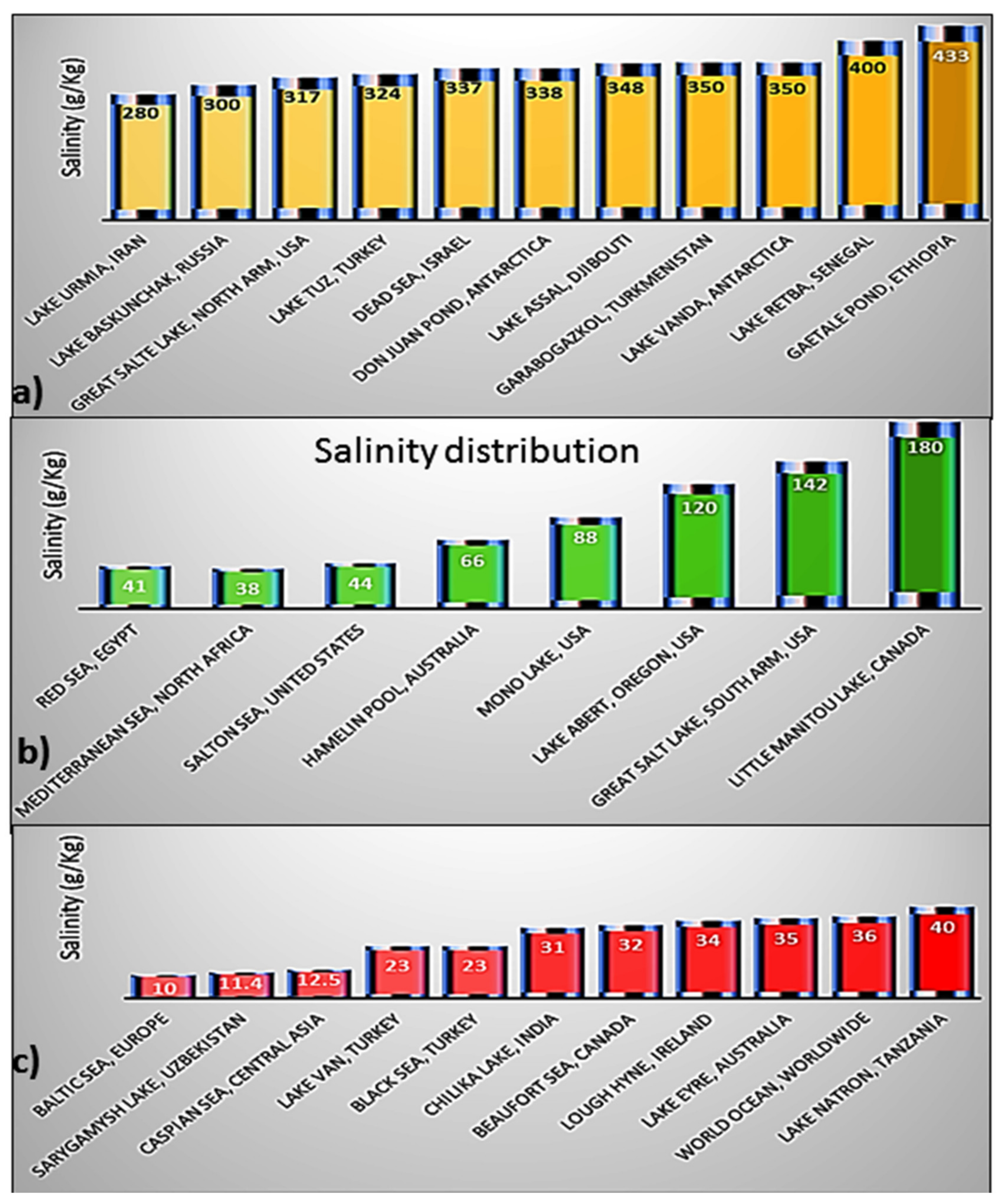
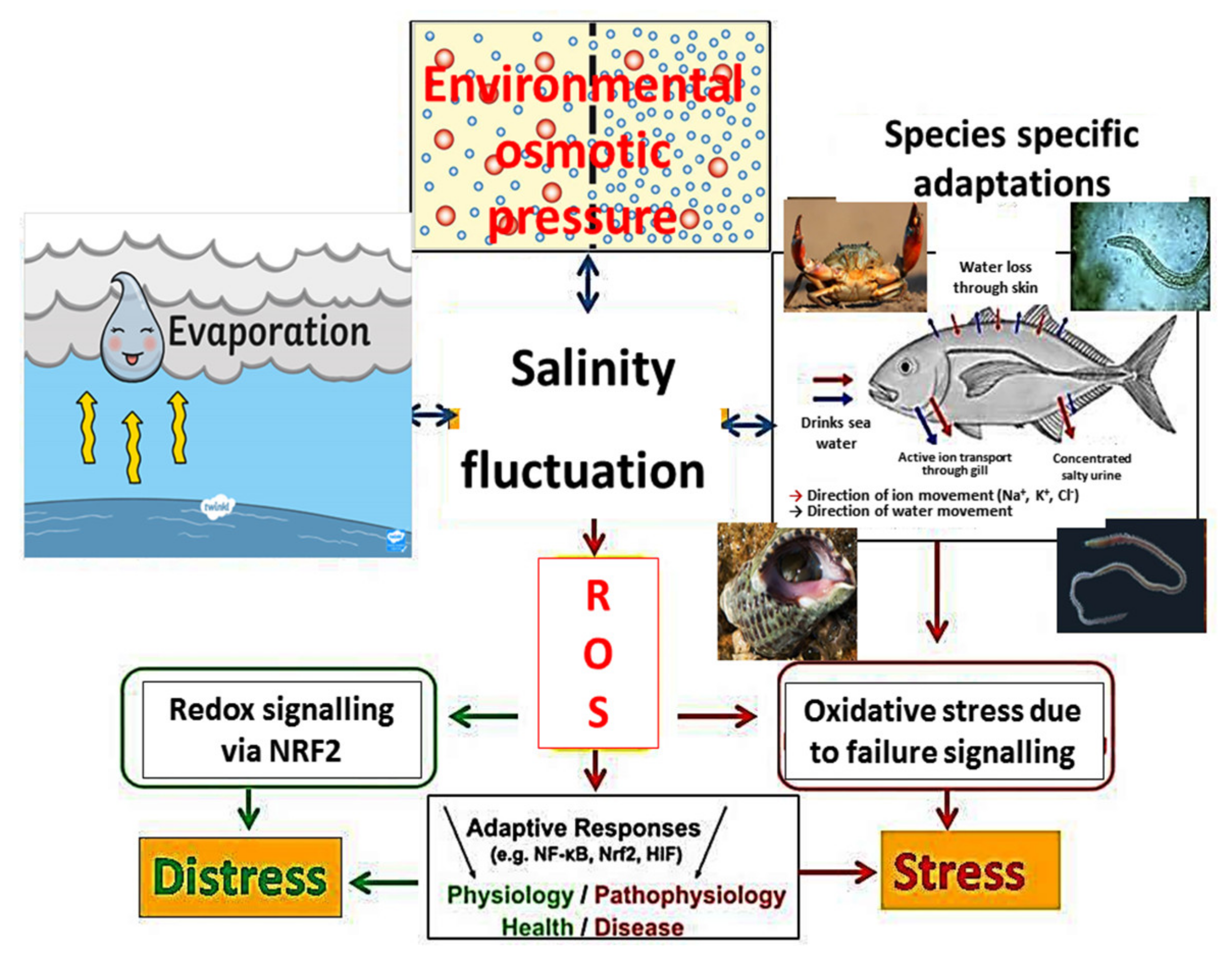
2. Influence of Anthropogenic Activities and Climatic Factors on the Salinity of Coastal Water

Salinity, among several abiotic factors—such as turbidity, dissolved oxygen, temperature, pH, total dissolved solids, etc.—contributes to the dynamics of coastal aquatic habitats and their biodiversity. The biological communities thriving in coastal aquatic ecosystems are progressively influenced by optimal levels of salinity. The molecular, morphological, physiological, biochemical, and ecological prospects of an organism are sustained by an optimal level of salinity in the water in which the organism survives. The osmotic concentration of an aquatic environment facilitates the distribution of the species in coastal habitats with respect to several auxiliary factors. These include the patterns of feeding and nesting, body fluid regulation, cellular homeostasis, regulation of osmotic genes, etc. [49,50,51]. The differential patterns of salinity shifts and the water levels recorded in the last few decades have led to devastating consequences for coastal regions. These regions experience erosion of the natural coastline, loss of biodiversity, contamination of freshwater biota due to saltwater invasion, etc. Further shifts in salinity levels in those coastal areas can disproportionately intensify the ramifications for the coastal ecosystems. In the present scenario, these coastal areas frequently experience variable salinity levels in absolute terms, mostly due to global climatic disturbances and anthropogenic events [52,53,54].
Today, the temperature patterns of the Earth are altered significantly and are expected to fluctuate even further over time under the influence of climatic changes induced by anthropogenic activities [55]. Such fluctuation in climate is estimated to affect the physicochemical parameters of water—especially the water levels in the ocean—and, thus, to affect the levels of water salinity [56,57]. The ocean contains nearly 97% of the total water and is responsible for 86% of the Earth’s total evaporation [58]. Therefore, various national and international programs are undertaken to monitor the fluctuations of water parameters, including salinity. According to the Intergovernmental Panel on Climate Change (IPCC), climatic changes such as increased global warming due to anthropogenic activity will affect organisms residing mostly in semi-arid and Mediterranean regions, due to a 25–30% increase in the rate of evaporation [59,60,61].
Higher salinity levels are observed where the rate of evaporation is high [62,63]. Similarly, lower salinity levels are observed where there is a high rate of precipitation and mixing of runoff water with the ocean [64,65]. The range of salinity varies from 34 to 36 ppt across the oceans, with some exceptions [66]. There are also some places where rainfall is limited but the warm wind evaporates the water and, thus, reduces the water level, ultimately resulting in increased salinity [67]. The Atlantic Ocean is one of the examples where such a case is observed. Similarly, a very high rate of evaporation is observed in the Mediterranean Sea, resulting in a salinity level of about 38 ppt [68]. Therefore, an increase in global warming can enhance the salinity levels of the Atlantic Ocean and Mediterranean Sea.
Some oceans have very low salinity, where there is heavy rain and the mixing of water from freshwater areas takes place. The coastal zones in the Antarctic and the Arctic can be affected by strong decreases in salinity due to ice melting and runoff [69], whereas the salinity levels of offshore Antarctic and Arctic waters have been observed to be 34 and 30 ppt, respectively. Thus, the increase in the Earth’s surface temperature due to various anthropogenic activities can melt glaciers and increase water levels. This ultimately reduces the salinity levels. The Baltic Sea in Northern Europe has low-salinity regimes accounting for ~10 ppt due to heavy mixing of freshwater into the sea [66]. Therefore, increased global warming can increase the evaporation rate and, subsequently, could increase the salinity levels in different water bodies [7].
A 2 ppt increase in salinity can increase the physicochemical characteristics of water, such as density and buoyancy [66,70]. The higher salinity of water can cause denser water to sink below less dense water, which can cause convective mixing of two water levels. The increase in salinity has an even greater effect at low temperatures, because the combination of these two factors causes the ocean water to increase its density; as a result, it could sink lower—especially towards the bottom [58]. Conversely, a decrease in the salinity of the water can increase the stratification effect and, thus, prevent the mixing of waters [71]. The elevated salinity under the high rate of evaporation causes the formation of salt fingers that can imbalance the equal distribution of salts in water. Similarly, low salinity causes a barrier layer that could can vertical exchanges between two layers of the water [58].
The unprecedented rate of anthropogenic activities has put substantial pressure on aquatic environments in general, and on the species residing within them in particular. Anthropogenic inputs may be categorized into hazardous solid wastes, gaseous emissions, and contaminated liquid discharge. Heavy metal contamination of water bodies due to mining, the release of industrial and household sewage discharge due to anthropization, agrochemical-led agricultural practices, poisonous gaseous emissions from industry, introduction of xenobiotic compounds, the use of antibiotics, etc., have significantly contributed to fluctuations in water quality parameters, leading to a stressful aquatic environment—especially in coastal regions. Furthermore, these anthropogenic events are direct or indirect causative factors of physicochemical changes in the coastal regions, thereby driving the shifts in salinity. The change in salinity levels subsequently influences the aquatic health of the coastal and marine ecosystems in multiple ways, e.g., resulting in differences in plasma concentrations, improper regulation of body fluids, variable cellular structure, ROS formation, generation of OS, etc. [72,73,74]. Despite the fluctuations in salinity levels, several investigations have reported that some species have become resilient to hypo/hypersaline environments to a certain extent and play a major role in biomonitoring [7,75]. The uncontrolled exponential increase in human populations and their related activities have been found to induce climatic changes globally.
Incessant anthropogenic activities of imprudent nature have been seen to be counterproductive for global climate change. The anthropogenic stimuli and natural factors have individually contributed to the dimensional shifting of ecosystems along coastal regions in every possible way. The intensive intervention of human activities has taken the edge off the long-term climatic disturbances. However, the anthropogenic vectors co-interact with climatic changes and exacerbate the alterations of abiotic factors in the coastal hydrological ecosystems by increasing or decreasing the rise in sea levels, the concentration of sediments, and fluctuating salinity levels. Global climatic changes are characterized by global increases in temperature, reduced rainfall, frequent cyclonic storms and droughts in the coastal regions, El Niño–Southern Oscillation, etc. [76,77]. Frequent drought-like conditions due to the irregular distribution of rainfall have led to interruptions of salinity, thereby causing salinization of the hydrological ecosystems along the coastal regions. The coastal regions are vulnerable to cyclonic storms and tsunamis, which gradually lead to an increase in the salinity of the groundwater aquifers and the freshwater ecosystems in the close vicinity of coastal regions [54,78,79]. Additionally, the climate-driven salinity instabilities have imposed severe detriments on the population and dynamics of biological communities. Hence, the above natural disturbances cause the loss of biodiversity, along with an acute impact on the socioeconomic conditions of human populations in the coastal areas.
The coastal regimes are home to species belonging to different taxa that are of ecological importance. Fluctuations in salinity in these regions have induced shifts in diverse species communities at the expense of higher energy expenditure, stimulating the stress physiology of the species by reducing their overall efficiency. These changes not only threaten the diverse species communities but also boost the biological invasion of new species and, subsequently, cause a decline in their ecological significance. Additionally, the livelihood of the humans inhabiting these areas is comprehensively affected due to climatic disturbances and/or anthropogenic inputs. Therefore, maintaining an optimal level of salinity for sustaining life and balancing the equilibrium—especially among the aquatic communities—is a constant need for ecologists, conservation biologists, and environmentalists.

3. Salinity and Responses of Animals from Different Phyla

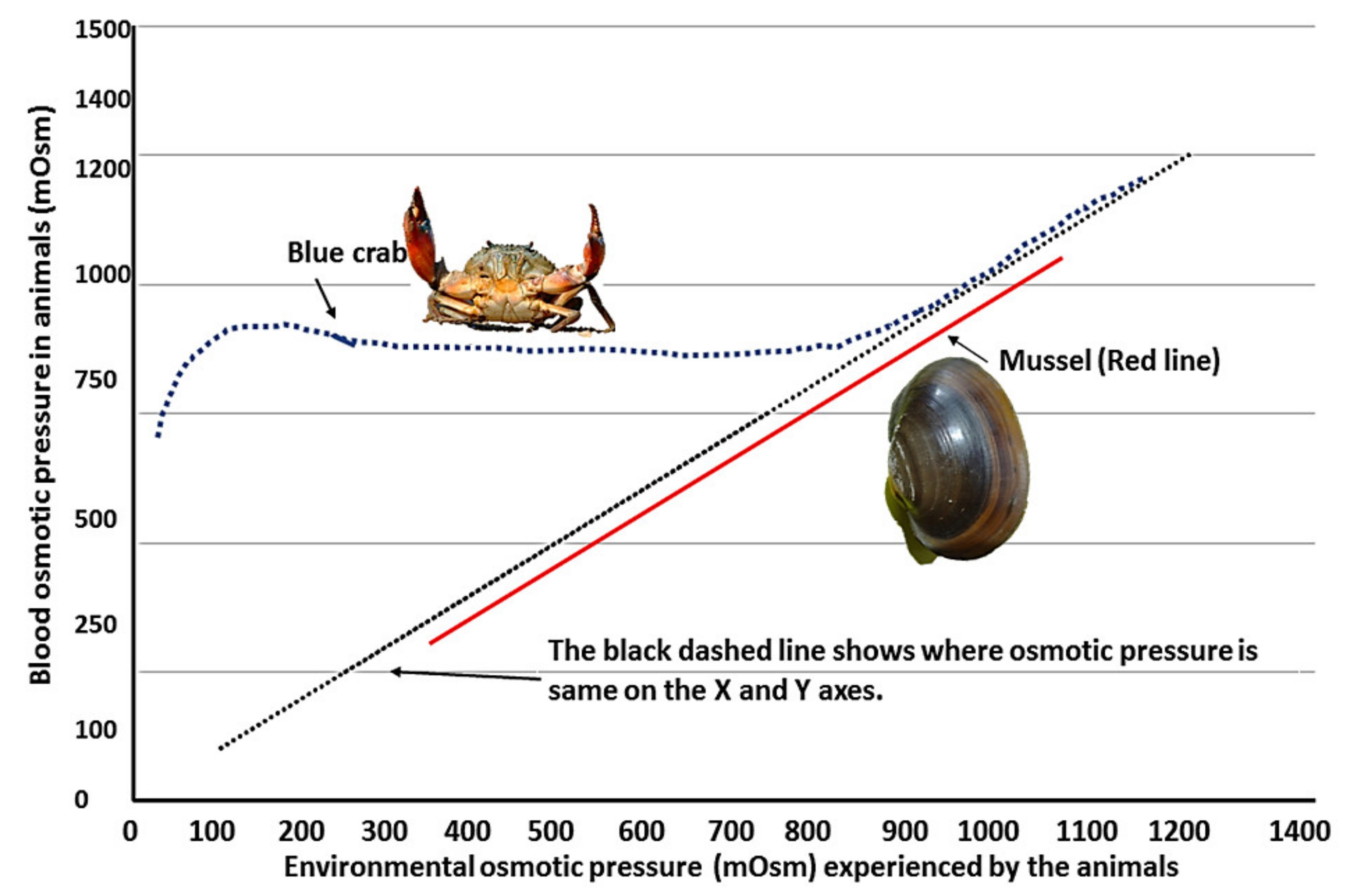
3.1. Physiology of Animals under Changing Salinity

Loss of biodiversity is observed in many wetlands due to salinization of water bodies, as the concentrations of salts and other inorganic ions increase for several reasons. Some of these causes include the breaking and silting out of the catchment areas of rivers and water bodies. The other processes that can lead to increases in salinity are o water cycles such as rain, evaporation, sea spray, glacial/interglacial cycles, etc. Adding to the above possible reasons, salinization can be caused by anthropogenic activities such as an influx of town/urban water drainage, demolition of foliage, exhaustive irrigation on land, regulation of rivers, de-icing, salting out, chemical processes, global warming that results in rising sea levels, or excessive mining works. Huge reductions in biodiversity indices that lead to unhealthy ecosystems can affect many aquatic organisms—especially ectotherms—and for this reason a lot of work on various aspects of various organisms has gradually been conducted from the 1960s to the year 2021 [7,80,81,82,83] (Table 1 and Table 2; Figure 3).
Salinization influences the life cycles of many animals. Activities such as the reproduction, spawning, growth, and migration of aquatic organisms are intensively influenced by salinity. For example, the micronutrient contents and gene regulation in stock broods of Atlantic salmon were linked to salinity. The growth of the fish is also influenced by seasonally induced salinity changes [84]. However, the influence of changing environmental salinity on the OS physiology in organisms has been scarcely addressed in the literature. The OS physiology begins with the consumption of O2 by the organisms, and the mitochondria act as the main hub for the generation of ROS. The effects of salinity on mitochondrial behavior have been studied by a few researchers [19,20] (Figure 4). It is worth mentioning that energy catabolism by kinases is very distinct in animals under varying levels of salinity [85,86,87,88,89,90,91,92,93,94,95]. Therefore, this could be instrumental in igniting many associated cellular pathways in animals to respond in a variety of ways to the changing salinity (Table 1 and Table 2). For example, mitogen-activated protein kinase action was observed in tilapia (Oreochromis mossambicus) and killifish (Fundulus heteroclitus) (Table 2).
Mitochondria act as the hub for the generation of active oxygen molecules [29], and the changes in the mitochondrial respiration and their respiratory chain enzymes are substantially modified by the changing salinity in the environment of aquatic organisms [6,19]. The osmoregulatory mechanisms induced by the changing salinity are associated with the respiratory behavior of mitochondria and are observed to be species-specific under hypo- and hypersaline conditions [19,20] (Figure 4). Different phyla can behave differently in response to the influence of the changing salinity on their blood or hemolymph osmotic pressure, which is influenced by environmental osmotic pressure. In turn, their OS responses vary under different salinity conditions (Figure 5).
Salinity can induce OS in organisms and, in contrast, the expression of antioxidant enzymes or responses in animals can contribute to augmented levels of amino acids [48,96,97]. Therefore, studying the intricate relationships between salinity and redox responses to balance or collapse the redox regulatory pathway is of paramount importance in aquatic systems. Since these phenomena are species-specific, a comparative literature review on the topic of OS responses to salinity in different phyla could fill the gaps in our understanding of this issue. Therefore, for the first time, we tried to consider all available information on the effects of salinity on the OS responses in organisms belonging to different phyla.

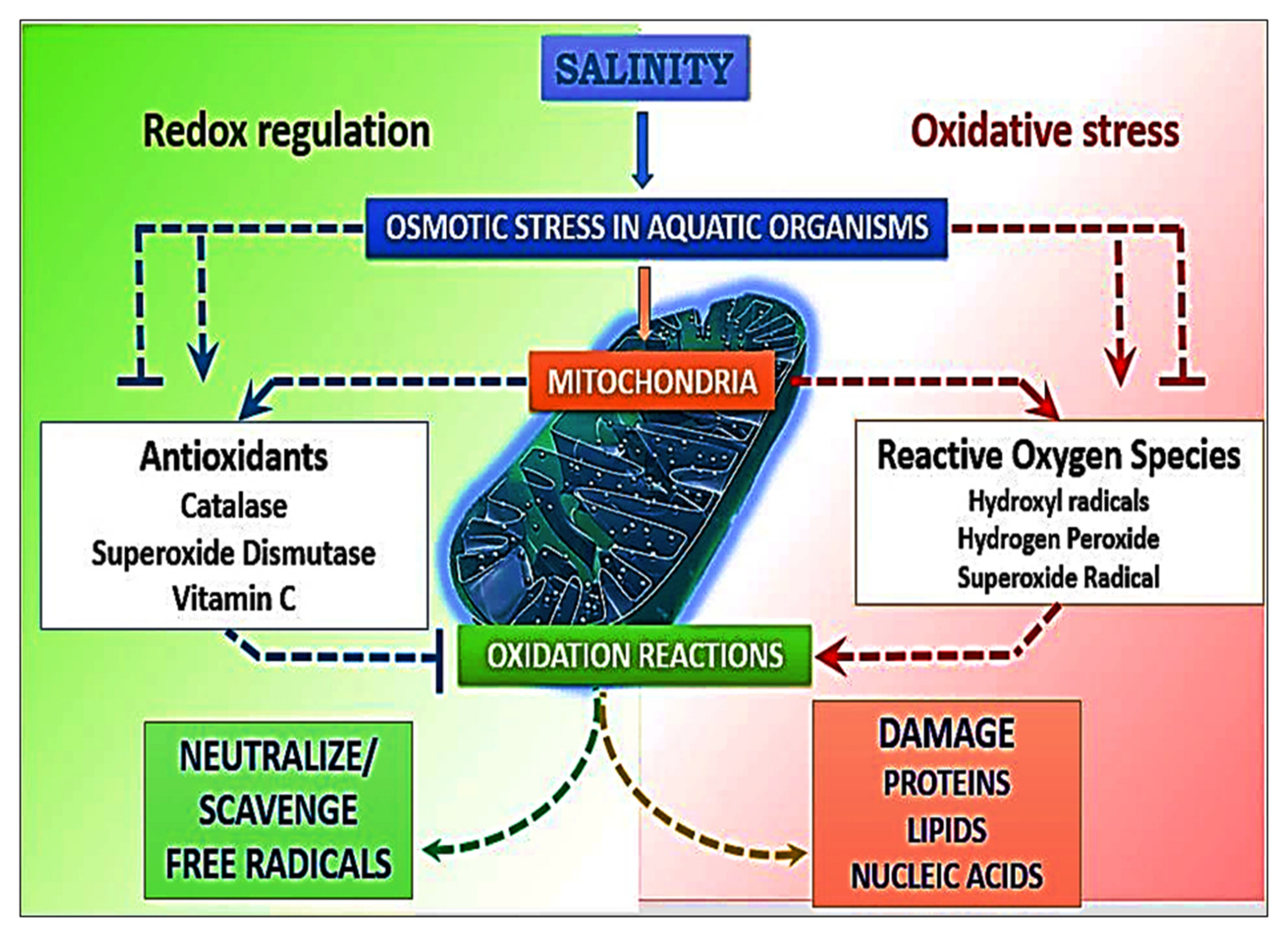
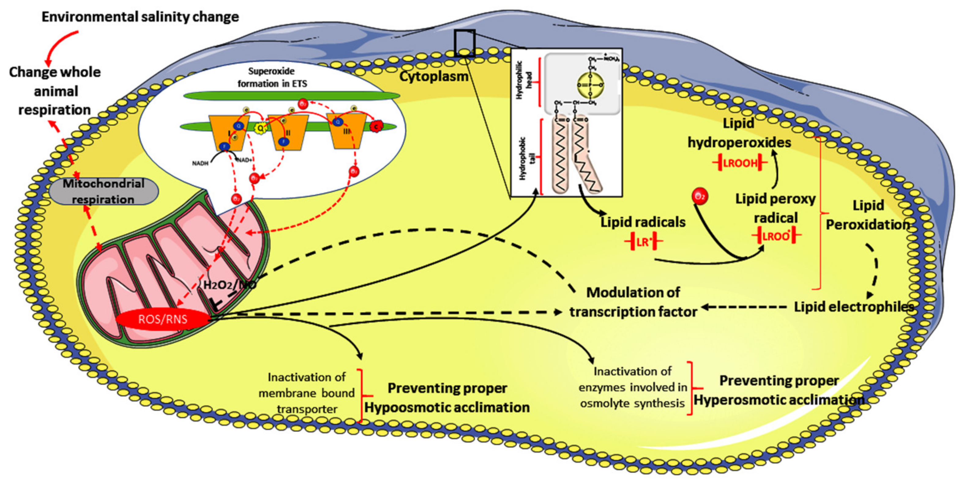
3.2. Stress Incurred in Animals under Salinity Stress

The osmotic discrepancy at the cellular level that is caused by the altered salinity can accelerate the physiological functions. For normal functioning under salinity stress—i.e., by maintaining the osmotic balance and the ionic gradient—the specialized systems (e.g., excretory organs) demand higher energy expenditure in animals [98]. In addition, maintenance of the ionic gradients along with the gaseous exchange is quite important and, under the circumstances of altered salinity, specific organs such as the gills, canals, or skin of aquatic animals play important roles in osmoregulation and the associated physiology [99]. Fluctuations in salinity can cause spikes in ROS accumulation and OS in animals. To protect against ROS-induced stress under higher salinity levels, animals develop redox regulation signaling systems that are species-specific (Figure 5).
The signaling systems in animals (e.g., nuclear factor erythroid 2-related factor) work in coordination with the redox-regulatory enzymes [100]. Investigations of the OS, ROS, redox, and associated physiology under salinity fluctuations are greatly associated with ecological studies, which have gained remarkable attention in the last few decades [101] (Figure 2 and Figure 3). The levels of ROS generation, enzyme activities, and the activity of small antioxidant molecules for redox regulation vary widely in different tissues and, furthermore, show variance in different species [102] (Table 1, Table 2 and Table 3). The altered physiology induced due to salinity shifts is correlated with the cellular responses and the different responses at the organismal level, which involve various physiological strategies. These include survival and reproduction, along with the moderated levels of transcription in different enzymes and corresponding variations in gene expression under various expected and unexpected saline environments.

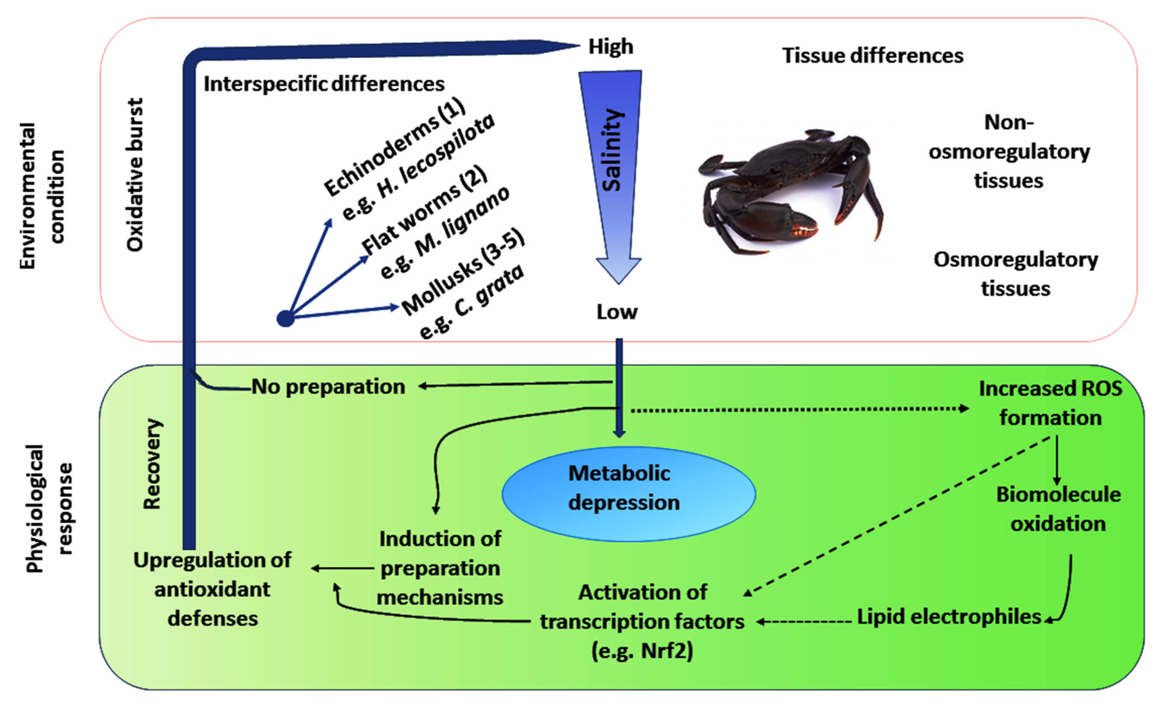
Responses of Animals to Primary and Drastic Salinization

Under the primary salinization condition, in hyperosmotic and hypo-osmotic environments, the OS physiology is activated (Figure 4). In particular, low salinity is responsible for augmenting the expression of the redox-regulatory enzymes in Dicentrarchus labrax [103]. The number of red blood cells and white blood cells increases under extreme salinity conditions, and this can also cause deformities in erythrocytes with higher hemoglobin levels in European seabass (Dicentrarchus labrax) [104]. The altered salinity stress can also lead to differentially regulated genes in the yellowfin sea bream (Acanthopagrus latus) [47]. Therefore, increases in environmental salinity can be stressful to many organisms.
Salinity can greatly influence water-intake capacity to compensate for water loss under hypersalinity-induced osmotic imbalance in two species of Culex mosquitoes [105]. In contrast to the above outcomes, elevated levels of antioxidant defense have been reported in Ruditapes decussates and Ruditapes philippinarum in response to lipid peroxidation induced by hypersalinity [43]. Thus, periodic changes or predicted alternations in salinity can lead to a controlled response in the organism, whereas the scenario may be different in response to unpredictable or drastic changes in salinity.
Specific strategies are often adapted by the inhabiting organisms to combat salinity stress under altered climatic/seasonal changes. For example, the seasonal variation in salinity in the Chilika Lagoon of Odisha, India shows 0–5 ppt in the rainy season, ~17 ppt in winter, and ~35 ppt in summer [6] (Table 1, Table 2 and Table 3). Various strategies—such as raising the level of antioxidants, preparation for OS, lowering metabolism or metabolic depression, low respiration rates, low food uptake, and electron leakage in the ETC (coupled with low ROS production)—are adopted to reduce the chance of cells experiencing OS. However, when the change in salinity is sudden and/or drastic, it pushes the inhabiting organisms towards oxidative burst. Therefore, such ecophysiological conditions need to be addressed comparatively as a function of phyla, because the mechanism of osmosensing in mitochondria acts as the primary hub for the generation of ROS (Figure 6).

3.3. Inclusive Responses of Organisms with Respect to Alterations in Salinity

Salinity in general modulates various biochemical and molecular events in helminthes. Marine nematodes are also not an exception to this rule. Recent work on the emerging marine nematode model Litoditis marina indicates the downregulation of multiple receptors responsible for neurotransmission and genes that regulate ion transport. Under hypersaline stress (30 ppt), the above receptors are downregulated. In contrast, the expression of genes that regulate unsaturated fatty acid biosynthesis, neuron-related tubulins, and intraflagellar transport is alleviated in the nematodes [106]. Therefore, redox status modulation and changes in OS physiology are apparent in nematodes under changing salinity.
Aquatic organisms, including helminthes, adjust themselves in terms of specific redox-regulatory strategies by maintaining different levels of redox-regulatory molecules to counteract the OS induced by salinity (Figure 3, Figure 4 and Figure 5; Table 1, Table 2 and Table 3). Lower organisms, such as ectotherms (e.g., Scylla serrata), experience OS under altered salinity. In particular, high salinity induces OS in S. serrata due to insufficient redox capacity. However, the response can be tissue- or organ-specific [6] (Table 2 and Table 3). Accordingly, the demand and supply of energy accompanied by redox regulation responses also vary among the animals [19,20,21].
It should be noted that salinity alone, or with multiple environmental stressors, can influence molecular mechanisms at the metabolomic, transcriptomic, proteomic, and genomic levels [107]. Therefore, salinity can potentially influence growth factors and reproduction at the biochemical and molecular levels, which may subsequently affect phylum-specific OS directly or indirectly in organisms and in a phylum-specific manner [19,104,108,109,110,111,112,113]. This may be due to switching of the signaling pathways that ultimately influence the OS physiology in aquatic animals under salinity stress (Table 2 and Table 3).

3.3.1. Rotifers

Rotifers are commercially cultivated to be served as food for the larvae of fish. Being microscopic in nature, rotifers generally reside in freshwater. Conversely, they are rarely found in marine water bodies. However, rotifer populations and OS mechanisms are adversely affected by increasing salinity, as is evident from a study conducted on the marine rotifer Brachionus plicatilis. The increase in levels of salinity up to 35 psu initiated a surge in the ROS levels and activated the activity of antioxidant enzymes such as SOD, CAT, and GST [114]. The effect of salinity on the rotifers and the comparative signaling pathways in marine and freshwater environments is an unexplored avenue for researchers.

3.3.2. Helminthes

Hypersaline conditions lead to dehydration in worms, as water efflux facilitates the uptake of organic osmolytes in the environment [115]. However, under experimental conditions, when organisms are exposed to artificial seawater, the absence of osmolytes in their cells is noticed. This urges organisms to synthesize free amino acids to recover from osmotic stress during the initial state of osmoregulation, as observed in the case of the copepod Tigriopus californicus [116] and the shrimp Penaeus aztecus [117]. However, similar studies on helminthes are scarce.
In a study on Macrostomum lignano, it was observed that an increase in their respiration rate occurred under hypersaline stress. This was coupled with the formation of new mitochondria to meet the energy demand for the required adjustment (Figure 6). On the other hand, the study also observed an increase in the production of superoxide radicals under hypersaline conditions in Macrostomum lignano. The formation of ROS can have adverse effects on phospholipids and other components of cell membranes by causing lipid peroxidation and forming hydroperoxides in helminthes.
In contrast, a decline in respiration rates as well as in the production and consumption of ATP was observed under hypo-osmotic stress in Procerodes littoralis [118]. This leads to a hypoxic state in the organism, which eventually results in metabolic shutdown due to the oxidative burst [42]. Thus, the strategies adopted by organisms to increase their redox-regulatory status, as explained in “preparation of OS”, seem to work in an effective manner in the above organisms.

3.3.3. Mollusks

Pulmonates are found to survive in both freshwater and seawater because they perform osmotic regulation, e.g., hypo-osmosis in freshwater and hyperosmosis in seawater [119]. Bivalves are able to tolerate wide fluctuations in salinity. The osmoregulation under extreme salinities is modulated by an increase in the calcium and sodium ion contents and shell valve closure. The closure of the shell valve leads to hypoxia and starvation under hypo-osmotic stress. Gradually, this leads to disturbances in the osmotic balance, which further allow the inflow of water into the cell, causing cellular rupture. Therefore, the environmental and bodily osmotic pressure is well mediated in mollusks (Figure 7).
In general, salinity modulates the expression of a vast number of enzymes and their respective genes in mollusks. Modulation of FoxO signaling, expression of tight junction proteins, and changes in immunomodulator titers under hyposalinity in Hong Kong oyster (Crassostrea hongkongensis) are a few examples. More specifically, hypersalinity-induced amino acid metabolism, along with signaling pathways such as AMPK and PI3K, is also recorded in the abovementioned oysters [120]. Similarly, both high (35 ppt) and low (10 ppt) salinity can induce increases in mRNA and amino acid levels in the hard clam (Meretrix lusoria), which possibly explains the upregulation of different proteins and genes under salinity stress in mollusks. The high energy dissipation cost of the salinity stress in mollusks is usually maintained, as observed in the Pacific abalone Haliotis discus hannai. The observed positive and strong correlation between cAMP activity, mRNA levels, and Na+/Ca+ ATPase in mollusks confirms this fact [121].
Changes in salinity and CO2 levels are potential stressors that can induce OS in bivalves such as Venerupis philippinarum [122], Ruditapes philippinarum [123], Pila globosa [124,125,126], and Crassostrea rhizophorae [127]. The activity of the ETC is increased, followed by an increase in the lipid peroxidation level, thereby accelerating the metabolism under low salinity stress. To mask the toxic effects of lipid peroxidation, redox-regulatory enzymes such as SOD, CAT, and GST are activated. Higher salinity directly inhibits the SOD and CAT activity levels; conversely, the CAT activity is found to increase under lower salinity [128]. Therefore, the majority of the studied mollusks experience OS under high salinity stress. Recent studies have also provided contrasting results indicating that low salinity may also upregulate the cellular ROS levels of the shallow water clam Scapharca subcrenata, ultimately elevating the activities of Mn, SOD, and GPx in [129].
Salinity and salicylic acid (the constituting substance of pharmaceutical and personal care products) in combination cause minimal cellular damage. However, the metabolism of mollusks is enhanced in the presence of salicylic acid under optimal salinity conditions, but it declines with the increase in salinity [130]. On the other hand, changes in salinity can also be tolerated by certain mollusks, since no elevation in the oxidation of lipids is observed under higher-salinity conditions. For example, no significant changes in the lipid peroxidation levels are observed under different salinity regimes, whereas in some cases—as observed in Ruditapes decussatus, R. philippinarum, Venerupis corrugata, and Crassostrea rhizophorae—low salinity induces higher levels of lipid peroxides and antioxidant enzymes such as SOD, CAT, and GST [44,122,127].
Salinity-induced hypoxia reduces oxidative phosphorylation by restricting the production of ATP (due to less oxygen concentration in the cells) and activates alternative pathways for generating ATP that use less energy, as observed in protein catabolism in Argopecten purpuratus [131] and in Ruditapes philippinarum [132], for example. Therefore, the alternative oxidase system is one of the strategies maintained in animals to bypass complex III of the electron transport chain and to reduce the surplus production of active oxygen molecules [29] (Figure 4).

3.3.4. Annelids

The marine taxon Oligochaeta can adopt both hypo-osmotic and hyperosmotic regulation [119]. Studies on the salinity-induced changes in annelids are very scarce. A study was conducted on the exposure of Neapolitan diopatra to salinity. Under the conditions of hypersalinity (i.e., at 42 g L−1), the lipid peroxidation levels were recorded to be the highest, whereas under hyposaline conditions (21 g L−1) the lipid peroxidation level was comparable to the lowest. This was consistent with the elevated levels of activity of the antioxidant enzyme SOD and the biotransformation of the enzyme GST, and the reverse was recorded in the case of exposure to salinity of 28 g L−1. The activity of CAT did not show any significant changes at varying salinity levels [37]. Therefore, an extensive study is essential to evaluate salinity-induced OS responses in annelids.

3.3.5. Arthropods

Salinity and O2 levels in the environment are usually inversely proportional to one another. This may be an external cause for why crustaceans are found to suffer from hypoxia under conditions of higher environmental salinity. As a result, they shift towards hyperoxia when exposed to low salinity and, hence, consume more oxygen. The osmotic stress conditions arising due to salinity shifts lead to the accumulation of different osmolytes in the tissues involved in the process of osmoregulation in shrimps. O2 consumption and osmotic imbalance regulate OS in animals, and crustaceans are not an exception to this rule; however, they can shift their niche to find a suitable salinity regime (Figure 7).
Delgado-Gaytán et al. [46] reported that the activity of the osmolyte betaine aldehyde dehydrogenase was increased in the gills and hepatopancreas of white shrimp at higher salinity [46], thereby leading to an increase in betaine glycine content in these tissues. Remarkably, this also affected the ammonia content, which showed a significant decreasing trend at higher salinity levels. The reported increase in the rate of O2 consumption at lower salinity levels should generate a higher energy budget, which further facilitates the passage of ions into the hemolymph from the habitat. In contrast, at higher salinity levels, the oxygen consumption is reduced, along with the lowering of the oxidative metabolism of the organism. The above phenomenon was confirmed by a study on South American crabs (originating from the sea but residing in freshwater). The species retained its ancestral osmoregulatory mechanism when exposed to higher salinities [133]. The adaptation to combat higher salinity stress was accompanied by a rise in hemolymph osmolality and a decline in V-H+-ATPase activity. Thus, energy demand and supply are highly correlated with the osmotic regulation and OS responses in crabs.
Experiments on S. serrata—a euryhaline crab—confirmed the reduced levels of lipid peroxidation in various tissues at lower salinity, accompanied by elevated levels of antioxidant enzymes such as SOD, CAT, and GPx in the gills and only SOD in the abdominal muscle tissues [6]. Thus, the mechanism in this crab suggests that the higher consumption of oxygen in the above tissues at lower salinity could lead to an increase in the production of ROS such as lipid peroxides, H2O2, and superoxide radicals, causing an increase in antioxidant levels. During the hypoxia state, when the species are subjected to higher salinity, it reduces the transport of electrons in the ETC [6,20]. Thus, euryhaline crabs may experience OS under both hypoxic and hyperoxic conditions that align with hyper- and hyposaline regimes in the environment.
This elicits the formation of ROS from electron carriers such as complex III ubiquinone. Among the tissues of crabs, the abdominal muscles were found to have lower metabolism with respect to the hepatopancreas and gills, leading to the conclusion of lower levels of superoxide radical production [6,134] (Table 2 and Table 3). Several other studies carried out on crustaceans such as Macrobrachium rosenbergii [135], M. malcolmsonii [136], Charybdis japonica [137], Neohelice granulate [138,139], Oronectus limosus [140], Carcinus maenas [141], and Litopenaeus vannamei [47] have reported similar increases in ROS production under salinity stress. Confirmation of the modulation of OS in arthropods under altered salinity can be obtained from the above examples.

3.3.6. Echinoderms

Within the span of evolutionary development, the echinoderms and ascidia have not been able to overcome the physiological adjustments in freshwater. The echinoderms are less adapted for adjusting to osmotic changes under salinity fluctuations. This is because they are devoid of the proper organs that can regulate osmotic balances. Thus, the coelomic fluid that is isosmotic to the environment is affected by salinity fluctuations [142,143]. Therefore, these organisms may become highly susceptible to OS under salinity stress.
The oxygen consumption rate is considered to be one of the most important parameters for energy production. In echinoderms such as Apostichopus japonicus (energy budget), Ophiophragmus filograneus (respiration), and Holothuria grisea (ionic gradients and hydration issues), the rate of oxygen consumption is altered under hypersaline conditions [144,145,146]. In Holothuria leucospilota, a decline in energy production is noticed under low saline exposure. Higher O2 consumption is observed in many organisms under low salinity. Higher O2 consumption is accompanied by high oxidative phosphorylation. The opposite trend is observed in H. leucospilota, setting up a new direction for further investigations.
The decline in the O2 consumption rate consequently leads to a reduction in metabolic activity. This indicates that adverse effects on specific tissues can potentially generate higher levels of ROS under low-salinity conditions [147]. Thus, metabolic depression may further induce elevated levels of antioxidant defenses that may help in the organisms’ recovery from oxidative bursts [48]. Echinoderms can be a good model to elicit the above fact.

3.3.7. Fishes

Fish can thrive in both freshwater and seawater. They adapt to both hypo-osmotic and hyperosmotic regulation in freshwater and seawater, respectively [119]. Salinity changes in inland freshwater and coastal wetlands extensively affect the wild habitats of freshwater species such as Catla catla, Labeo rohita, Cirrhinus mrigala, and Tenualosa ilisha in various ways [148]. For example, species specific to positive or negative growth rates may be observed under low-salinity conditions [149,150,151]. Thus, salinity fluctuations are one of the critical reasons behind the growth performance of fishes.
Specific fish species also show unique patterns in responsive OS events under salinity stress. Significant upregulation of Na+/K+ ATPaseα1, Na+/K+ ATPaseα2, and Na+/K+ ATPaseα3 was observed in the white-rimmed stingray Himantura sp. when exposed to saline stress. This fish also showed higher Na+/K+ ATPaseα3 mRNA levels and proteins of Na+/K+ ATPase in general under salinity stress [152]. Energy homeostasis can be maintained by a heavy rate of catabolism without changing the levels of glycogen in the livers of fish under salinity stress, as observed in the milkfish Chanos chanos. However, the Na+/K+ ATPase in the larvae of Takifugu obscurus increased significantly along with the levels of malondialdehyde and SOD activity with an increase in salinity [153]. This indicates the tolerance adaptation strategy in the fish under salinity stress [154].
Modulation of antioxidant enzymes and OS events under changing salinity is clear in fishes. Low salinity (5 ppt) stress for up to 144 h in the yellow-striped American perch (Perca flavescens) alleviates the expression of CuZn SOD, GPx, and GST, along with Hsp70 [155]. In addition to stenohaline fish, the physiological disturbances are also especially evident in euryhaline fish such as silver sea bream when exposed to salinity levels of 0, 6, 12, 33, 50, and 70 ppt [156]. The most important molecules that respond and play a vital role with respect to changing osmolality are aquaporin in general and aquaporin Ia in the gills, kidneys, intestines, and mantle tissues, which have now been correlated with the expression of regulatory redox and energy homeostasis genes [7].
Hypo-osmotic shock is associated with oxidative injury due to low antioxidant status in fish. Under lower salinity, the higher rate of oxygen consumption leads to the formation of surplus ROS and free radicals, and reduced antioxidant defense activity leads to an imbalance in the redox state [157,158]. In a detailed study on Pseudosciaena crocea, it was observed that salinity stress induces elevated expression of an antioxidant signaling marker in the nucleus (nuclear factor erythroid 2-related factor 2), which suggests that antioxidant genes were upregulated under low-salinity conditions [159,160]. The augmented expression of this nuclear marker in the nucleus is usually beneficial in cells, as it signals for antioxidant enzymes. Sometimes it includes subsequent events of apoptosis and tumors due to free radical damage. To maintain the optimal level of erythroid 2-related factor 2 in the nucleus, as a protective role, KEAP1 expression is activated as a control measure over its excess production. In addition, under salinity stress, the nuclear factor erythroid 2-related factor 2/Kelch-like ECH-associated protein 1 pathway plays a pivotal role in upregulating the redox regulation via the expression of redox-regulatory enzymes [100,161].
Under hypersaline conditions, OS was induced, as evidenced by elevated levels of lipid peroxidation and an inability of the antioxidant defense system to produce enough antioxidant molecules to eliminate ROS [162]. Salinity-induced stress leads to demand and supply of energy that allows animals to adapt to an optimal metabolic rate by activating the enzymes that take part in glycolysis and oxidative phosphorylation [163]. Different studies have established that salinity mainly affects the mitochondria of gills in fishes due to stress, which further generates ROS. In some species, such as Acipenser naccarii, the activated osmoregulatory process led to an increase in lipid peroxidation levels in the blood, which was not able to revert to its usual levels even after normal conditions were restored [164]. Although salinity has detrimental effects on OS physiology, the idea of dietary supplementation for fishes in adverse saline conditions could be an effective intervention to enhance their physiological mechanisms. Dietary supplementation of Aspergillus oryzae in the food of the Nile tilapia significantly enhanced the antioxidative enzyme pathway by increasing the activity of enzymes such as SOD, CAT, and GPx under increasing salinity stress. The dietary supplement was also considered to be effective in enhancing the non-specific immune responses and blood protein profiles [165]. Thus, a dietary substitution could be an effective implementation to overcome the adverse stress conditions caused by salinity in fishes.
It has also been observed that, along with multiple stressors—especially heavy metals—salinity can modulate the OS in various fish models. Studies carried out on the euryhaline killifish Fundulus heteroclitus as a function of zinc, copper, and nickel by Loro et al. [166], Blewett et al. [167], and Ransberry et al. [168], as well as by Glover et al. [169] on the galaxiid fish Galaxias maculatus, have observed that salinity extensively modulated the OS physiology. Thus, it can be concluded that fish, being higher-taxon organisms, can activate various signaling systems such as the nuclear factor erythroid 2-related factor 2/Kelch-like ECH-associated protein 1 system to control OS-mediated salinity, but sometimes the collapse of the system may lead to OS in the fish. Therefore, studying the signaling system seems important in the present context.
From the above discussion, it is clear that the OS responses in aquatic animals under changing salinity are species-specific. All of the organisms follow their own specific strategies for osmotic balance in relation to OS responses and the energy demand and supply chain of their cells (Figure 8). Therefore, when any specific aquatic models are used for research or aquaculture purposes, water salinity levels must be considered as one of the important physicochemical factors.

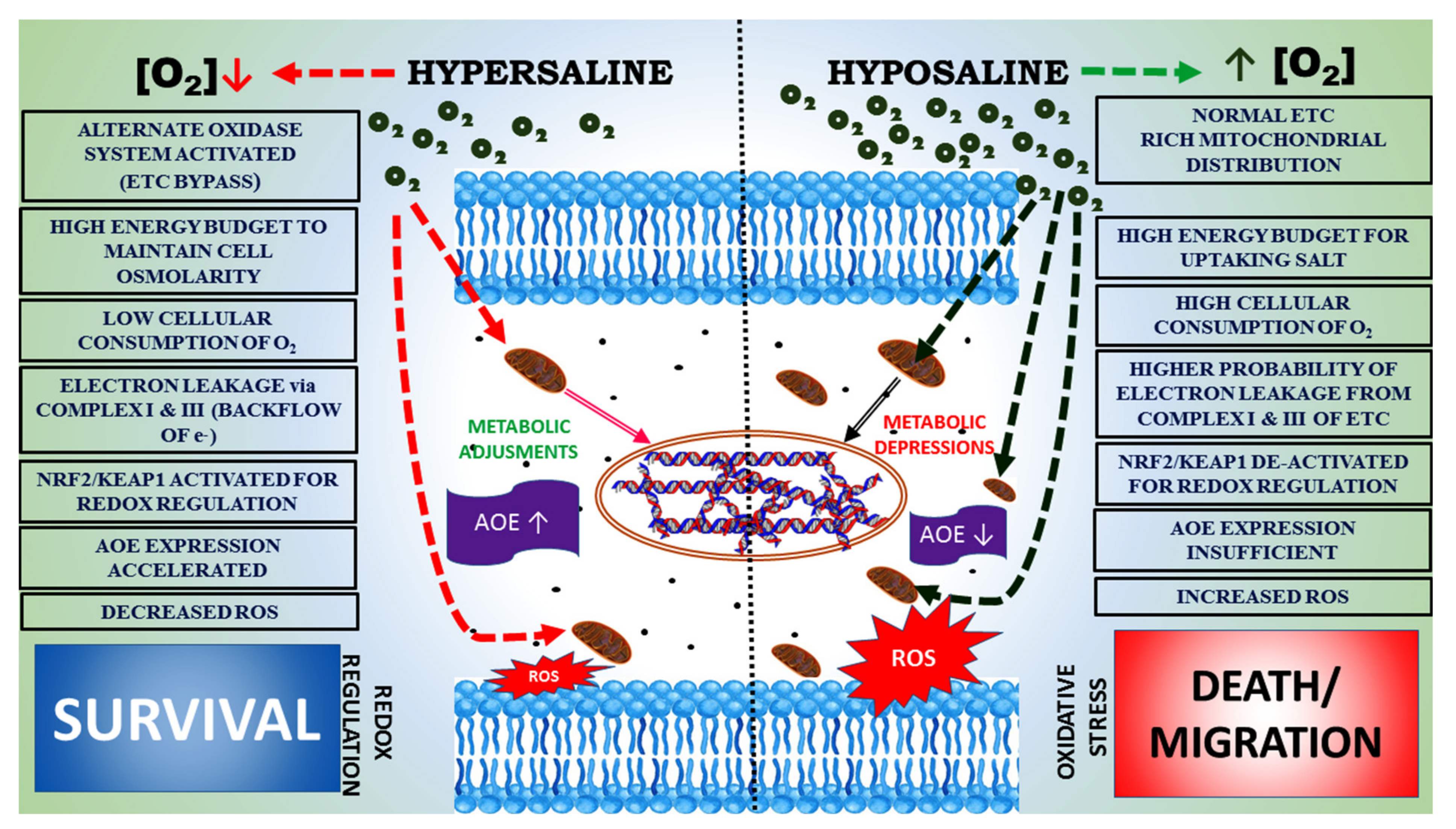
3.4. Signaling Pathways under Salinity Stress

High salinity increases water loss from the body and also facilitates the impairment of turgor pressure. As a result, osmolytes are taken in, as high salinity may be involved with the degradation of proteins and regulating cell volume through the regulation of associated genes and apoptosis [170,171,172,173]. For effective regulation of cell osmoregulation in the inhabiting organism, including fish under changing salinity conditions, signaling molecules such as phospholipase A2, calcium-sensing receptors, and cytokine receptors are activated. This aspect requires deep studies in aquatic models [174].
In this context, the role of respective hormones and receptors is well documented under changing salinity stress [175,176]. The protein phosphorylation events such as stimulated protein kinase signaling pathways that are activated by mitogens in different fishes were greatly influenced by the changes in salinity levels—for example, in tilapia [86], killifish [177], and turbot [87]. Similarly, several other factors—such as the activation of light chain kinase of myosin, focal adhesion kinase, and osmotic stress transcription factor—have been reported in different species i.e., the Japanese eel [91], killifish [87], and osmotic transcription factor 1 in Mozambique Tilapia [88], as systematically documented in this review (Table 3). This aptly indicates the activation of several signaling pathways that have evolved under changing salinity levels in aquatic models in general and in fish in particular.
Negative effects of salinity on signaling pathways have also been observed in some animals. The euryhaline bottom-dwelling goby Neogobius melanostomus can turn down its osmoregulatory activity at or above 25 ppt salinity. Thus, higher levels of blood plasma osmolality compete against the survival rate [178]. Higher energy expenses are borne by the animals due to activated levels of Na+/K+-ATPase activity when the animals shift from low to high salinity [179], and investigations in Fundulus heteroclitus [180] and Oreochromis mossambicus [181] validated the above fact experimentally. However, organisms try to use the signaling pathways judiciously to avoid harsh environmental conditions under changing salinity stress. This is because the adverse impact of salinity has already been established in invertebrates [20]. Simple seasonal changes in salinity may also impart such stress and activation of the redox sensing system to upregulate the associated enzyme responses (Table 2).
Aquatic organisms are reasonably sensitive to variations in salinity, which may be seasonal or drastic and unpredictable. Organisms from the lower phyla with comparative less motility capacity, such as Perna viridis, can experience more stress under salinity fluctuations [182]. Sometimes such organisms express lower redox-regulatory capacity in the form of lower activity of SOD, CAT, and GPx or high but insufficient expression of redox-regulatory enzymes. This may lead to the accumulation of cellular thiobarbituric-acid-reactive substances, as observed in the pearl cichlid (Geophagus brasiliensis), the three-spined stickleback (Gasterosteus aculeatus), and the Senegalese sole (Solea senegalensis). The regulation of OS physiology in many organisms—such as G. brasiliensis, G. aculeatus, S. senegalensis, and B. barbus is influenced by several stress factors, such as temperature and salinity, in combination or alone [183].
In the above organisms, the mitochondrial functions and energetics are changed accordingly, and this may occur under both low and high salinity stress, as both produce high ROS in different aquatic animals (Figure 3, Figure 4, Figure 5 and Figure 6; Table 1). This is also linked to elevated metabolic rates in animals, which are usually coupled with elevated levels of ROS generation in cells, as observed in M. lignano and Saccharomyces cerevisiae [32,48]. Therefore, the involvement of other environmental parameters such as O2 levels and temperature, which are directly linked to salinity, may contribute to the altered OS physiology in different phyla. However, lower organisms have developed fewer strategies to cope with such changes. Salinity, when combined with anthropogenic factors, causes more species-specific impacts on the inhabitants (Table 4), and knowing the sources of the anthropogenic factors and their impacts on the salinity of the habitats and the physiology of their inhabitants will be helpful for the management of aquaculture resources.

4. Conclusions and Future Prospects

Salinity is one of the major decisive environmental factors that modulate the physiology of the inhabitants of marine environments. Salinity of the environment is also modulated by various anthropogenic and climatic factors. Finally, the effects of environmental salinity on different physiological strategies—including growth and reproduction at the proteomic and genomic levels—have been studied in aquatic models using several genomic, proteomic, transcriptomic, and metabolomic tools over the last 50 years. The main goal may be to increase aquatic productivity to achieve “Zero Hunger” by 2030. Therefore, management of stress biology in general (and OS biology in particular) under salinity fluctuations as a function of phylum needs to be reviewed, because this information is scarce. However, OS biology is related to the physiology of aquatic organisms associated with their growth, production, and reproduction. Specific strategies for specific organisms need to be adapted from their stress response management under fluctuations in salinity. Organisms from lower phyla may respond sharply to a narrow range of salinity fluctuations, as they have fewer molecular adaptive mechanisms, while higher organisms such as fish may show better responses to salinity stress (Figure 9). According to their OS, their physiology is modulated and, hence, can be considered as markers for comparative studies between organisms for their stress tolerance. Responses at the mitochondrial level and energy expenditure are also coupled with salinity stress. The physiological adaptation strategies of marine aquatic organisms—including both vertebrates and invertebrates—in response to particular levels of environmental salinity are evident from significant studies at specific tissue levels and organismal levels. Meanwhile, comparative studies on the responses of different phyla to salinity remain unexplored. Studies on OS and redox regulation by enzymes and non-enzymes in aquatic organisms have been conducted by various researchers as a function of biotic factors—especially salinity, which is significant for the survival of the organisms belonging to various phyla—and these works have been validated by the molecular-level study of some factors that enhance the expression of antioxidant enzymes, such as the nuclear factor erythroid 2-related factor 2/Kelch-like ECH-associated protein 1 signaling system. This review systematically describes the OS physiology under changing salinity levels in different phyla for the first time and, thus, presents a clear picture of the different climatic and anthropogenic factors that sustain life underwater.

Author Contributions

Conceptualization, data curation, formal analysis, funding acquisition, investigation, methodology, project administration, resources, software, supervision, validation, visualization, writing—original draft, writing—review and editing, B.P.; conceptualization, writing—original draft, methodology, writing—review and editing, A.B.; writing—review and editing, revision, F.P., S.G.P., T.N.A. and K.D. All authors have read and agreed to the published version of the manuscript.

Funding

This work was generously supported by the funding to B.R.P. from the Science and Engineering Research Board, Department of Science and Technology, Govt. of India, New Delhi, India (No. ECR/2016/001984), and by the Department of Science and Technology, Government of Odisha (Grant letter number 1188/ST, Bhubaneswar, dated 01.03.2017, ST-(Bio)-02/2017). And funding Department of Science and Technology, Government of Odisha, India (1264/ST/BT-MISC-0034-2018) to AB to Abhipsa Bal under Biju Patnaik Research Fellowship to pursue Ph.D. course in Biotechnology is acknowledged.

Institutional Review Board Statement

Not applicable.

Informed Consent Statement

Not applicable.

Data Availability Statement

All data generated or analyzed during this study are included in this published article.

Acknowledgments

The authors duly acknowledge the use of the Central Instrumentation Facility of Odisha University of Agriculture and Technology, and especially the assistance of Sashikanta Dash in the analyses of samples.

Conflicts of Interest

The authors declare no conflict of interest.

Abbreviations

PSU: practical salinity unit; ppt: parts per thousand; ROS: reactive oxygen species; OS: oxidative stress; SOD: superoxide dismutase; CAT: catalase; GPx: glutathione peroxidase; GSH: reduced glutathione.

References

  1. Williams, W.D. Anthropogenic salinisation of inland waters. Hydrobiologia 2001, 466, 329–337. [Google Scholar] [CrossRef]
  2. Kefford, B.J.; Hickey, G.; Gasith, A.; Ben-David, E.; Dunlop, J.; Palmer, C.; Allan, K.; Choy, S.; Piscart, C. Global scale variation in the salinity sensitivity of riverine macroinvertebrates: Eastern Australia, France, Israel and South Africa. PLoS ONE 2012, 7, e35224. [Google Scholar] [CrossRef] [PubMed]
  3. Sowa, A.; Krodkiewska, M.; Halabowski, D.; Lewin, I. Response of the mollusc communities to environmental factors along an anthropogenic salinity gradient. Sci. Nat. 2019, 106, 60. [Google Scholar] [CrossRef] [Green Version]
  4. Robinson, R.A. The vapour pressure and osmotic equivalence of sea water. J. Mar. Biol. Assoc. UK 1954, 33, 449–455. [Google Scholar] [CrossRef] [Green Version]
  5. Haumann, F.A.; Gruber, N.; Münnich, M.; Frenger, I.; Kern, S. Sea-ice transport driving southern ocean salinity and its recent trends. Nature 2016, 537, 89–92. [Google Scholar] [CrossRef]
  6. Paital, B.; Chainy, G.B.N. Antioxidant defenses and oxidative stress parameters in tissues of mud crab (Scylla serrata) with reference to changing salinity. Comp. Biochem. Physiol. 2010, 151, 142–151. [Google Scholar] [CrossRef]
  7. Bal, A.; Panda, F.; Pati, S.G.; Das, K.; Agrawal, P.K.; Paital, B. Modulation of physiological oxidative stress and antioxidant status by abiotic factors especially salinity in aquatic organisms. Comp. Biochem. Physiol. C 2022, 241, 108971. [Google Scholar] [CrossRef]
  8. Lushchak, V.I.; Bagnyukova, T.V. Temperature increase results in oxidative stress in goldfish tissues. 1. Indices of oxidative stress. Comp. Biochem. Physiol. 2006, 143, 30–35. [Google Scholar] [CrossRef]
  9. Zeng, Y.; Zhao, C.; Kundzewicz, Z.W.; Lv, G. Distribution pattern of Tugai forests species diversity and their relationship to environmental factors in an arid area of China. PLoS ONE 2020, 15, e0232907. [Google Scholar] [CrossRef]
  10. Nasrolahi, A.; Havenhand, J.; Wrange, A.L.; Pansch, C. Population and life-stage specific sensitivities to temperature and salinity stress in barnacles. Sci. Rep. 2016, 6, 32263. [Google Scholar] [CrossRef]
  11. Vélez-Martín, A.; Davy, A.J.; Luque, C.J.; Castellanos, E.M. Disentangling elevation, annual flooding regime and salinity as hydrochemical determinants of halophyte distribution in non-tidal saltmarsh. Ann. Bot. 2020, 126, 277–288. [Google Scholar] [CrossRef] [PubMed]
  12. Sabat, P.; Narváez, C.; Peña-Villalobos, I.; Contreras, C.; Maldonado, K.; Sanchez-Hernandez, J.C.; Newsome, S.D.; Nespolo, R.; Bozinovic, F. Coping with salt water habitats: Metabolic and oxidative responses to salt intake in the rufous-collared sparrow. Front. Physiol. 2017, 8, 654. [Google Scholar] [CrossRef] [PubMed] [Green Version]
  13. El-Esawi, M.A.; Elansary, H.O.; El-Shanhorey, N.A.; Abdel-Hamid, A.M.E.; Ali, H.M.; Elshikh, M.S. Salicylic acid-regulated antioxidant mechanisms and gene expression enhance rosemary performance under saline conditions. Front. Physiol. 2017, 8, 716. [Google Scholar] [CrossRef] [PubMed]
  14. Hong, M.; Li, N.; Li, J.; Li, W.; Liang, L.; Li, Q.; Wang, R.; Shi, H.; Storey, K.B.; Ding, L. Adenosine monophosphate-activated protein kinase signaling regulates lipid metabolism in response to salinity stress in the red-eared slider turtle Trachemys scripta elegans. Front. Physiol. 2019, 10, 962. [Google Scholar] [CrossRef] [Green Version]
  15. Cilenti, A.; Provenzano, M.R.; Senesi, N. Characterization of dissolved organic matter from saline soils by fluorescence spectroscopy. Environ. Chem. Lett. 2005, 3, 53–56. [Google Scholar] [CrossRef]
  16. Munksgaard, N.C.; Wurster, C.M.; Bass, A.; Zagorskis, I.; Bird, M.I. First continuous shipboard δ18O and δD measurements in sea water by diffusion sampling—Cavity ring-down spectrometry. Environ. Chem. Lett. 2012, 10, 301–307. [Google Scholar] [CrossRef]
  17. Castaño-Sánchez, A.; Hose, G.C.; Reboleira, A.S.P.S. Salinity and temperature increase impact groundwater crustaceans. Sci. Rep. 2020, 10, 12328. [Google Scholar] [CrossRef]
  18. Mahmoud, E.; El Baroudy, A.; Ali, N.; Sleem, M. Soil amendment with nanoresidues from water treatment increases P adsorption in saline soils. Environ. Chem. Lett. 2020, 18, 171–179. [Google Scholar] [CrossRef]
  19. Paital, B.; Chainy, G.B.N. Effects of salinity on O2 consumption, ROS generation and oxidative stress status of gill mitochondria of the mud crab Scylla serrata. Comp. Biochem. Physiol. C 2012, 155, 228–237. [Google Scholar] [CrossRef]
  20. Paital, B.; Chainy, G.B.N. Modulation of expression of SOD isoenzymes in mud crab (Scylla serrata): Effects of inhibitors, salinity and season. J. Enzym. Inhib. Med. Chem. 2013, 28, 195–204. [Google Scholar] [CrossRef]
  21. Paital, B.; Chainy, G.B.N. Seasonal variability of antioxidant biomarkers in mud crabs (Scylla serrata). Ecotoxicol. Environ. Saf. 2013, 87, 33–41. [Google Scholar] [CrossRef] [PubMed]
  22. Dasgupta, N.; Ramalingam, C. Silver nanoparticle antimicrobial activity explained by membrane rupture and reactive oxygen generation. Environ. Chem. Lett. 2016, 14, 477–485. [Google Scholar] [CrossRef]
  23. Gaikwad, K.K.; Singh, S.; Lee, Y.S. Oxygen scavenging films in food packaging. Environ. Chem. Lett. 2018, 16, 523–538. [Google Scholar] [CrossRef]
  24. Ranjan, S.; Ramalingam, C. Titanium dioxide nanoparticles induce bacterial membrane rupture by reactive oxygen species generation. Environ. Chem. Lett. 2016, 14, 487–494. [Google Scholar] [CrossRef]
  25. Halliwell, B.; Gutteridge, J.M.C. Free Radicals in Biology and Medicine, 8th ed.; Oxford University Press: Oxford, UK, 2015. [Google Scholar] [CrossRef]
  26. Liu, J.; Jia, H.; Zhu, K.; Zhao, S.; Lichtfouse, E. Formation of environmentally persistent free radicals and reactive oxygen species during the thermal treatment of soils contaminated by polycyclic aromatic hydrocarbons. Environ. Chem. Lett. 2020, 18, 1329–1336. [Google Scholar] [CrossRef]
  27. Liu, J.; Gao, N.; Wen, X.; Jia, H.; Lichtfouse, E. Plant and algal toxicity of persistent free radicals and reactive oxygen species generated by heating anthracene-contaminated soils from 100 to 600 °C. Environ. Chem. Lett. 2021, 19, 2695–2703. [Google Scholar] [CrossRef]
  28. Netto, L.E.; Antunes, F. The roles of peroxiredoxin and thioredoxin in hydrogen peroxide sensing and in signal transduction. Mol. Cells 2016, 39, 65–71. [Google Scholar] [CrossRef] [Green Version]
  29. Turrens, J.F. Mitochondrial formation of reactive oxygen species. J. Physiol. 2003, 552, 335–344. [Google Scholar] [CrossRef]
  30. Liu, Y.; Wang, W.-N.; Wang, A.-L.; Wang, J.-M.; Sun, R.Y. Effects of dietary vitamin E supplementation on antioxidant enzyme activities in Litopenaeus vannamei (Boone, 1931) exposed to acute salinity changes. Aquaculture 2007, 265, 351–358. [Google Scholar] [CrossRef]
  31. Valko, M.; Leibfritz, D.; Moncol, J.; Cronin, M.T.; Mazur, M.; Telser, J. Free radicals and antioxidants in normal physiological functions and human disease. Int. J. Biochem. Cell Biol. 2007, 39, 44–84. [Google Scholar] [CrossRef]
  32. De Martinez, G.M.; Bianchini, A. Metallothionein-like proteinsin the bluecrab Callinectes sapidus: Effect of water salinity and ions. Comp. Biochem. Physiol. A 2009, 152, 366–371. [Google Scholar] [CrossRef] [PubMed]
  33. De Zoysa, M.; Whang, I.; Lee, Y.; Lee, S.; Lee, J.-S.; Lee, J. Transcriptional analysis of antioxidant and immune defense genes in disk abalone (Haliotis discus discus) during the thermal, low salinity and hypoxic stress. Comp. Biochem. Physiol. B 2009, 154, 387–395. [Google Scholar] [CrossRef] [PubMed]
  34. An, M.I.; Choi, C.Y. Activity of antioxidant enzymes and physiological responses in ark shell, Scapharca broughtonii, exposed to thermal and osmotic stress: Effects on hemolymph and biochemical parameters. Comp. Biochem. Physiol. B 2010, 155, 34–42. [Google Scholar] [CrossRef] [PubMed]
  35. Bayir, A.; Sirkecioglu, A.N.; Bayir, M.; Haliloglu, H.I.; Kocaman, E.M.; Aras, N.M. Metabolic responses to prolonged starvation, food restriction, and refeeding Salmo trutta: Oxidative stress and antioxidant defences. Comp. Biochem. Physiol. B 2011, 159, 191–196. [Google Scholar] [CrossRef]
  36. Jiang, W.; Fang, J.; Du, M.; Gao, Y.; Fang, J.; Jiang, Z. Integrated transcriptomics and metabolomics analyses reveal benzo[a]pyrene enhances the toxicity of mercury to the Manila clam, Ruditapes philippinarum. Ecotoxicol. Environ. Saf. 2021, 213, 112038. [Google Scholar] [CrossRef]
  37. Freitas, R.; Pires, A.; Velez, C.; Almeida, Â.; Wrona, F.J.; Soares, A.M.; Figueira, E. The effects of salinity changes on the Polychaete Diopatra neapolitana: Impacts on regenerative capacity and biochemical markers. Aquat. Toxicol. 2015, 163, 167–176. [Google Scholar] [CrossRef]
  38. Bertrand, C.; Zalouk-Vergnoux, A.; Giambérini, L.; Poirier, L.; Devin, S.; Labille, J.; Perrein-Ettajani, H.; Pagnout, C.; Châtel, A.; Levard, C.; et al. The influence of salinity on the fate and behavior of silver standardized nanomaterial and toxicity effects in the estuarine bivalve Scrobicularia plana. Environ. Toxicol. Chem. 2016, 35, 2550–2561. [Google Scholar] [CrossRef]
  39. Hossain, M.A.; Aktar, S.; Qin, J.G. Salinity stress response in estuarine fishes from the Murray Estuary and Coorong, South Australia. Fish Physiol. Biochem. 2016, 42, 1571–1580. [Google Scholar] [CrossRef]
  40. Lee, M.C.; Park, J.C.; Kim, D.H.; Kang, S.; Shin, K.; Park, H.; Han, J.; Lee, J.S. Interrelationship of salinity shift with oxidative stress and lipid metabolism in the monogonont rotifer Brachionus koreanus. Comp. Biochem. Physiol. A 2017, 214, 79–84. [Google Scholar] [CrossRef]
  41. Moreira, A.; Figueira, E.; Soares, A.M.V.M.; Freitas, R. Salinity influences the biochemical response of Crassostrea angulata to Arsenic. Environ. Pollut. 2016, 214, 756–766. [Google Scholar] [CrossRef]
  42. Rivera-Ingraham, G.A.; Nommick, A.; Blondeau-Bidet, E.; Ladurner, P.; Lignot, J.-H. Salinity stress from the perspective of the energy-redox axis: Lessons from a marine intertidal flatworm. Redox Biol. 2016, 10, 53–64. [Google Scholar] [CrossRef] [PubMed] [Green Version]
  43. Velez, C.; Figueira, E.; Soares, A.M.V.M.; Freitas, R. Combined effects of seawater acidification and salinity changes in Ruditapes philippinarum. Aquat. Toxicol. 2016, 176, 141–150. [Google Scholar] [CrossRef] [PubMed]
  44. Velez, C.; Figueira, E.; Soares, A.M.V.M.; Freitas, R. Native and introduced claims biochemical responses to salinity and pH changes. Sci. Total Environ. 2016, 566–567, 260–268. [Google Scholar] [CrossRef] [PubMed]
  45. Fuhrmann, M.; Delisle, L.; Petton, B.; Corporeau, C.; Pernet, F. Metabolism of the pacific oyster, Crassostrea gigas, is influenced by salinity and modulates survival to the Ostreid herpes virus OsHV-1. Biol. Open 2018, 7, bio028134. [Google Scholar] [CrossRef] [PubMed] [Green Version]
  46. Delgado-Gaytán, M.F.; Gómez-Jiménez, S.; Gámez-Alejo, L.A.; Rosas-Rodríguez, J.A.; Figueroa-Soto, C.G.; Valenzuela-Soto, E.M. Effect of salinity on the synthesis and concentration of glycine betaine in osmoregulatory tissues from juvenile shrimps Litopenaeus vannamei. Comp. Biochem. Physiol. A 2020, 240, 110628. [Google Scholar] [CrossRef]
  47. Lin, G.; Zheng, M.; Gao, D.; Li, S.; Fang, W.; Huang, J.; Xie, J.; Liu, J.; Liu, Y.; Li, Z.; et al. Hypoosmotic stress induced tissue-specific immune responses of yellowfin seabream (Acanthopagrus latus) revealed by transcriptomic analysis. Fish Shellfish. Immunol. 2020, 99, 473–482. [Google Scholar] [CrossRef]
  48. Rivera-Ingraham, G.A.; Lignot, J.H. Osmoregulation, bioenergetics and oxidative stress in coastal marine invertebrates: Raising the questions for future research. J. Exp. Biol. 2017, 220, LP-1760. [Google Scholar] [CrossRef] [Green Version]
  49. Paital, B.; Bal, A.; Rivera-Ingraham, G.A.; Jehan-Herve, L. Increasing frequency of large-scale die-off events in the Bay of Bengal: Reasoning, perspectives and future approaches. Indian J. Mar. Sci. 2018, 47, 2135–2146. [Google Scholar]
  50. Politis, S.N.; Mazurais, D.; Servili, A.; Zambonino-Infante, J.L.; Miest, J.J.; Tomkiewicz, J.; Butts, I.A.E. Salinity reduction benefits European eel larvae: Insights at the morphological and molecular level. PLoS ONE 2018, 13, e0198294. [Google Scholar] [CrossRef] [Green Version]
  51. Dominoni, D.M.; Halfwerk, W.; Baird, E.; Buxton, R.T.; Fernandez-Juricic, E.; Fristrup, K.M.; McKenna, M.F.; Mennitt, D.J.; Perkin, E.K.; Seymoure, B.M.; et al. Why conservation biology can benefit from sensory ecology. Nat. Ecol. Evol. 2020, 4, 502–511. [Google Scholar] [CrossRef]
  52. Chuang, M.F.; Cheng, Y.J.; Andersen, D.; Borzée, A.; Wu, C.S.; Chang, Y.M.; Yang, Y.; Jang, Y.; Kam, Y.C. Increasing salinity stress decreases the thermal tolerance of amphibian tadpoles in coastal areas of Taiwan. Sci. Rep. 2022, 12, 9014. [Google Scholar] [CrossRef] [PubMed]
  53. Jamei, M.; Karbasi, M.; Malik, A.; Abualigah, L.; Islam, A.R.M.T.; Yaseen, Z.M. Computational assessment of groundwater salinity distribution within coastal multi-aquifers of Bangladesh. Sci. Rep. 2022, 12, 11165. [Google Scholar] [CrossRef] [PubMed]
  54. Scarponi, D.; Nawrot, R.; Azzarone, M.; Pellegrini, C.; Gamberi, F.; Trincardi, F.; Kowalewski, M. Resilient biotic response to long-term climate change in the Adriatic Sea. Glob. Chang. Biol. Bioenergy 2022, 28, 4041–4053. [Google Scholar] [CrossRef] [PubMed]
  55. Perkins, S.E.; King, A.D.; Alexander, L. Increased simulated risk of the hot Australian summer of 2012/13 due to anthropogenic activity as measured by heat wave frequency and intensity. Bull. Am. Meteorol. Soc. 2014, 95, S34–S37. [Google Scholar]
  56. Toan, T.Q. Climate Change and Sea Level Rise in the Mekong Delta: Flood, Tidal Inundation, Salinity Intrusion, and Irrigation Adaptation Methods. In Coastal Disasters and Climate Change in Vietnam; Elsevier: Amsterdam, The Netherlands, 2014; pp. 199–218. [Google Scholar]
  57. Du, Y.; Zhang, Y.; Shi, J. Relationship between sea surface salinity and ocean circulation and climate change. Sci. China Earth Sci. 2019, 62, 771–782. [Google Scholar] [CrossRef]
  58. Schmitt, R.W. Salinity and the global water cycle. Oceanography 2008, 21, 12–19. Available online: https://www.jstor.org/stable/24860148 (accessed on 15 October 2022). [CrossRef]
  59. IPCC. Contribution of Working Group II to the Fourth Assessment Report of the Intergovernmental Panel on Climate Change. In Climate Change 2007: Impacts, Adaptation and Vulnerability; Cambridge University Press: Cambridge, UK, 2007. [Google Scholar]
  60. IPCC. Contribution of Working Group II to the Fifth Assessment Report of the Intergovernmental Panel on Climate Change. In Summary for Policymakers. Climate Change 2014: Impacts, Adaptation, and Vulnerability; Cambridge University Press: Cambridge, UK, 2014; pp. 1–32. [Google Scholar]
  61. Erol, A.; Randyr, T.O. Climatic change impacts on the ecohydrology of Mediterranean watersheds. Clim. Chang. 2012, 114, 319–341. [Google Scholar] [CrossRef]
  62. Al-Shammiri, M. Evaporation rate as a function of water salinity. Desalination 2002, 150, 189–203. [Google Scholar] [CrossRef]
  63. Obianyo, J.I. Effect of salinity on evaporation and the water cycle. Emerg. Sci. 2019, 3, 255–262. [Google Scholar] [CrossRef]
  64. Behara, A.; Vinayachandran, P.N.; Shankar, D. Influence of rainfall over eastern Arabian Sea on its salinity. J. Geophys. Res. Oceans 2019, 124, 5003–5020. [Google Scholar] [CrossRef]
  65. Weatherly, J.W.; Walsh, J.E. The effects of precipitation and river runoff in a coupled ice-ocean model of the Arctic. Clim. Dyn. 1996, 12, 785–798. [Google Scholar] [CrossRef]
  66. Sciencelearn. Ocean Salinity Science Learning Hub. 2022. Available online: https://www.sciencelearn.org.nz/resources/686-ocean-salinity (accessed on 27 September 2022).
  67. Murray, J. On the effects of winds on the distribution of temperature in the sea-and fresh-water lochs of the West of Scotland. Scott. Geogr. Mag. 1888, 4, 345–347. [Google Scholar] [CrossRef]
  68. Skliris, N.; Zika, J.D.; Herold, L.; Josey, S.A.; Marsh, R. Mediterranean Sea water budget long-term trend inferred from salinity observations. Clim. Dyn. 2018, 51, 2857–2876. [Google Scholar] [CrossRef] [Green Version]
  69. Karsten, U. Research note: Salinity tolerance of Arctic kelps from Spitsbergen. Psychol. Res. 2007, 55, 257–262. [Google Scholar] [CrossRef]
  70. Velings, N.M.; Mestdagh, M.M. Physico-chemical properties of alginate gel beads. Polym. Gels Netw. 1995, 3, 311–330. [Google Scholar] [CrossRef]
  71. Dierssen, H.M.; Smith, R.C.; Vernet, M. Glacial meltwater dynamics in coastal waters west of the Antarctic Peninsula. Proc. Natl. Acad. Sci. USA 2002, 99, 1790–1795. [Google Scholar] [CrossRef] [Green Version]
  72. Han, D.; Currell, M.J. Review of drivers and threats to coastal groundwater quality in China. Sci. Total Environ. 2022, 806, 150913. [Google Scholar] [CrossRef] [PubMed]
  73. Tang, X.; Xie, G.; Deng, J.; Shao, K.; Hu, Y.; He, J.; Zhang, J.; Gao, G. Effects of climate change and anthropogenic activities on lake environmental dynamics: A case study in Lake Bosten Catchment, NW China. J. Environ. Manag. 2022, 319, 115764. [Google Scholar] [CrossRef]
  74. Thai-Hoang, L.; Thong, T.; Loc, H.T.; Van, P.T.T.; Thuy, P.T.P.; Thuoc, T.L. Influences of anthropogenic activities on water quality in the Saigon River, Ho Chi Minh City. J. Water Health 2022, 20, 491–504. [Google Scholar] [CrossRef]
  75. Lorrain-Soligon, L.; Bichet, C.; Robin, F.; Brischoux, F. From the field to the lab: Physiological and behavioural consequences of environmental salinity in a coastal frog. Front. Physiol. 2022, 13, 919165. [Google Scholar] [CrossRef]
  76. Hapsari, K.A.; Jennerjahn, T.; Nugroho, S.H.; Yulianto, E.; Behling, H. Sea level rise and climate change acting as interactive stressors on development and dynamics of tropical peatlands in coastal Sumatra and South Borneo since the Last Glacial Maximum. Glob. Chang. Biol. 2022, 28, 3459–3479. [Google Scholar] [CrossRef] [PubMed]
  77. Cai, F.; Cao, C.; Qi, H.; Su, X.; Lei, G.; Liu, J.; Zhao, S.; Liu, G.; Zhu, K. Rapid migration of mainland China’s coastal erosion vulnerability due to anthropogenic changes. J. Environ. Manag. 2022, 319, 115632. [Google Scholar] [CrossRef]
  78. Martínez-Megías, C.; Rico, A. Biodiversity impacts by multiple anthropogenic stressors in Mediterranean coastal wetlands. Sci. Total Environ. 2022, 818, 151712. [Google Scholar] [CrossRef] [PubMed]
  79. Teunen, L.; De Jonge, M.; Malarvannan, G.; Covaci, A.; Belpaire, C.; Focant, J.F.; Blust, R.; Bervoets, L. Effect of abiotic factors and environmental concentrations on the bioaccumulation of persistent organic and inorganic compounds to freshwater fish and mussels. Sci. Total Environ. 2021, 799, 149448. [Google Scholar] [CrossRef]
  80. Herczeg, A.L.; Dogramaci, S.; Leaney, F.W.J. Origin of dissolved salts in a large, semi-arid groundwater system: Murray Basin, Australia. Mar. Freshw. Res. 2001, 52, 41–52. [Google Scholar] [CrossRef]
  81. Herbert, E.R.; Boon, P.; Burgin, A.J.; Neubauer, S.C.; Franklin, R.B.; Ardón, M.; Hopfensperger, K.N.; Lamers, L.P.M.; Gell, P. A global perspective on wetland salinization: Ecological consequences of a growing threat to freshwater wetlands. Ecosphere 2015, 6, art206. [Google Scholar] [CrossRef]
  82. Palmer, C.G.; Muller, W.J.; Gordon, A.K.; Scherman, P.A.; Davies-Coleman, H.D.; Pakhomova, L.; De Kock, E. The development of a toxicity database using freshwater macroinvertebrates, and its application to the protection of South African water resources. S. Afr. J. Sci. 2004, 100, 643–650. [Google Scholar]
  83. Cañedo-Argüelles, M.; Rieradevall, M. Disturbance caused by freshwater releases of different magnitude on the aquatic macroinvertebrate communities of two coastal lagoons. Estuar. Coast. Shelf Sci. 2010, 88, 190–198. [Google Scholar] [CrossRef]
  84. Skjærven, K.H.; Oveland, E.; Mommens, M.; Samori, E.; Saito, T.; Adam, A.-C.; Espe, M. Out-of-season spawning affects the nutritional status and gene expression in both Atlantic salmon female broodstock and their offspring. Comp. Biochem. Physiol. A 2020, 247, 110717. [Google Scholar] [CrossRef]
  85. Huang, Y.; Wu, D.; Li, Y.; Chen, Q.; Zhao, Y. Characterization and expression of arginine kinase 2 from Macrobrachium nipponense in response to salinity stress. Dev. Comp. Immunol. 2020, 113, 103804. [Google Scholar] [CrossRef]
  86. Loretz, C.A.; Pollina, C.; Hyodo, S.; Takei, Y.; Chang, W.; Shoback, D. cDNA cloning and functional expression of a Ca2+-sensing receptor with truncated C-terminal tail from the Mozambique Tilapia (Oreochromis mossambicus). J. Biol. Chem. 2004, 279, 53288–53297. [Google Scholar] [CrossRef] [PubMed]
  87. Marshall, W.S.; Ossum, C.G.; Hoffmann, E.K. Hypotonic shock mediation by pMAPK, JNK, PKC, FAK, OSR1 and SPAK in osmosensing chloride secreting cells of killifish opercular epithelium. J. Exp. Biol. 2005, 208, LP-1077. [Google Scholar] [CrossRef] [PubMed]
  88. Fiol, D.F.; Chan, S.Y.; Kültz, D. Regulation of osmotic stress transcription factor 1 (Ostf1) in tilapia (Oreochromis mossambicus) gill epithelium during salinity stress. J. Exp. Biol. 2006, 209, 3257. [Google Scholar] [CrossRef] [Green Version]
  89. Choi, C.Y.; An, K.W. Cloning and expression of Na+/K+- ATPase and osmotic stress transcription factor 1 mRNA in black porgy, Acanthopagrus schlegeli during osmotic stress. Comp. Biochem. Physiol. B 2008, 149, 91–100. [Google Scholar] [CrossRef]
  90. Tse, W.K.F.; Chow, S.C.; Wong, C.K.C. The cloning of eel osmotic stress transcription factor and the regulation of its expression in primary gill cell culture. J. Exp. Biol. 2008, 211, 1964–1968. [Google Scholar] [CrossRef] [Green Version]
  91. Chow, S.C.; Ching, L.Y.; Wong, A.M.F.; Wong, C.K.C. Cloning and regulation of expression of the Na+-Cl taurine transporter in gill cells of freshwater Japanese eels. J. Exp. Biol. 2009, 212, 3205. [Google Scholar] [CrossRef] [PubMed] [Green Version]
  92. Breves, J.P.; Hasegawa, S.; Yoshioka, M.; Fox, B.K.; Davis, L.K.; Vijayan, M.M. Acute salinity challenges in Mozambique and Nile Tilapia: Differential responses of plasma prolactin growth hormone and branchial expression of ion transporters. Gen. Comp. Endocrinol. 2010, 167, 135–142. [Google Scholar] [CrossRef] [PubMed]
  93. McGuire, A.; Aluru, N.; Takemura, A.; Weil, R.; Wilson, J.M.; Vijayan, M.M. Hyperosmotic shock adaptation by cortisol involves upregulation of branchial osmotic stress transcription factor 1 gene expression in Mozambique Tilapia. Gen. Comp. Endocrinol. 2010, 165, 321–329. [Google Scholar] [CrossRef]
  94. Tse, W.K.; Lai, K.P.; Takei, Y. Osmotic stress transcription factor 1b (Ostf1b/TSC 222) triggers hyperosmotic responses of different ion transporters in medaka gill and human embryonic kidney cells via the JNK signalling pathway. Int. J. Biochem. Cell Biol. 2011, 43, 1764–1775. [Google Scholar] [CrossRef]
  95. Hwang, P.P.; Chou, M.Y. Zebrafish as an animal model to study ion homeostasis. Pflug. Arch. 2013, 465, 1233–1247. [Google Scholar] [CrossRef] [Green Version]
  96. Yancey, P.H. Organic osmolytes as compatible, metabolic and counteracting cytoprotectants in high osmolarity and other stresses. J. Exp. Biol. 2005, 208, 2819–2830. [Google Scholar] [CrossRef] [PubMed] [Green Version]
  97. Salin, K.; Auer, S.K.; Rey, B.; Selman, C.; Metcalfe, N.B. Variation in the link between oxygen consumption and ATP production, and its relevance for animal performance. Proc. R. Soc. B 2015, 282, 10–25. [Google Scholar] [CrossRef] [PubMed]
  98. Bradley, T.J. Animal Osmoregulation; Oxford University Press: Oxford, UK, 2008. [Google Scholar] [CrossRef]
  99. Belli, N.M.; Faleiros, R.O.; Firmino, K.C.; Leone, F.A.; Mc Namara, J.C.; Furriel, R.P. Na+, K+ ATPase activity and epithelial interfaces in gills of the freshwater shrimp Macrobrachium amazonicum (Decapoda, Palaemonidae). Comp. Biochem. Physiol. A 2009, 152, 431–439. [Google Scholar] [CrossRef] [PubMed]
  100. Mishra, P.; Paital, B.; Jena, S.; Swain, S.S.; Kumar, S.; Yadav, M.K.; Chainy, G.B.N.; Samanta, L. Possible activation of NRF2 by Vitamin E/Curcumin against altered thyroid hormone induced oxidative stress via NFĸB/AKT/mTOR/KEAP1 signalling in rat heart. Sci. Rep. 2019, 9, 7408. [Google Scholar] [CrossRef] [Green Version]
  101. McGraw, K.J.; Cohen, A.A.; Costantini, D.; Hõrak, P. The ecological significance of antioxidants and oxidative stress: A marriage between mechanistic and functional perspectives. Funct. Ecol. 2010, 24, 947–949. [Google Scholar] [CrossRef]
  102. Sanz, A.; Trenzado, C.E.; Castro, H.B.; López-Rodríguez, M.J.; de Figueroa, J.T. Relationship between brain and liver oxidative state and maximum lifespan potential of different fish species. Comp. Biochem. Physiol. Part A Mol. Integr. Physiol. 2013, 16, 358–364. [Google Scholar] [CrossRef]
  103. Zhang, M.; Li, L.; Liu, Y.; Gao, X. Effects of a sudden drop in salinity on Scapharca subcrenata antioxidant defenses and metabolism determined using LC-MS non-targeted metabolomics. Sci. Rep. 2020, 10, 7324. [Google Scholar] [CrossRef]
  104. Islam, M.J.; Kunzmann, A.; Thiele, R.; Slater, M.J. Effects of extreme ambient temperature in European seabass, Dicentrarchus labrax acclimated at different salinities: Growth performance, metabolic and molecular stress responses. Sci. Total Environ. 2020, 735, 139371. [Google Scholar] [CrossRef]
  105. Patrick, M.L.; Bradley, T.J. The physiology of salinity tolerance in larvae of two species of Culex mosquitoes: The role of compatible solutes. J. Exp. Biol. 2000, 203, 821–830. [Google Scholar] [CrossRef]
  106. Xie, Y.; Zhang, P.; Zhang, L. Genome-wide transcriptional responses of marine nematode Litoditis marina to hyposaline and hypersaline stresses. Front. Physiol. 2021, 12, 672099. [Google Scholar] [CrossRef]
  107. Hawkins, L.J.; Storey, K.B. Advances and applications of environmental stress adaptation research. Comp. Biochem. Physiol. Part A Mol. Integr. Physiol. 2020, 240, 110623. [Google Scholar] [CrossRef]
  108. Cutler, C.P.; Cramb, G. Branchial expression of an aquaporin 3 (AQP-3) homologue is downregulated in the European eel Anguilla anguilla following seawater acclimation. J. Exp. Biol. 2002, 205, 2643–2651. [Google Scholar] [CrossRef] [PubMed]
  109. Perry, S.F.; Shahsavarani, A.; Georgalis, T.; Bayaa, M.; Furimsky, M.; Thomas, S.L.Y. Channels, pumps and exchangers in the gill and kidney of freshwater fishes: Their role in ionic and acid-base regulation. J. Exp. Zool. A Comp. Exp. Biol. 2003, 300A, 53–62. [Google Scholar] [CrossRef] [PubMed]
  110. Santos, C.R.A.; Estêvão, M.D.; Fuentes, J.; Cardoso, J.C.; Fabra, M.; Passos, A.L.; Detmers, F.J.; Deen, P.M.T.; Cerdà, J.; Power, D.M. Isolation of a novel aquaglyceroporin from a marine teleost (Sparus auratus): Function and tissue distribution. J. Exp. Biol. 2004, 207, 1217–1227. [Google Scholar]
  111. Evans, D.H.; Piermarini, P.M.; Choe, K.P. The multifunctional fish gill: Dominant site of gas exchange, osmoregulation, acid-base regulation, and excretion of nitrogenous waste. Physiol. Rev. 2005, 85, 97–177. [Google Scholar] [CrossRef]
  112. Ern, R.; Esbaugh, A.J. Effects of salinity and hypoxia-induced hyperventilation on oxygen consumption and cost of osmoregulation in the estuarine red drum (Sciaenops ocellatus). Comp. Biochem. Physiol. A Mol. Integr. Physiol. 2018, 222, 52–59. [Google Scholar] [CrossRef]
  113. Fang, Y.; Chan, V.K.; Hines, C.W.; Stiller, K.T.; Richards, J.G.; Brauner, C.J. The effects of salinity and photoperiod on aerobic scope, hypoxia tolerance and swimming performance of coho salmon (Oncorhynchus kisutch) reared in recirculating aquaculture systems. Comp. Biochem. Physiol. A Mol. Integr. Physiol. 2019, 231, 82–90. [Google Scholar] [CrossRef]
  114. Han, J.; Lee, K.W. Influence of salinity on population growth, oxidative stress and antioxidant defense system in the marine monogonont rotifer Brachionus plicatilis. Comp. Biochem. Physiol. B Biochem. Mol. Biol. 2020, 250, 110487. [Google Scholar] [CrossRef]
  115. Pequignat, E. A kinetic autoradiographic study of the direct assimilation of amino acids and glucose by organs of the mussel Mytilus edulis. Mar. Biol. 1973, 19, 227–244. [Google Scholar] [CrossRef]
  116. Burton, R.S. Incorporation of 14C-bicarbonate into the free amino acid pool during hyperosmotic stress in an intertidal copepod. J. Exp. Zool. 1986, 238, 55–61. [Google Scholar] [CrossRef]
  117. Bishop, J.S.; Burton, R.S. Amino acid synthesis during hyperosmotic stress in Penaeus aztecus post larvae. Comp. Biochem. Physiol. A Mol. Integr. Physiol. 1993, 106, 49–56. [Google Scholar] [CrossRef]
  118. McAllen, R.; Walker, D.; Taylor, A. The environmental effects of salinity and temperature on the oxygen consumption and total body osmolality of the marine flatworm Procerodes littoralis. J. Exp. Mar. Biol. Ecol. 2002, 268, 103–113. [Google Scholar] [CrossRef]
  119. Khlebovich, V.V. Levels of Homeostasis, 2nd ed.; Priroda: Moscow, Russia, 2007; pp. 61–65. [Google Scholar]
  120. Xiao, S.; Wong, N.K.; Li, J.; Lin, Y.; Zhang, Y.; Ma, H.; Mo, R.; Zhang, Y.; Yu, Z. Analysis of in situ transcriptomes reveals divergent adaptive response to hyper- and hypo-salinity in the Hong Kong oyster, Crassostrea hongkongensis. Front. Physiol. 2018, 9, 1491. [Google Scholar] [CrossRef] [PubMed] [Green Version]
  121. Jia, Y.; Liu, X. Expression of Na+/K+-ATPase was affected by salinity change in Pacific abalone Haliotis discus hannai. Front. Physiol. 2018, 9, 1244. [Google Scholar] [CrossRef] [PubMed] [Green Version]
  122. Carregosa, V.; Velez, C.; Soares, A.M.; Figueira, E.; Freitas, R. Physiological and biochemical responses of three Veneridae clams exposed to salinity changes. Comp. Biochem. Physiol. B Biochem. Mol. Biol. 2014, 177, 1–9. [Google Scholar] [CrossRef]
  123. Matozzo, V.; Binelli, A.; Parolini, M.; Previato, M.; Masiero, L.; Finos, L.; Bressan, M.; Marin, M.G. Biomarker responses in the clam Ruditapes philippinarum and contamination levels in sediments from seaward and landward sites in the Lagoon of Venice. Ecol. Indic. 2012, 19, 191–205. [Google Scholar] [CrossRef]
  124. Panda, F.; Pati, S.G.; Bal, A.; Das, K.; Samanta, L.; Paital, B. Control of invasive apple snails and their use as pollutant ecotoxic indicators: A review. Environ. Chem. Lett. 2021, 19, 4627–4653. [Google Scholar] [CrossRef]
  125. Panda, F.; Pati, S.G.; Bal, A.; Mathur, S.; Nirmaladevi, R.; Paital, B. Temporal morphometric analyses of Pila globosa in India for its use in aquaculture and food industry. J. Basic Appl. Zool. 2021, 82, 17. [Google Scholar] [CrossRef]
  126. Pati, S.G.; Panda, F.; Samanta, L.; Paital, B. Spatio-temporal changes in oxidative stress physiology parameters in apple snail Pila Globosa as a function of soil Mg, Ca, organic carbon and aquatic physico-chemical factors. Environ. Geochem. Health 2022. [Google Scholar] [CrossRef]
  127. da Silva, A.Z.; Zanette, J.; Ferreira, J.F.; Guzenski, J.; Marques, M.R.F.; Bainy, A.C.D. Effects of salinity on biomarker responses in Crassostrea rhizophorae (Mollusca, Bivalvia) exposed to diesel oil. Ecotoxicol. Environ. Saf. 2005, 62, 376–382. [Google Scholar] [CrossRef]
  128. Pati, S.G.; Panda, F.; Jena, S.; Sahoo, D.K.; Paital, B. Effects of soil trace metals, organic carbon load and physicochemical stressors on active oxygen species metabolism in Scylla Serrata sampled along the bay of Bengal in Odisha State, India. Front. Environ. Sci. 2022, 10, 1733. [Google Scholar] [CrossRef]
  129. Mo, Z.; Li, L.; Ying, L.; Xiaolong, G. Effects of sudden drop in salinity on osmotic pressure regulation and antioxidant defense mechanism of Scapharca subcrenata. Front. Physiol. 2020, 11, 884. [Google Scholar] [CrossRef] [PubMed]
  130. Freitas, R.; Silvestro, S.; Coppola, F.; Meucci, V.; Battaglia, F.; Intorre, L.; Soares, A.M.V.M.; Pretti, C.; Faggio, C. Combined effects of salinity changes and salicylic acid exposure in Mytilus galloprovincialis. Sci. Total Environ. 2020, 715, 136804. [Google Scholar] [CrossRef] [PubMed]
  131. Navarro, J.M.; Gonzalez, C.M. Physiological responses of the Chilean scallop Argopecten purpuratus to decreasing salinities. Aquaculture 1998, 167, 315–327. [Google Scholar] [CrossRef]
  132. Coughlan, B.M.; Moroney, G.A.; Pelt, F.N.A.M.; O’Brien, N.M.; Davenport, J.; O’Halloran, J. The effects of salinity on the Manila clam (Ruditapes philippinarum) using the neutral red retention assay with adapted physiological saline solutions. Mar. Pollut. Bull. 2009, 58, 1680–1684. [Google Scholar] [CrossRef]
  133. Bozza, D.C.; Freire, C.A.; Prodocimo, V. Osmo-ionic regulation and carbonic anhydrase, Na+/K+-ATPase and V-H+-ATPase activities in gills of the ancient freshwater crustacean Aegla schmitti (Anomura) exposed to high salinities. Comp. Biochem. Physiol. A Mol. Integr. Physiol. 2019, 231, 201–208. [Google Scholar] [CrossRef]
  134. Liu, Z.M.; Zhu, X.L.; Lu, J.; Cai, W.J.; Ye, Y.P.; Lv, Y.P. Effect of high temperature stress on heat shock protein expression and antioxidant enzyme activity of two morphs of the mud crab Scylla paramamosain. Comp. Biochem. Physiol. A Mol. Integr. Physiol. 2018, 223, 10–17. [Google Scholar] [CrossRef]
  135. Dandapat, J.; Chainy, G.B.N.; Janardhana, R.K. Dietary vitamin-E modulates antioxidant defence system in giant freshwater prawn, Macrobrachium rosenbergii. Comp. Biochem. Physiol. C Toxicol. Pharmacol. 2000, 127, 101–115. [Google Scholar] [CrossRef]
  136. Arun, S.; Subramanian, P. Antioxidant enzymes activity in subcellular fraction of freshwater prawns M. malcolmsonii and M. lamarrei lamarrei. Appl. Biochem. Biotechnol. 1998, 75, 187–192. [Google Scholar] [CrossRef]
  137. Pan, L.; Zhang, H. Metallothionein, antioxidant enzymes and DNA strand breaks as biomarkers of Cd exposure in a marine crab, Charybdis japonica. Comp. Biochem. Physiol. C Toxicol. Pharmacol. 2006, 144, 67–75. [Google Scholar] [CrossRef]
  138. Maciel, F.E.; Rosa, C.E.; Santos, E.A.; Monserrat, J.M.; Nery, L.E.M. Daily variations in oxygen consumption, antioxidant defenses, and lipid peroxidation in the gills and hepatopancreas of an estuarine crab. Can. J. Zool. 2004, 82, 1871–1877. [Google Scholar] [CrossRef]
  139. de Oliveira, U.O.; Belló-Klein, A.; Kucharski, L.C. Oxidative balance and immunodetection of antioxidant enzymes in the hepatopancreas of the crab Chasmagnathus granulata subjected to anoxia and reoxygenation. Can. J. Zool. 2006, 84, 677–684. [Google Scholar] [CrossRef]
  140. Borković, S.S.; Pavlović, S.Z.; Kovačević, T.B.; Štajn, A.Š.; Petrović, V.M.; Saičić, Z.S. Antioxidant defence enzyme activities in hepatopancreas, gills and muscle of Spiny cheek crayfish (Orconectes limosus) from the river Danube. Comp. Biochem. Physiol. C Toxicol. Pharmacol. 2008, 147, 122–128. [Google Scholar] [CrossRef] [PubMed]
  141. Rodrigues, A.P.; Oliveira, P.C.; Guilhermino, L.; Guimarães, L. Effects of salinity stress on neurotransmission, energy metabolism, and anti-oxidant biomarkers of Carcinus maenas from two estuaries of the NW Iberian Peninsula. Mar. Biol. 2012, 159, 2061–2074. [Google Scholar] [CrossRef]
  142. Binyon, J. Osmotic and hydrostatic permeability of the integument of the starfish Asterias rubens. J. Mar. Biol. Assoc. UK 1980, 60, 627–630. [Google Scholar] [CrossRef]
  143. Shirley, T.C.; Stickle, W.B. Responses of Leptasterias hexactis (Echinodermata: Asteroidea) to low salinity. Mar. Biol. 1982, 69, 155–163. [Google Scholar] [CrossRef]
  144. Talbot, T.D.; Lawrence, J.M. The effect of salinity on respiration, excretion, regeneration and production in Ophiophragmus filograneus (Echinodermata: Ophiuroidea). J. Exp. Mar. Biol. Ecol. 2002, 275, 1–14. [Google Scholar] [CrossRef]
  145. Yuan, X.; Yang, H.; Wang, L.; Zhou, Y.; Gabr, H.R. Effects of salinity on energy budget in pond-cultured sea cucumber Apostichopus japonicus (Selenka) (Echinodermata: Holothuroidea). Aquaculture 2010, 306, 348–351. [Google Scholar] [CrossRef]
  146. Castellano, G.C.; Santos, I.A.; Freire, C.A. Maintenance of ionic gradients and tissue hydration in the intertidal sea cucumber Holothuria grisea under hypo- and hyper-salinity challenges. J. Mar. Biol. Assoc. UK 2018, 98, 325–332. [Google Scholar] [CrossRef]
  147. Yu, Z.; Zhou, Y.; Yang, H.; Hu, C. Bottom culture of the sea cucumber Apostichopus japonicus Selenka (Echinodermata: Holothuroidea) in a fish farm, southern China. Aquac. Res. 2014, 45, 1434–1441. [Google Scholar] [CrossRef]
  148. Paul, B.K.; Rashid, H. (Eds.) Chapter Five—Salinity Intrusion and Impacts. In Climatic Hazards in Coastal Bangladesh; Butterworth-Heinemann: Boston, MA, USA, 2017; pp. 153–182. [Google Scholar] [CrossRef]
  149. Bray, W.A.; Lawrence, A.L.; Leung-Trujillo, J.R. The effect of salinity on growth and survival of Penaeus vannamei, with observations on the interaction of IHHN virus and salinity. Aquaculture 1994, 122, 133–146. [Google Scholar] [CrossRef]
  150. Chen, J.C.; Chen, W.C. Temperature tolerance of Haliotis diversicolor supertexta at different salinity and temperature levels. Comp. Biochem. Physiol. A 1999, 124, 73–80. [Google Scholar] [CrossRef]
  151. Imsland, A.K.; Foss, A.; Gunnarsson, S.; Berntssen, M.H.; FitzGerald, R.; Bonga, S.W.; Ham, E.V.; Nævdal, G.; Stefansson, S.O. The interaction of temperature and salinity on growth and food conversion in juvenile turbot (Scophthalmus maximus). Aquaculture 2001, 198, 353–367. [Google Scholar] [CrossRef]
  152. Ip, Y.K.; Hiong, K.C.; Wong, S.Z.; Ching, B.; Chen, X.L.; Soh, M.M.; Chng, Y.R.; Ong, J.L.; Wilson, J.M.; Chew, S.F. Branchial Na(+):K(+):2Cl(-) cotransporter 1 and Na(+)/K(+)-ATPase α-subunit in a brackish water-type ionocyte of the euryhaline freshwater white-rimmed stingray, Himantura signifer. Front. Physiol. 2013, 4, 362. [Google Scholar] [CrossRef] [PubMed] [Green Version]
  153. Wang, J.; Hou, X.; Xue, X.; Zhu, X.; Chen, Y.; Yang, Z. Interactive effects of temperature and salinity on the survival, oxidative stress, and Na+/K+- ATPase activity of newly hatched obscure puffer (Takifugu obscurus) larvae. Fish Physiol. Biochem. 2019, 45, 93–103. [Google Scholar] [CrossRef] [PubMed]
  154. Chang, C.H.; Huang, J.J.; Yeh, C.Y.; Tang, C.H.; Hwang, L.Y.; Lee, T.H. Salinity effects on strategies of glycogen utilization in livers of euryhaline milkfish (Chanos chanos) under hypothermal stress. Front. Physiol. 2018, 9, 81. [Google Scholar] [CrossRef] [PubMed] [Green Version]
  155. Eissa, N.; Wang, H.P.; Yao, H.; Shen, Z.G.; Shaheen, A.A.; Abou-ElGheit, E.N. Expression of Hsp70, Igf1, and three oxidative stress biomarkers in response to handling and salt treatment at different water temperatures in yellow perch, Perca flavescens. Front. Physiol. 2017, 8, 683. [Google Scholar] [CrossRef] [PubMed] [Green Version]
  156. Deane, E.E.; Luk, J.C.; Woo, N.Y. Aquaporin 1a expression in gill, intestine, and kidney of the euryhaline silver sea bream. Front. Physiol. 2011, 2, 39. [Google Scholar] [CrossRef] [Green Version]
  157. Skomal, G.B.; Mandelman, J.W. The physiological response to anthropogenic stressors in marine elasmobranch fishes: A review with a focus on the secondary response. Comp. Biochem. Physiol. A Mol. Integr. Physiol. 2012, 162, 146–155. [Google Scholar] [CrossRef]
  158. Kim, J.H.; Park, H.J.; Kim, K.W.; Hwang, I.K.; Kim, D.H.; Oh, C.W.; Lee, J.S.; Kang, J.C. Growth performance, oxidative stress and non-specific immune responses in juvenile sablefish Anoplopoma fimbria by changes of water temperature and salinity. Fish Physiol. Biochem. 2017, 43, 1421–1431. [Google Scholar] [CrossRef]
  159. Wang, L.; Gallagher, E.P. Role of Nrf2 antioxidant defense in mitigating cadmium-induced oxidative stress in the olfactory system of zebrafish. Toxicol. Appl. Pharmacol. 2013, 266, 177–186. [Google Scholar] [CrossRef] [PubMed]
  160. Sahin, K.; Yazlak, H.; Orhan, C.; Tuzcu, M.; Akdemir, F.; Sahin, N. The effect of lycopene on antioxidant status in rainbow trout (Oncorhynchus mykiss) reared under high stocking density. Aquaculture 2014, 418, 132–138. [Google Scholar] [CrossRef]
  161. Katoh, Y.; Iida, K.; Kang, M.I.; Kobayashi, A.; Mizukami, M.; Tong, K.I.; McMahon, M.; Hayes, J.D.; Itoh, K.; Yamamoto, M. Evolutionary conserved N-terminal domain of Nrf2 is essential for the Keap1-mediated degradation of the protein by proteasome. Arch. Biochem. Biophys. 2005, 433, 342–350. [Google Scholar] [CrossRef] [PubMed]
  162. Zeng, L.; Ai, C.X.; Wang, Y.H.; Zhang, J.S.; Wu, C.W. Abrupt salinity stress induces oxidative stress via the Nrf2-Keap1 signaling pathway in large yellow croaker Pseudosciaena crocea. Fish Physiol. Biochem. 2017, 43, 955–964. [Google Scholar] [CrossRef]
  163. Hu, D.; Pan, L.; Zhao, Q.; Ren, Q. Transcriptomic response to low salinity stress in gills of the Pacific white shrimp, Litopenaeus vannamei. Mar. Genom. 2015, 24, 297–304. [Google Scholar] [CrossRef]
  164. Martínez-Álvarez, R.M.; Hidalgo, M.C.; Domezain, A.; Morales, A.E.; García-Gallego, M.; and Sanz, A. Physiological changes of sturgeon Acipenser naccarii caused by increasing environmental salinity. J. Exp. Biol. 2002, 205, 3699–3706. [Google Scholar] [CrossRef] [PubMed]
  165. Shukry, M.; Abd EL-Kader, M.F.; Hendam, B.M.; Dawood, M.; Farrag, F.A.; Aboelenin, S.M.; Soliman, M.M.; Abdel-Latif, H.M. Dietary Aspergillus oryzae modulates serum biochemical indices, immune responses, oxidative stress and transcription of HSP 70 and cytokine genes in Nile Tilapia exposed to salinity stress. Animals 2021, 11, 1621. [Google Scholar] [CrossRef]
  166. Loro, V.L.; Jorge, M.B.; Silva, K.R.; Wood, C.M. Oxidative stress parameters and antioxidant response to sublethal waterborne zinc in a euryhaline teleost Fundulus heteroclitus: Protective effects of salinity. Aqua. Toxicol. 2012, 110, 187–193. [Google Scholar] [CrossRef]
  167. Blewett, T.A.; Ransberry, V.E.; McClelland, G.B.; Wood, C.M. Investigating the mechanisms of Ni uptake and sub-lethal toxicity in the Atlantic killifish Fundulus heteroclitus in relation to salinity. Environ. Pollut. 2016, 211, 370–381. [Google Scholar] [CrossRef]
  168. Ransberry, V.E.; Blewett, T.A.; Mc Clelland, G.B. The oxidative stress response in freshwater-acclimated killifish (Fundulus heteroclitus) to acute copper and hypoxia exposure. Comp. Biochem. Physiol. C Toxicol. Pharmacol. 2016, 179, 11–18. [Google Scholar] [CrossRef]
  169. Glover, C.N.; Urbina, M.A.; Harley, R.A.; Lee, J.A. Salinity- dependent mechanisms of copper toxicity in the galaxiid fish, Galaxias maculatus. Aqua. Toxicol. 2016, 174, 199–207. [Google Scholar] [CrossRef]
  170. Hoffmann, E.K.; Lambert, I.H.; Pedersen, S.F. Physiology of cell volume regulation in vertebrates. Physiol. Rev. 2007, 89, 193–277. [Google Scholar] [CrossRef] [PubMed]
  171. Yang, Z.; Xu, G.; Ge, X.; Liu, B.; Xu, P.; Song, C.; Zhou, Q.; Zhang, H.; Zhang, W.; Shan, F. The effects of crowding stress on the growth, physiological response, and gene expression of the Nrf2-keapsignaling pathway in blunt snout bream (Megalobrama amblycephala) reared under in-pond raceway conditions. Comp. Biochem. Physiol. Part A Mol. Integr. Physiol. 2019, 231, 19–29. [Google Scholar] [CrossRef]
  172. Gómez-Angelats, M.; Cidlowski, J.A. Invited review: Cell volume control and signal transduction in apoptosis. J. Toxicol. Pathol. 2002, 30, 541–551. [Google Scholar] [CrossRef] [Green Version]
  173. Gutierre, S.M.M.; Vitule, J.R.S.; Freire, C.A.; Prodocimo, V. Physiological tools to predict invasiveness and spread via estuarine bridges: Tolerance of Brazilian native and worldwide introduced freshwater fishes to increased salinity. Mar. Freshw. Res. 2014, 65, 425–436. [Google Scholar] [CrossRef]
  174. Kültz, D. Osmotic, Ionic and Nitrogenous-Waste Balance|Osmosensing. In Encyclopedia of Fish Physiology; Academic Press: San Diego, CA, USA, 2011; pp. 1373–1380. [Google Scholar] [CrossRef]
  175. Kültz, D. 2—Osmosensing. In Euryhaline Fishes; McCormick, S.D., Farrell, A.P., Brauner, C.J., Eds.; Academic Press: Cambridge, MA, USA, 2012; pp. 45–68. [Google Scholar] [CrossRef]
  176. Foskett, J.K.; Bern, H.A.; Machen, T.E.; Conner, M. Chloride cells and the hormonal control of teleost fish osmoregulation. J. Exp. Biol. 1983, 106, 255–281. [Google Scholar] [CrossRef]
  177. Kültz, D.; Avila, K. Mitogen-activated protein kinases are in vivo transducers of osmosensory signals in fish gill cells. Comp. Biochem. Physiol. Part B Biochem. Mol. Biol. 2001, 129, 821–829. [Google Scholar] [CrossRef]
  178. Behrens, J.W.; van Deurs, M.; Christensen, E.A.F. Evaluating dispersal potential of an invasive fish by the use of aerobic scope and osmoregulation capacity. PLoS ONE 2017, 12, e0176038. [Google Scholar] [CrossRef] [Green Version]
  179. Laverty, G.; Skadhauge, E. Adaptation of teleosts to very high salinity. Comp. Biochem. Physiol. A Mol. Integr. Physiol. 2012, 163, 1–6. [Google Scholar] [CrossRef]
  180. Karnaky, K.J. Structure and function of the chloride cell of Fundulus heteroclitus and other teleosts. Am. Zool. 1986, 26, 209–224. [Google Scholar] [CrossRef] [Green Version]
  181. Kültz, D.; Onken, H. Long-term acclimation of the teleost Oreochromis mossambicus to various salinities: Two different strategies in mastering hypertonic stress. Mar. Biol. 1993, 117, 527–533. [Google Scholar] [CrossRef]
  182. Verlecar, X.N.; Jena, K.B.; Chainy, G.B.N. Biochemical markers of oxidative stress in Perna viridis exposed to mercury and temperature. Chem. Biol. Interact. 2007, 167, 219–226. [Google Scholar] [CrossRef] [PubMed]
  183. Chainy, G.B.N.; Paital, B.; Dandapat, J. An overview of seasonal changes in oxidative stress and antioxidant defence parameters in some invertebrate and vertebrate species. Scientifica 2016, 2016, 6126570. [Google Scholar] [CrossRef] [PubMed] [Green Version]
  184. Khamidov, M.; Ishchanov, J.; Hamidov, A.; Donmez, C.; Djumaboev, K. Assessment of soil salinity changes under the climate change in the Khorezm region, Uzbekistan. Int. J. Environ. Res. Public Health 2022, 19, 8794. [Google Scholar] [PubMed]
  185. Mitra, A.; Banerjee, K.; Sengupta, K.; Gangopadhyay, A. Pulse of climate change in Indian sundarbans: A myth or reality? Natl. Acad. Sci. Lett. 2009, 32, 19–25. [Google Scholar]
  186. Banerjee, K.; Gatti, R.C.; Mitra, A. Climate change-induced salinity variation impacts on a Stenoecious mangrove species in the Indian sundarbans. Ambio 2017, 46, 492–499. [Google Scholar] [CrossRef] [Green Version]
  187. Moss, B.; Kosten, S.; Meerhoff, M.; Battarbee, R.W.; Jeppesen, E.; Mazzeo, N.; Havens, K.; Lacerot, G.; Liu, Z.; De Meester, L.; et al. Allied attack: Climate change and eutrophication. Inland Waters 2011, 1, 101–105. [Google Scholar] [CrossRef] [Green Version]
  188. Guerrero, R.A.; Acha, E.M.; Framiñan, M.B.; Lasta, C.A. Physical oceanography of the Río de la Plata Estuary, Argentina. Cont. Shelf Res. 1997, 17, 727–742. [Google Scholar] [CrossRef]
  189. Morcillo, M.; Chico, B.; Mariaca, L.; Otero, E. Salinity in marine atmospheric corrosion: Its dependence on the wind regime existing in the site. Corros. Sci. 2000, 42, 91–104. [Google Scholar] [CrossRef]
  190. Chang, X.; Wang, S.; Chen, H.; Guan, X. Analysis of the main factors affecting salinity in Wuliangsuhai lake. IOP Conf. Ser. Earth Environ. Sci. 2021, 658, 12045. [Google Scholar] [CrossRef]
  191. Eric, D. Salt weathering: A selective review. Geol. Soc. Lond. Spec. Publ. 2002, 205, 51–64. [Google Scholar] [CrossRef]
  192. Mol, L.; Viles, H.A. Geoelectric investigations into sandstone moisture regimes: Implications for rock weathering and the deterioration of San Rock Art in the Golden Gate Reserve, South Africa. Geomorphology 2010, 118, 280–287. [Google Scholar] [CrossRef]
  193. Wedekind, W.; López-Doncel, R.; Dohrmann, R.; Kocher, M.; Siegesmund, S. Weathering of volcanic tuff rocks caused by moisture expansion. Environ. Earth Sci. 2013, 69, 1203–1224. [Google Scholar] [CrossRef] [Green Version]
  194. Blanke, B.; Arhan, M.; Speich, S. Salinity changes along the upper limb of the Atlantic thermohaline circulation. Geophys. Res. Lett. 2006, 33, 1–4. [Google Scholar] [CrossRef] [Green Version]
  195. Powell, T.M.; Cloern, J.E.; Huzzey, L.M. Spatial and temporal variability in South San Francisco Bay (USA). I. Horizontal distributions of salinity, suspended sediments, and phytoplankton biomass and productivity. Estuar. Coast. Shelf Sci. 1989, 28, 583–597. [Google Scholar] [CrossRef]
  196. Liu, C.; Liang, X.; Ponte, R.M.; Vinogradova, N.; Wang, O. Vertical redistribution of salt and layered changes in global ocean salinity. Nat. Commun. 2019, 10, 3445. [Google Scholar]
  197. Oguchi, C.T.; Yu, S. A review of theoretical salt weathering studies for stone heritage. Prog. Earth Planet Sci. 2021, 8, 32. [Google Scholar]
  198. Camuffo, D.; Delmonte, M.; Sabbioni, C. Origin and growth mechanisms of the sulfated crusts on urban limestone. Water Air Soil Pollut. 1983, 19, 351–359. [Google Scholar] [CrossRef]
  199. Knacke, O.; von Erdberg, R. The crystallisation pressure of sodium sulphate decahydrate. Ber. Bunsenges. Phys. Chem. 1975, 79, 653–657. [Google Scholar] [CrossRef]
  200. Akter, S.; Ahmed, K.R.; Marandi, A.; Schüth, C. Possible factors for increasing water salinity in an embanked coastal island in the Southwest Bengal Delta of Bangladesh. Sci. Total Environ. 2020, 713, 136668. [Google Scholar] [CrossRef]
  201. Kakeh, J.; Gorji, M.; Mohammadi, M.H.; Asadi, H.; Khormali, F.; Sohrabi, M. Effect of biocrusts on profile distribution of soil water content and salinity at different stages of evaporation. J. Arid Environ. 2021, 191, 104514. [Google Scholar] [CrossRef]
  202. Badaruddin, S.; Werner, A.D.; Morgan, L.K. Water table salinization due to seawater intrusion. Water Resour. Res. 2015, 51, 8397–8408. [Google Scholar] [CrossRef]
  203. Song, S.; Li, Z.; Li, C.; Yu, Z. The response of spring phytoplankton assemblage to diluted water and upwelling in the eutrophic Changjiang (Yangtze River) estuary. Acta Oceanol. Sin. 2017, 36, 101–110. [Google Scholar] [CrossRef]
Figure 1. Distribution of various salinity regimes in different water bodies across the globe: Salinity (ppt) distribution in the ranges. (a) Salinity distribution in the range from 180 to 433 ppt, (b) Salinity distribution in the range from 38 to 142 ppt and (c) Salinity distribution in the range from 10–36 ppt.
Water 14 04026 g001
Figure 2. Influence of environmental salinity in different habitats. Salinity distribution and its cumulative impact on different aquatic animals—especially on fish and fisheries—are found to be highly relevant. Different zones, such as deep sea or mangrove habitats, have specific fish and fisheries values that are highly influenced by the salinity regime.
Water 14 04026 g002
Figure 3. Works in the last 60 years on different aspects and effects of salinity in organisms: Literature from PubMed with specific terminologies was examined to collect overall information on the work types. The results indicate that work on salinity stress in animals has begun to understand the influence of salinity on overall physiology (e.g., growth, morphology, development, etc.) and has reached the target level of rapamycin and nuclear factor erythroid 2-related factor 2 expression, which regulates the expression of the redox-regulatory enzymes or for redox sensing.
Water 14 04026 g003
Figure 4. Responses of mitochondria to the changing salinity in relation to hypoxia or hyperoxia: Hyposalinity with hyperoxia can modulate mitochondrial respiration, which can lead to the accumulation of reactive oxygen species following normal oxidative stress pathways, i.e., high rates of respiration lead to high rates of reactive oxygen species generation due to electron leakage via complex I and III enzymes. The normal electron leakage and lipid radical production in the chain may lead to more accumulation of reactive oxygen species, which can only be controlled by or recovered under high redox adaptation or by shifting the animals to isosmotic environments. Under special conditions, mitochondria can activate alternate oxidase systems to reduce electron leakage and reactive oxygen species generation. Associated signaling or metabolic pathways, as shown in the figure, are found to be regulated in cells under hypo- or hypersaline conditions. The photos of the species were collected from Google Images with a creative common attribution license.
Water 14 04026 g004
Figure 5. An overall outline of the oxidative stress responses and redox signaling in animals under salinity stress: Environmental osmotic stress can induce the generation of reactive oxygen species, but at the same time it also induces redox signaling. If the latter system is deactivated, it can lead to the alleviated expression of redox-regulating enzymes, resulting in elevated oxidative stress. Mitochondria play a pivotal role in this process.
Water 14 04026 g005
Figure 6. Responses of mitochondria to changing salinity: When mitochondria experience changes in the osmotic pressure in the cell due to environmental ionic strength, it may lead to electron transport chain dysfunction, thereby resulting in more electron accumulation in the intermembrane space. Thus, superoxide radicals are generated and are converted into H2O2 by the enzyme Mn-superoxide dismutase. The hydrophobic or hydrophilic nature of the mitochondrial membrane can allow or restrict inflow of lipids that leads to their oxidation. This establishes the first steps to generate oxidative stress, because lipid radicals are produced in the chain following their further oxidation.
Water 14 04026 g006
Figure 7. Effects of salinity on maintaining environmental and bodily osmotic pressure: The bodily osmotic pressure rises sharply under hypersaline states in osmoconformers such as mussels, while the reverse is observed in osmoregulators such as crabs, for example, which can regulate their bodly osmotic pressure up to 1000 mOsm under environmental osmotic conditions. However, higher environmental osmotic pressure under hypersalinity misbalances the osmoregulation process in many aquatic animals, irrespective of their osmoconformer status or regulatory capacity. The photo of the species was collected from Google Images with a creative common attribution license.
Water 14 04026 g007
Figure 8. Redox-regulatory responses in different phyla under salinity stress: Different organisms have comparatively different adaptive mechanisms to control bodily osmotic pressure. The final osmotic tissue pressure under low or high salinity invariably leads to high reactive oxygen species in all lower invertebrates, including mollusks, worms, and echinoderms. NrF2 signaling to sense the high ROS-induced redox regulation via antioxidant enzyme expression may be able to control the reactive oxygen species and oxidative stress, and other animals may undergo low metabolic adaptations such as metabolic depression that can help the animals to produce low amounts of reactive oxygen species and achieve high rates of survival under salinity stress.
Water 14 04026 g008
Figure 9. Redox regulation under salinity stress in animals: Low or high salinity can induce oxidative stress in aquatic animals, mainly due to the lower activity levels of the redox-regulatory enzymes. Hypersaline stress is associated with elevated oxidative stress in animals and explained through the high energy demand that needs speedy oxidative phosphorylation coupled with a high rate of the electron transport chain, which may lead to high electron leakage at complex I and III enzymes to produce more reactive oxygen species. Activation of the alternate oxidase system to leak fewer electrons via complex enzymes under hyperoxia (low salinity) is one of the strategies that may produce low amounts of reactive oxygen species under low salinity stress, as observed in the ectotherm Scylla serrata [6,19,20,21,29]. Metabolic adjustments in both osmoconformers and osmoregulators can be seen to protect against salinity stress, or the animal may simply migrate to an area with more favorable salinity (animals in inserts). Elevating the number of mitochondria is also one of the main strategies observed to meet the energy demand under salinity stress. Involvement of the nuclear factor erythroid 2-related factor 2)/Kelch-like ECH-associated protein 1 redox signaling system for redox sensing and upregulation of the redox-regulatory enzymes is now established. Balancing the above pathways enables some animals to survive and gives them the capacity to face challenges under a wide range of salinities (euryhaline), while some simply migrate to a safer salinity zone because such animals can tolerate very low fluctuations in salinity (stenohaline). Some open-access photos were collected from Google Images.
Water 14 04026 g009
Table 1. Low-salinity-modulated physiological responses (oxidative stress) in freshwater fish.
Physiological/Biochemical IndexSalinityResponses ↑ ↓Remark
Feed intake−0.94↓ ↓↓Increases metabolic depression
Gain in BW−0.97↓ ↓ ↓Modulate circulation, immunity, and blood functions such as O2 and CO2-carrying capacity.
Total length−0.89↓ ↓ ↓
RBC count−0.96↓ ↓ ↓
Hemoglobin−0.99↓ ↓ ↓
Thrombocytes0.38
Lymphocytes−0.80↓↓↓
Monocytes0.38
Eosinophils−0.75↓ ↓
Basophils−0.72↓ ↓
Neutrophils0.80↑ ↑ ↑
LPx in the ARO0.93↑ ↑ ↑Markers for tissue-level damage
LPx in the brain0.92↑ ↑ ↑
LPx in the gills0.95↑ ↑ ↑
LPx in the liver0.95↑ ↑ ↑
LPx in the muscle0.95↑↑↑
Data were gathered on the response of a freshwater hardy fish (Heteropneustes fossilis) to low salinity (1 to 9 ppt) stress. The blood parameters were increased, perhaps to increase the rate of respiration and circulation efficiency under saline stress, whereas almost all tissues of the fish experienced oxidative stress in the form of lipid peroxide accumulation. The symbol represents an increase in the value under low salinity stress, while is used to indicate downregulation of the value. Single, double, or triple arrows are used when the correlation coefficient with the low salinity changes was found to be low, medium, or high, respectively. RBC: red blood cell; LPx: lipid peroxidation level, as a marker of stress; ARO: accessory respiratory organ.
Table 2. Cellular energetic responses of different aquatic animals under salinity stress.
Kinases in Signaling PathwaysSpecies in Which the Kinase Response is Observed
Mitogen-activated protein kinaseTilapia (Oreochromis mossambicus)
Killifish (Fundulus heteroclitus)
Turbot (Scophthalmus maximus)
Myosin light-chain kinaseJapanese eel (Anguilla japonica)
Focal adhesion kinaseKillifish (Fundulus heteroclitus)
Osmotic stress transcription factor 1Mozambique tilapia (Oreochromis mossambicus)
Blackhead seabream (Acanthopagrus schlegelii)
Japanese eel (Anguilla japonica)
Mozambique tilapia (Oreochromis mossambicus)
Nile tilapia (Oreochromis niloticus)
Medaka, the Japanese rice fish (Oryzias latipes)
Zebra fish (Danio rerio)
Any type of stress generally induces energy metabolism. In the ratio between energy demand and supply, whether remaining constant or increased, the former always has a higher value under stress in general and under salinity stress in particular. High salinity and low salinity cause osmoconformers and osmoregulators, or saline or freshwater animals, respectively, to need extra energy, for which different kinases are activated to meet the demand, as mentioned in the table. Modified based on the work of Bal et al. [7].
Table 3. Redox responses to counter OS in animals under salinity stress.
ResponseSpeciesOsmoregulation
Hypo/Hyper/Iso
SalinityRespiration RateROS
Responses
Redox Enzyme Level
Extracellular responsesM. liganoHyper/isoLow salinity (O2)
H2O2 other ROS
C. aestuariiHyper/isoHigh salinitySOD
N. granulataHyper/hypoHigh salinity--
S. serrataHyper/hypoSalinity SOD
CAT
B. koreanusNDHigh salinity--GST activity
A. naccariiNDHigh salinity--SOD
CAT
GPx
Intracellular responsesA. microstomaNDLow salinity--CAT
SOD insignificant
A. tamarensisNDHigh salinity--SOD
CAT insignificant
C. gigasOsmoconformerWide rangeNo effectNASOD not affected CAT at high salinity
D. labraxNDLow salinity--CAT
L. vannameiNDBoth high and low salinity--SOD
CAT
A. schlegeliNDLow salinity----SOD CAT
S. broughtoniiNDLow salinity----SOD CAT
S. planaOsmoconformerLow salinity--TBARS GPx
GST
CAT
D.
neopolitana
OsmoconformerLow salinity----CAT
V. decussateOsmoconformerWide range of salinity--LPO highGSH
SOD high at low salinity for all
V. corrugataNDChange in salinity LPO highGSH
CAT high at low salinity for all
V. philippinarumNDChange in salinity LPO highGSH high
H. discus discusOsmoconformerLower salinity----
C. angulataOsmoconformerLower salinity----No change
R. philippinarumOsmoconformerLower salinity----SOD
C. gigasOsmoconformerLower salinity----No change
R. decussatesOsmoconformerHigher salinity--High LPO at extreme salinitySOD
CAT
GST
R. philippinarumNDChanging salinity-- SOD
CAT
GST no change
The classification was performed based on respiration rates, levels of active oxygen molecules, and the activity or expression of redox-regulatory enzymes. The symbols and are used to denote elevated and reduced levels of the parameters, respectively. SOD: superoxide dismutase; CAT: catalase; GSH: the reduced glutathione; ROS: reactive oxygen species; LPO: lipid peroxide; GPx: glutathione peroxidase; GST: glutathione-S-transferase; ND: not detected; NA: not available. Modified based on the work of Bal et al. [7].
Table 4. Effects of anthropogenic factors on changes in salinity in coastal areas.
Anthropogenic or Climatic FactorArea of InvestigationImpact on SalinityReasons for Salinity AlterationReference
Temperature and rainfallKhorezm, UzbekistanIncreased salinityDecreased groundwater resources[184]
Glacial meltingIndian SundarbansDecreased salinity in the Hooghly estuaryFreshwater input from Himalayan glaciers[185]
Regional warmingCentral Gangetic Delta, IndiaIncreasing salinityHigh rate of evaporation[186]
Global warmingNAEnhanced salinizationNutrient loading, evaporative up-concentration of nutrients in reduced water volumes[187]
Wind directionCoast of Tarragona (Spain), Uruguayan coastIncreases atmospheric salinityEntrainment of marine aerosols from the sea[187,188,189]
PrecipitationWuliangsuhai Lake, ChinaHigh salinityConcentrations of salts increase with low precipitation[190]
MoistureSouth Africa, Mexico, Germany, and HungaryIncreases salinityRock weathering releases mineral salts[191,192,193]
Thermohaline circulationAcross the Atlantic to 47° NSalinity increases of 0.38 psuCooling results in a density increase[194]
Horizontal distributionEquator (between 20–30 degrees north)34 ppt salinityHigh humidity and less precipitation[188,195]
Vertical distribution300–500 m depth33–37 ppt salinityNA[196]
Gaseous pollutants MadridIncreased salinityCrystallization and hydration pressure; salt weathering[197]
Sulfate/sulfuric acidMadridIncreased salinityCrystallization and hydration pressure; carbonate and silicate rock[197,198,199]
Nitrate/nitric acidNAIncreased salinityDecreased nitrate reduction, organic waste, and deicing salts[197]
Ca2+, Mg2+, SO42−, HCO3, Na+, and ClSouthwest Bengal Delta of BangladeshHigh salinity in both ground and surface water (7.5 to 8 ppt)Saltwater shrimp cultivation, embankments, and excessiveirrigation[200,201]
BiocrustsAlagol Lake, Northern IranIncreased salinity up to 50%Lack of biocrust induces high rates of evaporation[202]
Manmade/naturalYangtze River, East China SeaGroundwater salinizationSeawater intrusion[202,203]
Publisher’s Note: MDPI stays neutral with regard to jurisdictional claims in published maps and institutional affiliations.

