Article

Removal of Amoxicillin Antibiotic from Polluted Water by a Magnetic Bionanocomposite Based on Carboxymethyl Tragacanth Gum-Grafted-Polyaniline

by
Seyedeh Soghra Mosavi
1,
Ehsan Nazarzadeh Zare
1,*,
Hossein Behniafar
1 and
Mahmood Tajbakhsh
2
1
School of Chemistry, Damghan University, Damghan 36716-45667, Iran
2
Department of Organic Chemistry, Faculty of Chemistry, University of Mazandaran, Babolsar 47416-13534, Iran
*
Author to whom correspondence should be addressed.
Water 2023, 15(1), 202; https://doi.org/10.3390/w15010202
Submission received: 13 December 2022 / Revised: 26 December 2022 / Accepted: 31 December 2022 / Published: 3 January 2023
(This article belongs to the Section Wastewater Treatment and Reuse)

Abstract

:
Removal of antibiotics from contaminated water is very important because of their harmful effects on the environment and living organisms. This study describes the preparation of a bionanocomposite of carboxymethyl tragacanth gum-grafted-polyaniline and γFe2O3 using an in situ copolymerization method as an effective adsorbent for amoxicillin antibiotic remediation from polluted water. The prepared materials were characterized by several analyses. The vibrating sample magnetometer and thermal gravimetric analysis showed that the carboxymethyl tragacanth gum-grafted-polyaniline@ γFe2O3 bionanocomposite has a magnetization saturation of 25 emu g−1 and thermal stability with a char yield of 34 wt%, respectively. The specific surface area of bionanocomposite of about 8.0794 m2/g was obtained by a Brunauer–Emmett–Teller analysis. The maximum adsorption capacity (909.09 mg/g) of carboxymethyl tragacanth gum-grafted-polyaniline@ γFe2O3 was obtained at pH 7, an agitation time of 20 min, a bioadsorbent dose of 0.005 g, and amoxicillin initial concentration of 400 mg/L. The Freundlich isotherm and pseudo-second-order kinetic models were a better fit with the experimental data. The kinetic model showed that chemical adsorption is the main mechanism for the adsorption of amoxicillin on the bioadsorbent. In addition, the maximum adsorption capacity for amoxicillin compared to other reported adsorbents showed that the prepared bionanocomposite has a higher maximum adsorption capacity than other adsorbents. These results show that carboxymethyl tragacanth gum-grafted-polyaniline@ γFe2O3 would be a favorable bioadsorbent for the remediation of amoxicillin from contaminated water.

1. Introduction

Pharmaceuticals, particularly antibiotics, represent a concerning new class of pollutants in the environment, not to mention their effects on human health with regard to several infectious illnesses [1,2]. Most of these chemicals are found in surface water at elevated amounts [3]. Their bioresistant character and ability to elude traditional sewage treatment procedures are strongly tied together. It harms the ecology by making bacteria resistant and interfering naturally with the growth, development, and movement of a variety of microorganisms [4]. One of the biggest obstacles to a sustainable water future is the presence of this material in the aquatic environment, particularly in arid nations where water recycling is crucial. As a result of their extensive use in both human and animal medications, antibiotics play a significant role in water contamination. They are employed to treat bacterial infections and are extremely resistant to being inactivated until they have completed their intended function, which causes their incomplete metabolism in the organism [5]. More than 90% of medications taken orally don’t decompose, thus they become active compounds. Since antibiotics are highly soluble in water, conventional treatment procedures cannot eliminate them, which poses a significant obstacle to their removal [6].
Amoxicillin is a commonly used antibiotic whose structure is based on a β-lactam antibiotic categorized as penicillin, which is the cause of its great bacterial resistance to many microorganisms [7]. Systemic, bacterial, and gastrointestinal illnesses are treated with amoxicillin in both human and veterinary medicine. It is widely recognized that amoxicillin is utilized in modern medicine, and its ecotoxicity contributes to the danger of medical wastewater. Due to the difficulty of breaking down this antibiotic, the residue is eliminated in the urine and feces. Consuming too much amoxicillin creates resistant bacteria because it accumulates in the body and feeds the organisms [8]. Consequently, an effective technique to remove it is essential before it is released into the aquatic environment.
The most common techniques for removing antibiotics include electrochemical degradation [9], Fenton oxidation process [10], UV radiation [11,12], ozonation [13], membrane filtration [14,15], photolysis [16], biological degradation [17], and adsorption [18]. The most appealing technology, nevertheless, is the adsorption process, which has a flexible and straightforward design, is simple to use, is inexpensive, and is highly effective [19,20]. The adsorbent used in industrial applications should be able to quickly absorb the target material and be ecologically benign [21]. The choice of an appropriate adsorbent for antibiotic elimination was the subject of several investigations. In order to effectively remove amoxicillin from water, a range of micro/nanostructures were deemed acceptable adsorbents, either as single phases or composites. Several works have reported the use of various adsorbents for the removal of amoxicillin from aqueous solutions. For example, a metal–organic framework (MIL-53(Al)) was prepared with a hydrothermal technique and used as an adsorbent for the removal of amoxicillin antibiotics from water [22]. It was reported that the adsorption capacity of MIL-53 is 758.5 mg g−1 in experimental conditions due to its high surface area. In another work, an adsorbent based on NH4Cl-induced activated carbon was employed for the removal of amoxicillin from water [23]. It was expressed that the removal percentage of amoxicillin is above 99%, owing to the high specific surface area (1029 m2/g) of NH4Cl-induced activated carbon. Amoxicillin removal from water was reported by an adsorbent based on activated carbon produced from pomegranate peel/iron nanoparticles [24]. The high removal percentage (97.9%) was obtained at a pH of 5 during a contact time of 30 min. A green magnetic adsorbent based on functional CoFe2O4-modified biochar was used for amoxicillin removal from water. It was reported that the maximum adsorption capacity (99.99 mg/g) is obtained at a pH of 7 and at ambient temperature [25].
Of those, given their biodegradability, availability, reasonable cost, and minimal toxicity to biological systems, biopolymer-based composites have recently been used often as an ecologically friendly adsorbent to remove water contaminants. Natural polymer hydrogels were created using guar gum, karaya gum, xanthan gum, ghatti gum, and tragacanth gum. These natural polymers were capable of efficiently capturing pollutants in their three-dimensional (3D) network. As a result, the amount of interaction between contaminants and the hydrogel’s surface functional groups is increased, improving adsorption effectiveness. They also have several industrial uses because of their capacity to hydrate in cold or hot water, either by stabilizing emulsion systems or by gel formation.
Tragacanth gum (TG), as a colorless and odorless natural polymer, is a highly complex heterogeneous anionic polysaccharide that forms from the stems and branches of Astragalus gummier and other Asian Astragalus species [26]. Within a few weeks, the exudate can be recovered after it has solidified into flakes or coils of ribbon [27]. Tragacanth gum is a substance that is found in Iran, India, Afghanistan, and Turkey. Neutral and anionic sugars, such as D-galacturonic acid, D-galactose, D-xylose, L-arabinose, L-fucose, and d-glucose are found in TG [26,28]. This natural polymer is widely used in the food industry, in medicine, and in cosmetics [29]. In addition, it is inexpensive, readily accessible, and has great solubility, strong thermal stability, and a long shelf life [30]. The existence of the hydroxyl- and carboxylic-acid-reactive functional groups in the TG can be used as chelating sites for the removal of pollutants from water. One method for improving the adsorption capacity of natural polymers is copolymerization with functional monomers and adding inorganic fillers.
The aforementioned advantages can be strengthened by developing bionanocomposite materials that contain polyaniline and magnetic nanoparticles, such as γFe2O3. Consequently, in this study, a three-step oxidative polymerization procedure using ammonium persulfate oxidizer was used to create an antibacterial magnetic bionanocomposite based on carboxymethyl tragacanth gum-grafted-polyaniline, and then various aspects of it were examined. The bionanocomposite was subsequently applied for amoxicillin antibiotic removal from polluted water.

2. Materials and Methods

2.1. Materials and Instruments

Distilled aniline, ammonium persulfate, monochloroacetic acid, hydrochloric acid, iron(III) chloride hexahydrate, iron(II) chloride tetrahydrate, and other solvents were supplied by Merck Company (Darmstadt, Germany). Tragacanth gum (TG) with high-quality in translucent flakes was purchased from Rad Kimia-Garan Company (Tehran, Iran).
The chemical structure of prepared materials was studied by Fourier transform infrared (FTIR, Equinox 55, Bruker, Leipzig, Germany), elemental analysis (CHNSO, ECS 4010, NC technologies) and energy-dispersive X-ray (EDX, MIRA 3-XMU, Tescan, Kohoutovice, Brno-Kohoutovice Czech Republic). The crystallinity and morphology of the samples were assessed by X-ray diffraction (XRD, D8 Advance X-ray diffractometer, Bruker, Leipzig, Germany) and a field emission scanning electron microscope (FESEM, MIRA 3-XMU, Tescan, Kohoutovice, Brno-Kohoutovice Czech Republic). The specific surface area of the samples was studied by the Brunauer–Emmett–Teller (BET, Belsorp mini II, MicrotracBEL, Osaka, Japan) technique. The thermal behavior of the samples was investigated by thermogravimetric analysis (TGA, L81A1750, Linseis, Selb, Germany).

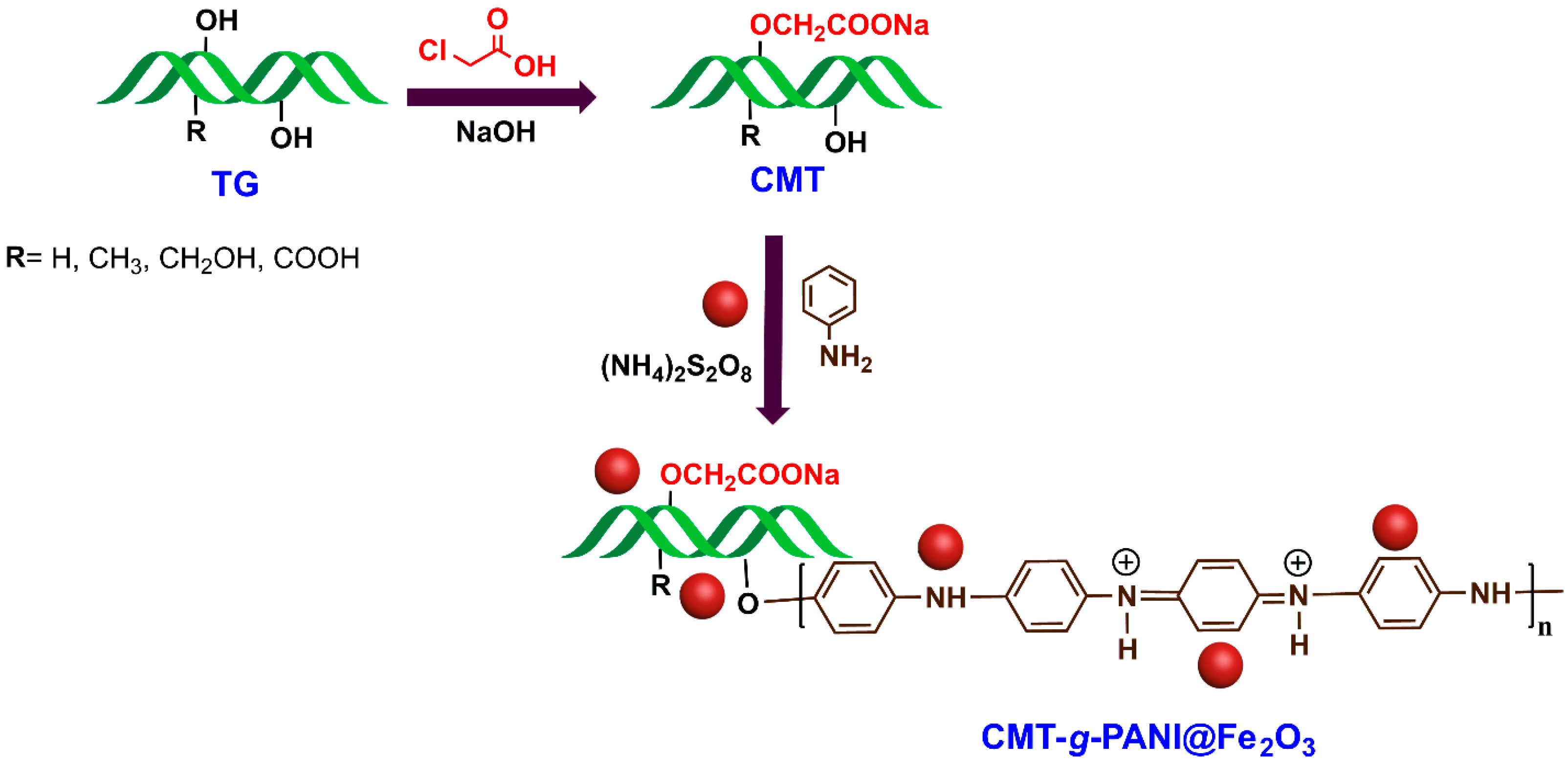
2.2. Preparation of Carboxymethyl Tragacanth Gum (CMT)

The carboxymethyl tragacanth gum (CMT) was synthesized according to our previous literature with slight modifications [31]. We fully dissolved 1 g of powdered tragacanth gum (TG) in a mixture of water/ethanol (85 mL/15 mL). Then, 1.2 g of NaOH in 10 mL of distilled water was added to the reaction mixture. The solution was kept at 50 °C for 30 min under a magnetic stirrer (Heidolph, Schwabach, Germany). Afterward, 1.3 g of monochloroacetic acid in 10 mL of distilled water was added to the reaction solution, and the final solution was stirred for 4 h at 50 °C. After cooling, the solution was poured into a double volume of ethanol or methanol. The resultant CMC sediment was separated using filter paper and dried at 40 °C (Figure 1).

2.3. Synthesis of γFe2O3

The co-precipitation technique was employed for the γ-Fe2O3 nanoparticles synthesis [32]. A solution of NaOH (1.0 M) was added to 100 mL of deionized water, and the reaction mixture was stirred magnetically for 15 min under an inert atmosphere. Afterward, a solution of iron(III) and iron(II) salts was dropped into the previous solution, and the final solution was kept under vigorous stirring at ambient temperature for 70 min. The resulting brown precipitate was separated and washed several times with distilled water and ethanol. Lastly, the precipitate was dried and the obtained powder was calcined at 300 °C for 2 h to gain the γ-Fe2O3 nanoparticles.

2.4. Preparation of Carboxymethyl Tragacanth Gum-Grafted-Polyaniline@γFe2O3

In a 250 mL round-bottom flask, 0.62 g of carboxymethyl tragacanth gum (CMT) was dissolved in 50 mL of distilled water. Then, 1.5 mL of HCl was added to the solution, and the flask was kept under N2 gas at 0–5 °C. Subsequently, 10 wt% (an optimal amount) of γFe2O3 nanoparticles were dispersed in 20 mL of distilled water for 30 min using an ultrasonic bath and then added to the above solution. After that, within 30 min, 3 g of ammonium persulfate in 10 mL of distilled water was added to the solution. The flask was then placed on a magnetic stirrer for 12 h under the aforementioned conditions after 1.25 mL of aniline monomer was added. The nanocomposite was separated by a magnet and rinsed with 10 mL of N-methyl pyrrolidone, water, and acetone before they were dried at 50 °C (Figure 1).

2.5. Adsorption Experiment

The carboxymethyl tragacanth gum-grafted-polyaniline@γFe2O3 (CMT-g-PANI@Fe2O3) bionanocomposite’s ability to remove amoxicillin antibiotic from aqueous solutions was tested in a few different ways. The calibration curve of amoxicillin was prepared by defining the absorbance at 228 nm with a series of standard solutions (1−8 mg/L) achieved from reducing the stock solution at pH 7. Then, the amoxicillin initial or equilibrium concentrations were measured with the calibration curve [22].
Amoxicillin’s initial concentration in an aqueous solution, solution pH, the amount of adsorbent, the agitation time, and other important parameters were all examined for their effects on adsorption capacity. The pH was changed from 4 to 9 using HCl (0.1 N) and NaOH (0.1 N). The optimal adsorption conditions were then investigated using a range of CMT-g-PANI@Fe2O3 biosorbent doses (0.005–0025 g), contact periods (5–30 min), and initial amoxicillin concentrations (50–400 ppm). By comparing the outcomes of the experimental data with those predicted by the Freundlich and Langmuir models, the adsorption isotherms were also explored. To assess the adsorption kinetics, the pseudo-first-order and pseudo-second-order models were also applied. The experimental tests were carried out three times, and an average of the results was provided. A UV-visible spectrometer was used to assess the amoxicillin concentration. Amoxicillin’s capacity and adsorption efficiency onto CMT-g-PANI@Fe2O3 bionanocomposite were calculated using Equations (1) and (2), respectively [20].
R % = C i C e C i × 100
Q e = C i C e m × V
where Ci and Ce are the amoxicillin initial and the equilibrium concentrations in the solutions (mg/L), correspondingly. m is the weight (g) of CMT-g-PANI@Fe2O3 bionanocomposite and V is the solution volume (L).

2.6. Isotherm Study

Langmuir and Freundlich’s isotherm models are used to evaluate the maximum adsorption capacity and equilibrium adsorption isotherms. The Langmuir isotherm model measured the single-layer adsorption of contaminants onto the adsorbent surface, whereas the Freundlich isotherm model measured the multilayer adsorption of pollutants on the adsorbent surface. The mathematical expression of Langmuir’s (Equation (3)) and Freundlich’s (Equation (4)) isotherm models are as follows [22,33]:
C e Q e = 1 K L Q max + 1 Q max C e
LnQ e = LnK F + 1 n LnC e
where, Ce, Qe, and Qmax are the equilibrium concentration (mg/L), the equilibrium, and maximum adsorption capacity (mg/g) correspondingly; KL (L/mg) is the Langmuir constant calculated from the plot between Ce/Qe and Ce; KF (L/mg) is the Freundlich constant calculated from the plot between Ln Qe and Ln Ce. n is a parameter to define the adsorption process favorability; once n > 1, the amoxicillin adsorption onto bioadsorbent is anticipated at high concentrations.

2.7. Kinetic Study

The famous kinetics models were used for studying the effect of time on the adsorption process. The mathematical expression of pseudo-first-order (PFO, Equation (5)) and pseudo-second-order (PSO, Equation (6)) models are shown as follows [22,33]:
Ln Q e Q t = LnQ e k 1 t  
t Q t = 1 k 2 Q e 2 + 1 Q e t  
where Qt (mg/g), and Qe (mg/g) are the adsorption capacity at time t and equilibrium, correspondingly. k1 (1/min) and k2 (g/mg⋅min) are the rate constants of the PFO and PSO, correspondingly.

2.8. Desorption and Reusability

To examine the desorption and reusability of the CMT-g-PANI@Fe2O3 bionanocomposite, the amoxicillin adsorbed onto CMT-g-PANI@Fe2O3 bionanocomposite was floated in ethanol and stirred at ambient temperature for 1 h. After that, the bionanocomposite was separated by a magnet. The quantity of released amoxicillin in the elution medium was measured afterward employing a UV-visible spectrophotometer. The following equation was employed to obtain the desorption percentage.
% D = A B × 100  
where A is the amoxicillin desorbed (mg) in the medium and B is the amoxicillin adsorbed (mg) on the CMT-g-PANI@Fe2O3 bionanocomposite.

3. Results and Discussion

Currently, the removal of antibiotic drugs from polluted water due to their destructive effect on humans and other living beings is very important. In this regard, we employed an antibacterial magnetic nanobiosorbent based on a carboxymethyl tragacanth gum-grafted-polyaniline and γFe2O3 nanoparticles (CMT-g-PANI@Fe2O3) for the elimination of amoxicillin from contaminated water.

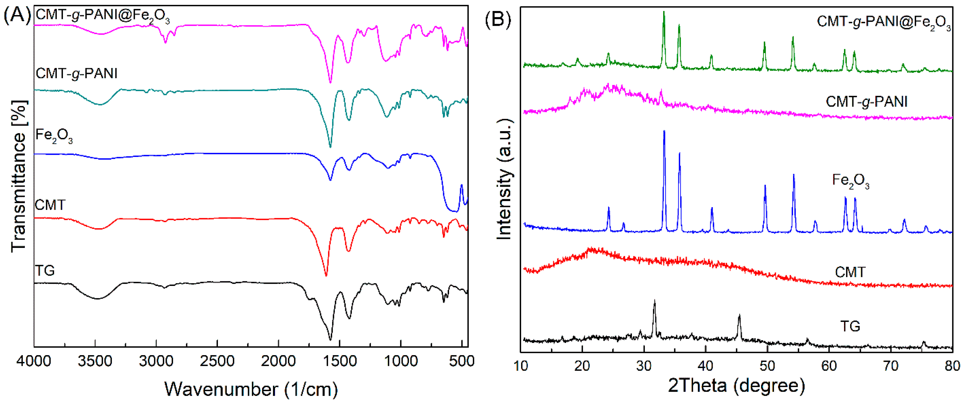
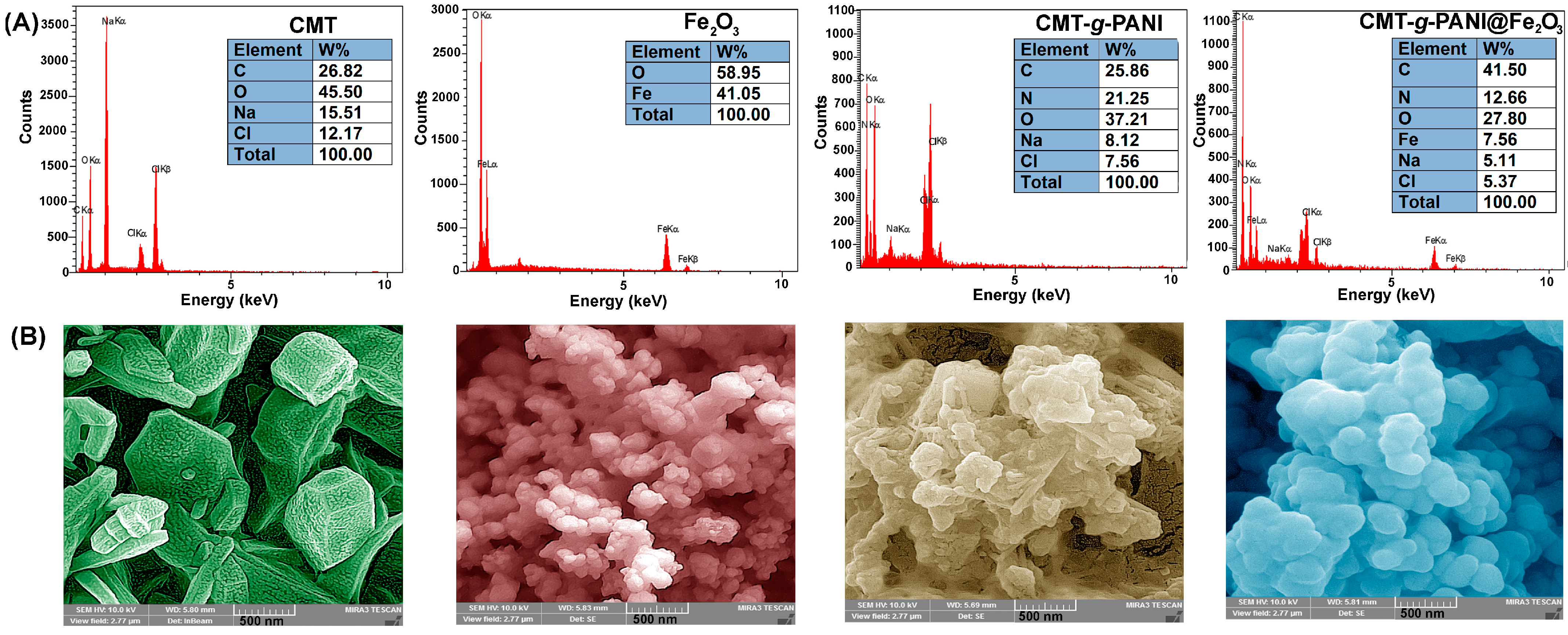
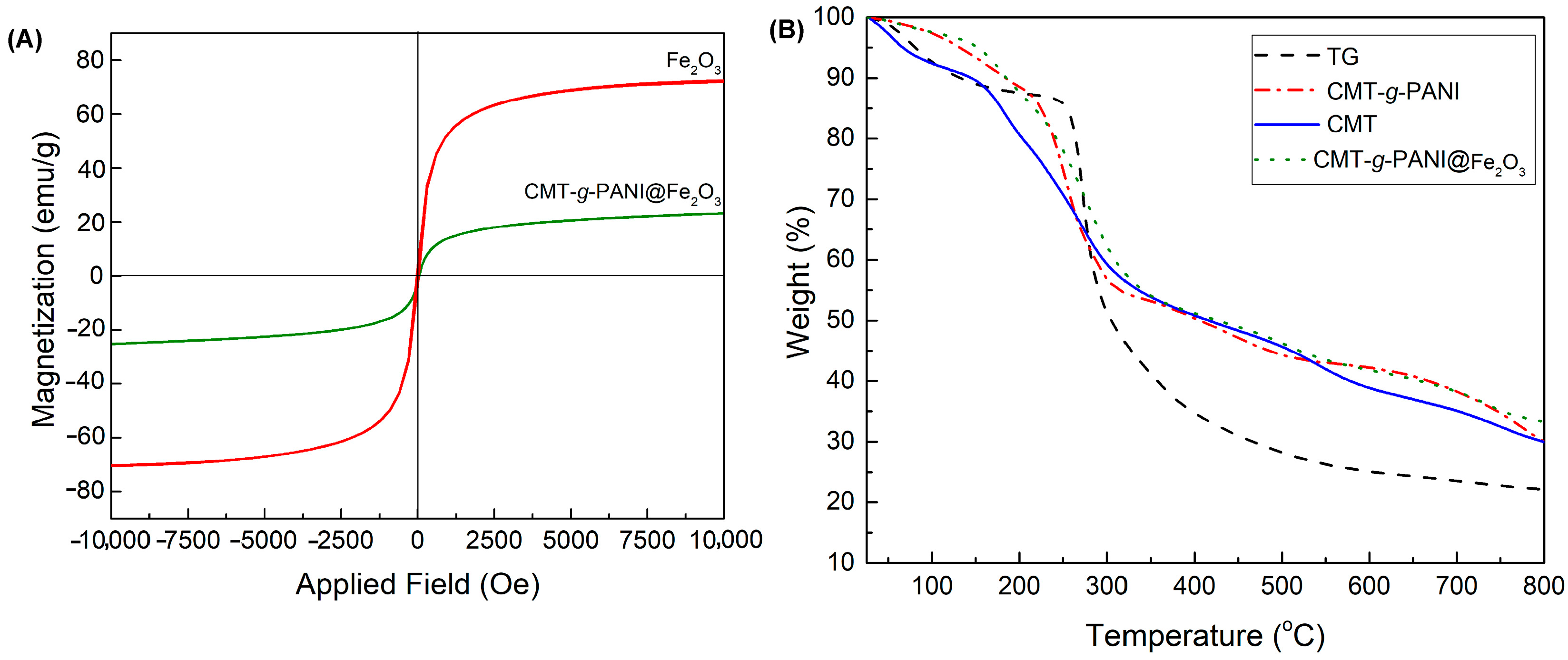
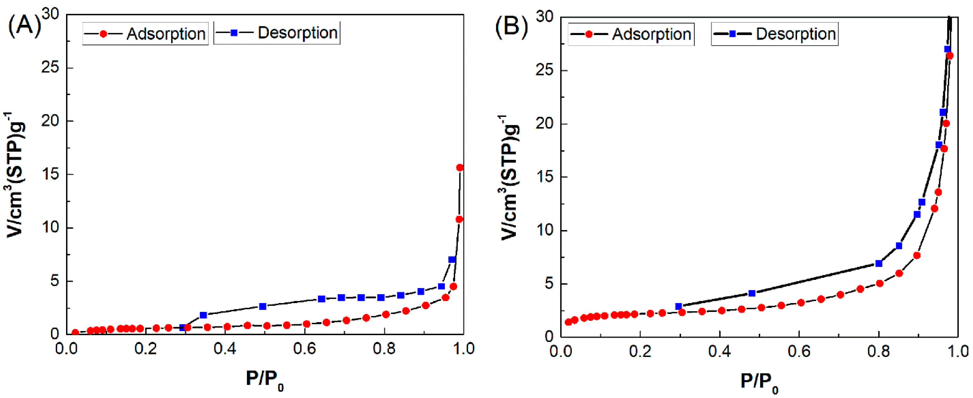
3.1. Nanobiosorbent Characterization

FTIR spectra are employed for studying the chemical structure of prepared materials and are shown in Figure 2A. In the FTIR spectrum of the TG, the absorption bands at 3470 cm−1, 2934 cm−1, and 1740 cm−1 are related to the stretching vibrations of the OH, –CH, and ester carbonyl groups, respectively [34,35]. In addition, the absorption band at 1604 cm−1 is due to the existence of the carboxylate anion of d-galacturonic acid. In the FTIR spectrum of the CMT, the absorption band at 1622 cm−1 is associated with the –COO asymmetric vibrations which overlapped with the carbonyl groups of acidic and ester existing in the TG parts of the CMT [31,34]. Moreover, the absorption band that appeared at 1433 cm1 is related to the bending vibrations of –CH2. The FTIR spectrum of γFe2O3 nanoparticles displayed an absorption band at 550 cm−1 that was related to the stretching vibrations of Fe–O. Moreover, the bands at 1620 cm−1 and 3400 cm−1 were associated with the –OH stretching vibrations on the surface of the γFe2O3 nanoparticles [36,37]. The FTIR spectrum of CMT-g-PANI displayed a broad absorption band at ~3205 cm−1 and is related to the stretching vibrations of the N–H and OH of PANI and CMT, respectively. The absorption bands at 3050 cm−1 and 2949 cm−1 are attributed to aromatic and aliphatic C–H in the PANI and CMT, respectively. The two bands appearing at 622 cm−1 and 1470 cm−1 were related to the asymmetric vibrations of the –COO and stretching vibration of the benzenoid ring, respectively [38]. The presence of characteristic absorption bands of CMT, PANI, and Fe2O3 in the FTIR spectrum of CMT-g-PANI@Fe2O3 revealed that the bioadsorbent was synthesized.
The XRD analysis was employed for the investigation of the crystallinity nature of the samples as shown in Figure 2B. The XRD pattern of CMT showed an amorphous nature as compared to the XRD pattern of TG [31,39]. This crystallinity reduction may be related to the the –OH group’s replacement by the –COO groups in the CMT [34]. A crystalline nature with diffraction peaks located at 2theta = 23°, 27°, 34°, 35°, 41°, 49°, 53°, 57°, 62°, and 63° was observed in the XRD pattern of Fe2O3 [39]. Synthesized Fe2O3 has an ordered cubic structure, which is consistent with JCPDS file no. 39-1346 [40]. XRD patterns comparison of CMT-g-PANI and CMT-g-PANI@Fe2O3 showed that the crystallinity of CMT-g-PANI improved in the presence of Fe2O3.
The elemental analysis of TG, CMT, and CMT-g-PANI was performed, and the percentage mass contents of the elements are shown in Table 1. In CMT, the oxygen content reaches 66.27% as compared to TG which means that the substitution of –COO was carried out in the TG, successfully. Correspondingly, in CMT-g-PANI, the oxygen content decreased to 47.71%. This could be related to the grafting reaction between CMT and PANI. Furthermore, the presence of nitrogen content of 8.03% in CMT-g-PANI is related to the PANI.
EDX examination was also applied for the chemical structure study of the prepared samples as shown in Figure 3A. The existence of C, O, Cl, and Na elements in the EDX spectrum of CMT confirmed the preparation of CMT and the substitution of –COONa in the TG backbone. The existence of a tiny amount of Cl is related to chlorine removal from monochloroacetic acid due to the nucleophilic substitution reaction between the hydroxyl of TG and monochloroacetic acid. The presence of Fe and O elements in Fe2O3 and C, N, O, Cl, and Na elements in the CMT-g-PANI expressed that the Fe2O3 and CMT-g-PANI@Fe2O3 were prepared. In addition, the presence of C, O, N, Fe, Cl, and Na in CMT-g-PANI@Fe2O3 showed that the bionancomposite was prepared successfully.
Figure 3B shows the FESEM micrographs of CMT, Fe2O3, CMT-g-PANI, and CMT-g-PANI@Fe2O3. The FESEM image of CMT shows polyhedral particles with microsize. It appears that the chemical modification of TG leads to the formation of polyhedral microparticles. The nano-spherical and amorphous structures were observed in the FESEM images of Fe2O3 and CMT-g-PANI. The presence of Fe2O3 nanoparticles lead to a granular structure in the FESEM image of CMT-g-PANI@Fe2O3.
VSM analysis was employed to evaluate of magnetic properties of Fe2O3 and CMT-g-PANI@Fe2O3 as shown in Figure 4A. The magnetization saturation (Ms) values of Fe2O3 and CMT-g-PANI@Fe2O3 were 70 emu g−1 and 25 emu g−1, respectively, and showed superparamagnetic properties for both. In addition, the Ms value of Fe2O3 decreased with the coating of Fe2O3 by CMT-g-PANI.
TG analysis was applied to study the thermal stability of prepared materials as seen in Figure 4B. The TGA thermogram of TG displays two steps of mass loss from 50 to 178 °C and from 200 to 580 °C. The first mass loss (10 wt%) was related to the removal of moisture absorbed in the TG, and the second mass loss (65 wt%) corresponded to the branched heterogeneous structure degradation of the TG [34,39]. The char yield of TG at 800 °C was 25 wt%. Four mass losses at 30–150 °C, 150–300 °C, 300–550 °C, and 550–780 °C were observed in the TGA thermogram of CMT. The first (10 wt%) and second (32 wt%) mass losses were related to the moisture evaporation and combination of the saccharide ring degradation, the C–O–C breaking, and the CO2 elimination from the CMC, respectively [34,39]. The two latter mass losses were attributed to the CMT backbone breaking [39]. The char yield of CMC at 800 °C was 33 wt%. In the TGA thermogram of CMT-g-PANI, four mass losses were observed. The first mass loss (10 wt%) at 5–200 °C was caused by the evaporation of water and solvent entrapment in the copolymer chains [41]. The second mass loss (25 wt%) at 200–300 °C probably corresponded to the loss of hydrochloric acid, saccharide ring, and the CO2 elimination from the CMT-g-PANI. The third mass loss (10 wt%) at 300–500 °C and the fourth mass loss (20 wt%) at 500–750 °C was ascribed to the breakdown of sugar units in the CMT structure and PANI, respectively. The char yield of CMT-g-PANI at 800 °C was 34 wt%. Compared with CMT-g-PANI, the thermogram of the CMT-g-PANI@Fe2O3 showed good thermal stability owing to the existence of Fe2O3 nanoparticles in the CMT-g-PANI matrix.
Brunauer–Emmett–Teller (BET) analysis was applied to define the specific surface area of materials and evaluate the effect of the presence of Fe2O3 nanoparticles within the CMT-g-PANI matrix, as seen in Figure 5. The specific surface area (as,BET) values of CMT-g-PANI@Fe2O3 and Fe2O3 were 8.0794 m2/g and 2.8278 m2/g, respectively. This confirms that the CMT-g-PANI@Fe2O3 had better surface properties in terms of surface area and micropore area compared to Fe2O3. Thus, the presence of Fe2O3 together with CMT-g-PANI led to an improvement in the as,BET value of CMT-g-PANI@Fe2O3 which could be effective in antibiotic adsorption by bioadsorbent.

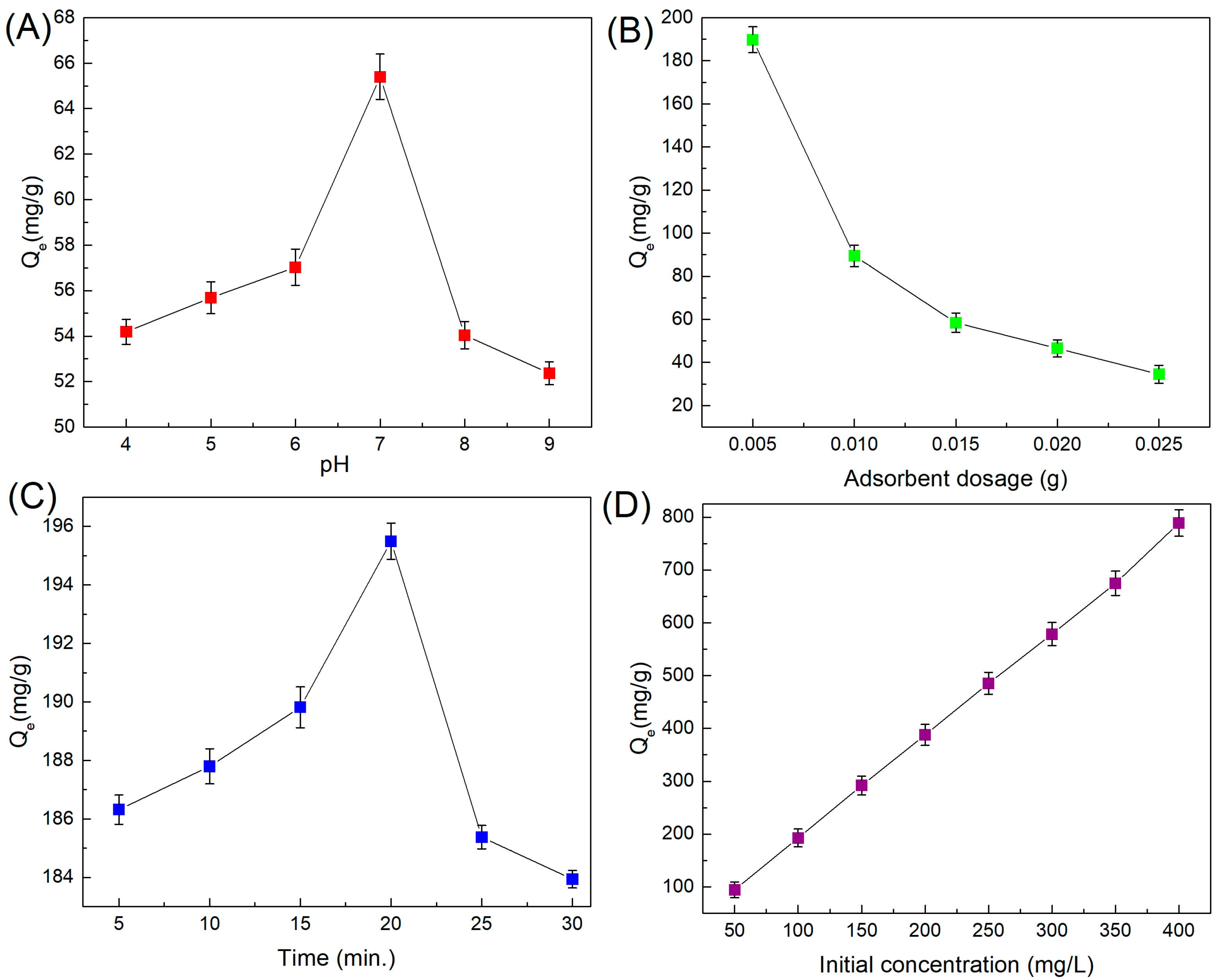
3.2. Optimization of Effective Parameters for Amoxicillin Adsorption

Solution pH: It is well known that the changes in the medium pH can directly affect the removal percentage of pollutants from an aqueous solution. In this regard, a solution pH range of 4–9 for finding the best pH for amoxicillin adsorption by the CMT-g-PANI@Fe2O3 was examined (Figure 6A). Results revealed that by growing the pH values from 4 to 7, the Qe increased from 54.18 mg/g to 65.39 mg/g and then decreased at high pH (8 and 9). At pH 7, amoxicillin antibiotic occurs as zwitterions, while at pH 4 and 5, it performs in cationic species, and at pH 8 and 9, it appears in anionic species. Thus, amoxicillin removal was efficient under pH 7 (neutral). The hydroxyl, carboxyl, and amine groups in the copolymer together with a high surface-to-volume ratio of Fe2O3 in the CMT-g-PANI@Fe2O3 adsorbent lead to the rapid diffusion of amoxicillin molecules into the adsorbent and interaction with its functional groups for efficient amoxicillin.
Amount of biosorbent: Adsorption examinates were carried out with changing quantities (0.005–0.025 g) of the CMT-g-PANI@Fe2O3 at pH 7 (optimal) to investigate the association between the adsorbent amount and its adsorption capability for amoxicillin. According to Figure 6B, the Qe decreased from 189.73 to 34.50 mg/g as the adsorbent amount increased from 0.005 g to 0.025 g. At a lower adsorbent amount, the higher the amount of amoxicillin adsorbed by the CMT-g-PANI@Fe2O3. The optimal quantity of CMT-g-PANI@Fe2O3. for subsequent examinations was found to be 0.005 g.
Agitation time: The agitation time effect on the Qe of the CMT-g-PANI@Fe2O3 adsorbent for the removal of amoxicillin was examined (Figure 6C). Results showed that the Qe increased up to 195.49 mg/g by increasing the agitation time from 5 to 20 min at pH 7 with an adsorbent dose of 0.005 g (optimal). Furthermore, the Qe decreased slightly to 183.93 mg/g once the agitation time reached 30 min. Consequently, an agitation time of 20 min was chosen to be the perfect time for subsequent examinations. Generally, at the beginning of the adsorption process, numerous vacant sites of adsorbent are vacant to interact with amoxicillin molecules in solution. The functional group’s interaction of the adsorbent and the amoxicillin grow stronger once the agitation time is increased to 20 min. At longer time frames (>20 min), there was no more improvement in Qe, which was probably related to the existence of adsorbent active sites and the equilibrium state approach.
Amoxicillin initial concentration: The association between the amoxicillin initial concentration and the Qe of the CMT-g-PANI@Fe2O3 adsorbent was studied by altering the amoxicillin concentration from 50 to 400 mg/L at the optimal pH (7.0), dose (0.005 g), and agitation time (20 min). Figure 6D shows that the amoxicillin initial concentration influences the Qe of CMT-g-PANI@Fe2O3 adsorbent. With increasing the amoxicillin initial concentration from 50 to 400 mg/L, Qe of adsorbent with an intensity rose to 788.83 mg/g with a steep slope. Thus, the Qe increases once the amoxicillin initial concentration is increased, whereas the adsorbent amount remains constant, and this pattern holds until the amoxicillin initial concentration reaches an equilibrium level.

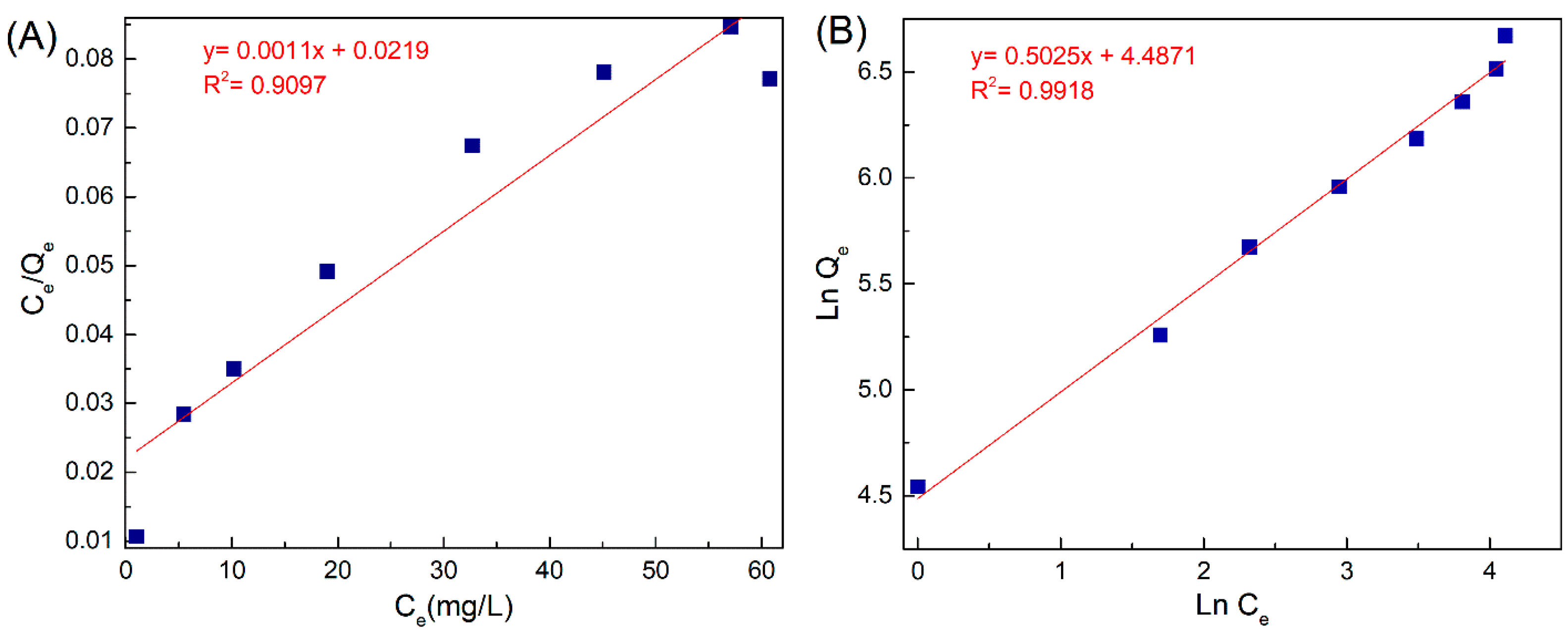
3.3. Adsorption Isotherm

To find the interaction between amoxicillin and CMT-g-PANI@Fe2O3 bioadsorbent, the isotherm study was employed. It is well known that Langmuir and Freundlich’s isotherm models are used to evaluate the maximum adsorption capacity and equilibrium adsorption isotherms. Figure 7A,B demonstrates the Langmuir and Freundlich isotherm. The obtained parameters of isotherm models are tabulated in Table 2. Consistent with obtained correlation coefficient (R2) of isotherm models, the Freundlich isotherm was found to be more reliable with the experimental data than the Langmuir model. This result shows that the amoxicillin adsorbed over the CMT-g-PANI@Fe2O3 bioadsorbent surface as a multilayer. In addition, the obtained “n” in the Freundlich model is an important parameter to define the adsorption process favorability. The value of n > 1 in Table 2 shows that the amoxicillin adsorbed over the CMT-g-PANI@Fe2O3 bioadsorbent surface is desirable at high concentrations.
The comparison of Qmax for amoxicillin to other adsorbents reported in recent years showed that the CMT-g-PANI@Fe2O3 biosorbent has a higher maximum adsorption capacity (909.09 mg∙g) than other adsorbents (Table 3). This could be related to the presence of Fe2O3 nanoparticles and numerous active sites, for example, hydroxyl, carboxylate, and amine groups in the nanocomposite, which can effectively interact with amoxicillin (via electrostatic interaction and hydrogen bonding), led to eliminating the amoxicillin.

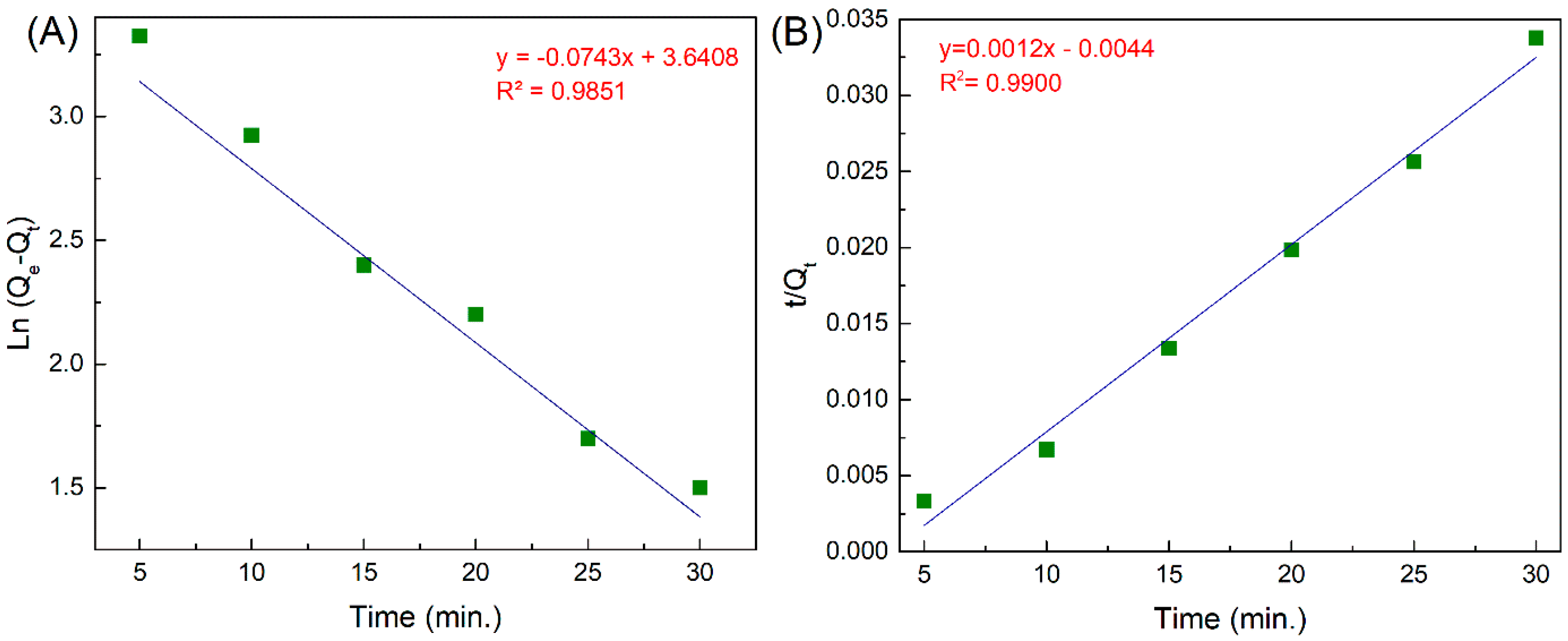
3.4. Adsorption Kinetic

For discovering and evaluating possible adsorption routes, equilibrium time, and adsorption rate-limiting phase of amoxicillin adsorbed over the CMT-g-PANI@Fe2O3 adsorbent used the PFO and PSO kinetics models. In addition, kinetic models are employed to define the adsorption process mechanism, for example, surface and chemical adsorption. The PFO is based on the adsorbent capacity and employed once adsorption happens via diffusion in a border layer, whereas the PSO expresses that the chemical adsorption procedure is the main controlling procedure. Figure 8A,B and Table 4 exhibit kinetic linear plots, and their calculated parameters. According to R2, the difference between the estimated Qe and observed Qe values, the PSO is more appropriate for considering the amoxicillin adsorption kinetics on the CMT-g-PANI@Fe2O3. Consequently, chemical adsorption is the main mechanism in the adsorption of amoxicillin on the adsorbent and it displays that, in addition to amoxicillin molecules, the CMT-g-PANI@Fe2O3 adsorbent is involved in the adsorption procedure.

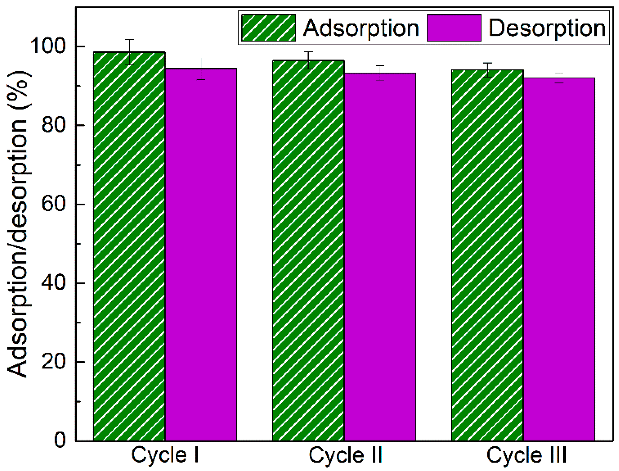
3.5. Desorption and Reusability

The reusability of adsorbent is a significant factor to create the adsorption procedure economically. The reusability of adsorbent can be effective for its use in numerous consecutive cycles without considerable performance debility. In the present work, we evaluated the amoxicillin desorption and reusability of CMT-g-PANI@Fe2O3 bioadsorbent in three consecutive cycles, and an ethanolic solution was investigated. In this regard, amoxicillin adsorbed onto the CMT-g-PANI@Fe2O3 was performed in optimal conditions and was immersed into ethanol (10 mL) under stirring at room temperature for 1 h to desorb amoxicillin. Afterward, CMT-g-PANI@Fe2O3 was collected by a magnet, washed with distilled water, and then dried for successive adsorption/desorption processes. Then, the released amoxicillin amount in the elution medium was measured using a UV-visible spectrophotometer. Figure 9 demonstrates that the adsorption percentage decreased from 98.59 % to 94.03%, and the desorption percentage decreased from 94.41% to 92.001% after three consecutive cycles, these results show that the CMT-g-PANI@Fe2O3 could continue to remove amoxicillin after three consecutive cycles of adsorption/desorption process without considerably losing adsorption capacity.

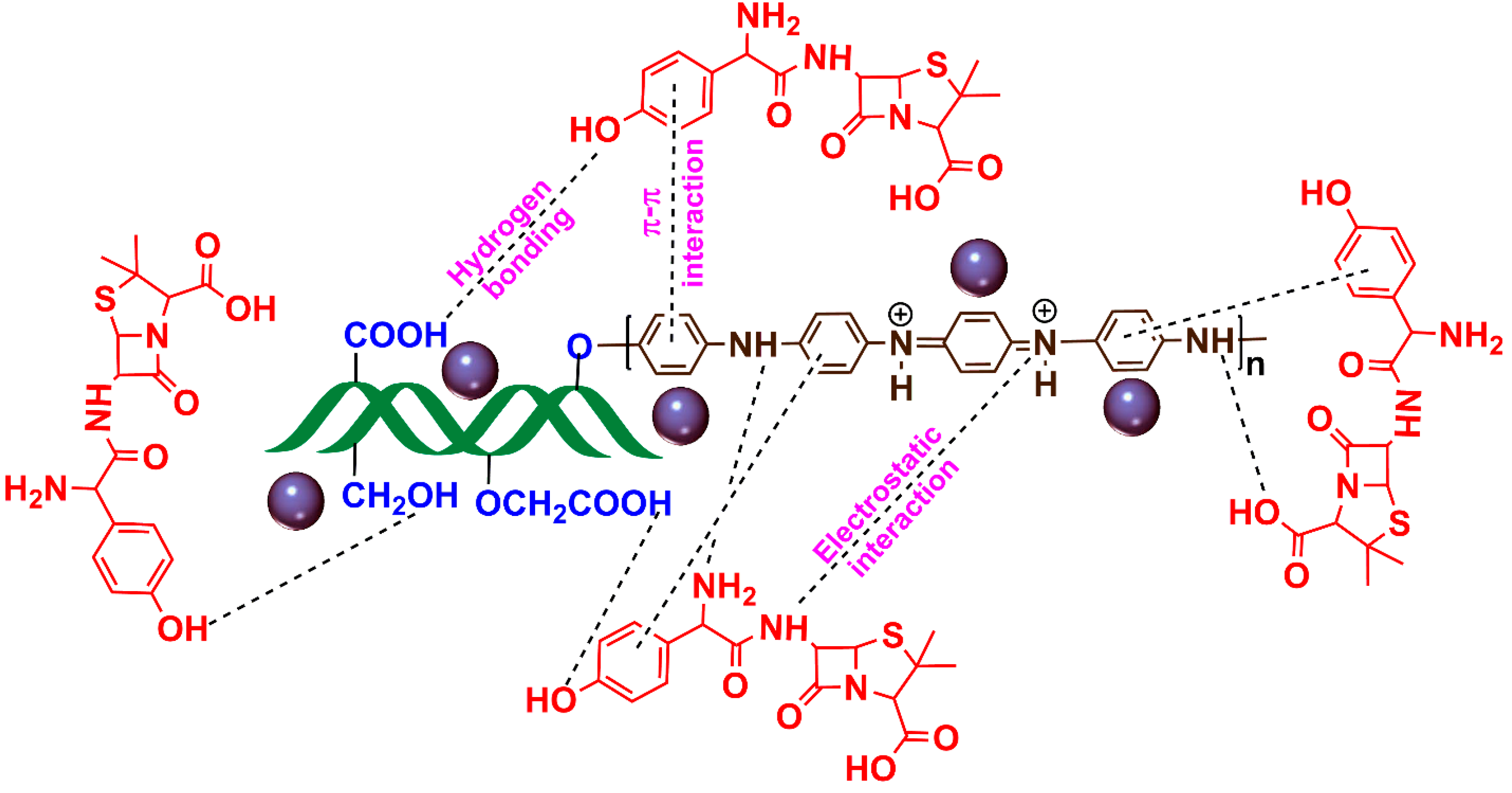
3.6. Suggested Mechanism of Amoxicillin Adsorption

The pollutant adsorption mechanism by the adsorbent depends on the functional groups and morphology of the adsorbent, which can be created a synergistic effect in increasing the pollutant adsorption by the adsorbent. Considering that the CMT-g-PANI@Fe2O3 bioadsorbent has amine, carboxylate, and hydroxyl functional groups, it can create intermolecular interactions such as hydrogen bonding, electrostatic and π–π interactions with the amoxicillin antibiotic, and cause its efficient adsorption on the CMT-g-PANI@Fe2O3 bioadsorbent as shown in Figure 10.

4. Conclusions

A magnetic bionanocmposite was synthesized by an in situ copolymerization method from carboxy methyl tragacanth gum in the presence of aniline monomer and γ Fe2O3 nanoparticles (CMT-g-PANI@Fe2O3). The CMT-g-PANI@Fe2O3 bionanocmposite was characterized by several analyses and employed as a bioadsorbent for the removal of amoxicillin antibiotic from contaminated water. A granular structure with good thermal stability (char yield 34 wt%), magnetization saturation (25 emu g−1), and a specific surface area (8.0794 m2/g) observed for CMT-g-PANI@Fe2O3 bionanocmposite. The maximum adsorption capacity (909.09 mg/g) was found at pH 7, an agitation time of 20 min, an adsorbent dose of 0.005 g, and an amoxicillin initial concentration of 400 mg/L. The Freundlich isotherm and pseudo-second-order kinetic models fit closely with the experimental data. The adsorption/desorption process showed that the CMT-g-PANI@Fe2O3 could continue to remove amoxicillin after three consecutive cycles without considerably losing adsorption capacity. The intermolecular interactions such as hydrogen bonding, electrostatic, and π–π interactions between amoxicillin antibiotic and CMT-g-PANI@Fe2O3 were suggested for the adsorption mechanism of amoxicillin antibiotic by bionanocomposite.

Author Contributions

Conceptualization, E.N.Z.; methodology, S.S.M.; software, E.N.Z.; validation, E.N.Z., H.B. and M.T.; formal analysis, S.S.M.; investigation, E.N.Z., H.B. and M.T.; data curation, S.S.M.; writing—original draft preparation, E.N.Z.; writing—review and editing, E.N.Z., H.B. and M.T.; supervision, E.N.Z., H.B. and M.T.; project administration E.N.Z.; funding acquisition, S.S.M. All authors have read and agreed to the published version of the manuscript.

Funding

This research received no external funding.

Data Availability Statement

Data is available upon request.

Acknowledgments

The authors are grateful for the support of Damghan University’s financial support.

Conflicts of Interest

The authors declare no conflict of interest.

References

  1. Ahmadi, S.A.R.; Kalaee, M.R.; Moradi, O.; Nosratinia, F.; Abdouss, M. Core–shell activated carbon-ZIF-8 nanomaterials for the removal of tetracycline from polluted aqueous solution. Adv. Compos. Hybrid. Mater. 2021, 4, 1384–1397. [Google Scholar] [CrossRef]
  2. Li, Z.; Xie, W.; Yao, F.; Du, A.; Wang, Q.; Guo, Z.; Gu, H. Comprehensive electrocatalytic degradation of tetracycline in wastewater by electrospun perovskite manganite nanoparticles supported on carbon nanofibers. Adv. Compos. Hybrid. Mater. 2022, 5, 2092–2105. [Google Scholar] [CrossRef]
  3. Zare, E.N.; Fallah, Z.; Le, V.T.; Doan, V.D.; Mudhoo, A.; Joo, S.W.; Vasseghian, Y.; Tajbakhsh, M.; Moradi, O.; Sillanpää, M.; et al. Remediation of pharmaceuticals from contaminated water by molecularly imprinted polymers: A review. Environ. Chem. Lett. 2022, 20, 2629–2664. [Google Scholar] [CrossRef] [PubMed]
  4. Limousy, L.; Ghouma, I.; Ouederni, A.; Jeguirim, M. Amoxicillin removal from aqueous solution using activated carbon prepared by chemical activation of olive stone. Environ. Sci. Pollut. Res. 2017, 24, 9993–10004. [Google Scholar] [CrossRef] [PubMed]
  5. Naeini, A.H.; Kalaee, M.R.; Moradi, O.; Khajavi, R.; Abdouss, M. Synthesis, characterization and application of Carboxylmethyl cellulose, Guar gam, and Graphene oxide as novel composite adsorbents for removal of malachite green from aqueous solution. Adv. Compos. Hybrid. Mater. 2022, 5, 335–349. [Google Scholar] [CrossRef]
  6. Zhu, J.; Tian, M.; Zhang, Y.; Zhang, H.; Liu, J. Fabrication of a novel “loose” nanofiltration membrane by facile blending with Chitosan–Montmorillonite nanosheets for dyes purification. Chem. Eng. J. 2015, 265, 184–193. [Google Scholar] [CrossRef]
  7. Bebu, A.; Szabó, L.; Leopold, N.; Berindean, C.; David, L. IR, Raman, SERS and DFT study of amoxicillin. J. Mol. Struct. 2011, 993, 52–56. [Google Scholar] [CrossRef]
  8. Andreozzi, R.; Canterino, M.; Marotta, R.; Paxeus, N. Antibiotic removal from wastewaters: The ozonation of amoxicillin. J. Hazard. Mater. 2005, 122, 243–250. [Google Scholar] [CrossRef]
  9. Sánchez-Montes, I.; Fuzer Neto, J.R.; Silva, B.F.; Silva, A.J.; Aquino, J.M.; Rocha-Filho, R.C. Evolution of the antibacterial activity and oxidation intermediates during the electrochemical degradation of norfloxacin in a flow cell with a PTFE-doped β-PbO2 anode: Critical comparison to a BDD anode. Electrochim. Acta 2018, 284, 260–270. [Google Scholar] [CrossRef] [Green Version]
  10. Ioannou-Ttofa, L.; Raj, S.; Prakash, H.; Fatta-Kassinos, D. Solar photo-Fenton oxidation for the removal of ampicillin, total cultivable and resistant E. coli and ecotoxicity from secondary-treated wastewater effluents. Chem. Eng. J. 2019, 355, 91–102. [Google Scholar] [CrossRef]
  11. Rodríguez-Chueca, J.; Della Giustina, S.V.; Rocha, J.; Fernandes, T.; Pablos, C.; Encinas, Á.; Barceló, D.; Rodríguez-Mozaz, S.; Manaia, C.M.; Marugán, J. Assessment of full-scale tertiary wastewater treatment by UV-C based-AOPs: Removal or persistence of antibiotics and antibiotic resistance genes? Sci. Total Environ. 2019, 652, 1051–1061. [Google Scholar] [CrossRef] [PubMed]
  12. Alizadeh, E.; Baseri, H. Photocatalytic degradation of Sumatriptan Succinate by ZnO, Fe doped ZnO and TiO2-ZnO nanocatalysts. Mater. Chem. Horizons 2022, 1, 7–21. [Google Scholar] [CrossRef]
  13. Alsager, O.A.; Alnajrani, M.N.; Abuelizz, H.A.; Aldaghmani, I.A. Removal of antibiotics from water and waste milk by ozonation: Kinetics, byproducts, and antimicrobial activity. Ecotoxicol. Environ. Saf. 2018, 158, 114–122. [Google Scholar] [CrossRef]
  14. Cheng, D.; Ngo, H.H.; Guo, W.; Liu, Y.; Chang, S.W.; Nguyen, D.D.; Nghiem, L.D.; Zhou, J.; Ni, B. Anaerobic membrane bioreactors for antibiotic wastewater treatment: Performance and membrane fouling issues. Bioresour. Technol. 2018, 267, 714–724. [Google Scholar] [CrossRef] [PubMed]
  15. Ghanbari, R.; Amanat, N. Approaches of Membrane Modification for Water Treatment. Mater. Chem. Horizons. 2022, 1, 153–167. [Google Scholar] [CrossRef]
  16. Neghi, N.; Krishnan, N.R.; Kumar, M. Analysis of metronidazole removal and micro-toxicity in photolytic systems: Effects of persulfate dosage, anions and reactor operation-mode. J. Environ. Chem. Eng. 2018, 6, 754–761. [Google Scholar] [CrossRef]
  17. Tiwari, B.; Sellamuthu, B.; Ouarda, Y.; Drogui, P.; Tyagi, R.D.; Buelna, G. Review on fate and mechanism of removal of pharmaceutical pollutants from wastewater using biological approach. Bioresour. Technol. 2017, 224, 1–12. [Google Scholar] [CrossRef] [Green Version]
  18. Nasseh, N.; Barikbin, B.; Taghavi, L.; Nasseri, M.A. Adsorption of metronidazole antibiotic using a new magnetic nanocomposite from simulated wastewater (isotherm, kinetic and thermodynamic studies). Compos. Part B Eng. 2019, 159, 146–156. [Google Scholar] [CrossRef]
  19. Movagharnezhad, N.; Ehsanimehr, S.; Najafi Moghadam, P. Synthesis of Poly (N-vinylpyrrolidone)-grafted-Magnetite Bromoacetylated Cellulose via ATRP for Drug Delivery. Mater. Chem. Horizons. 2022, 1, 89–98. [Google Scholar] [CrossRef]
  20. Mehdizadeh, A.; Najafi Moghadam, P.; Ehsanimehr, S.; Fareghi, A.R. Preparation of a New Magnetic Nanocomposite for the Removal of Dye Pollutions from Aqueous Solutions: Synthesis and Characterization. Mater. Chem. Horizons. 2022, 1, 23–34. [Google Scholar] [CrossRef]
  21. Akter, T.; Bañuelos, J.L.; Andrade, D.; Bañuelos, D.I.; Saupe, G.B. Rapid Adsorption Mechanism of Methylene Blue onto a Porous Mixed Ti-Nb Oxide. Mater. Chem. Horizons. 2022, 1, 49–67. [Google Scholar] [CrossRef]
  22. Imanipoor, J.; Mohammadi, M.; Dinari, M.; Ehsani, M.R. Adsorption and desorption of amoxicillin antibiotic from water matrices using an effective and recyclable MIL-53 (Al) metal–organic framework adsorbent. J. Chem. Eng. Data 2020, 66, 389–403. [Google Scholar] [CrossRef]
  23. Moussavi, G.; Alahabadi, A.; Yaghmaeian, K.; Eskandari, M. Preparation, characterization and adsorption potential of the NH4Cl-induced activated carbon for the removal of amoxicillin antibiotic from water. Chem. Eng. J. 2013, 217, 119–128. [Google Scholar] [CrossRef]
  24. Ali, I.; Afshinb, S.; Poureshgh, Y.; Azari, A.; Rashtbari, Y.; Feizizadeh, A.; Hamzezadeh, A.; Fazlzadeh, M. Green preparation of activated carbon from pomegranate peel coated with zero-valent iron nanoparticles (nZVI) and isotherm and kinetic studies of amoxicillin removal in water. Environ. Sci. Pollut. Res. 2020, 27, 36732–36743. [Google Scholar] [CrossRef]
  25. Chakhtouna, H.; Benzeid, H.; Zari, N.; Bouhfid, R. Functional CoFe2O4-modified biochar derived from banana pseudostem as an efficient adsorbent for the removal of amoxicillin from water. Sep. Purif. Technol. 2021, 266, 118592. [Google Scholar] [CrossRef]
  26. Taghavizadeh Yazdi, M.E.; Nazarnezhad, S.; Mousavi, S.H.; Sadegh Amiri, M.; Darroudi, M.; Baino, F.; Kargozar, S. Gum Tragacanth (GT): A Versatile Biocompatible Material beyond Borders. Molecules 2021, 26, 1510. [Google Scholar] [CrossRef] [PubMed]
  27. Verbeken, D.; Dierckx, S.; Dewettinck, K. Exudate gums: Occurrence, production, and applications. Appl. Microbiol. Biotechnol. 2003, 63, 10–21. [Google Scholar] [CrossRef] [PubMed]
  28. Tischer, C.A.; Iacomini, M.; Gorin, P.A.J. Structure of the arabinogalactan from gum tragacanth (Astralagus gummifer). Carbohydr. Res. 2002, 337, 1647–1655. [Google Scholar] [CrossRef] [PubMed]
  29. Nejatian, M.; Abbasi, S.; Azarikia, F. Gum Tragacanth: Structure, characteristics and applications in foods. Int. J. Biol. Macromol. 2020, 160, 846–860. [Google Scholar] [CrossRef]
  30. Zare, E.N.; Makvandi, P.; Tay, F.R. Recent progress in the industrial and biomedical applications of tragacanth gum: A review. Carbohydr. Polym. 2019, 212, 450–467. [Google Scholar] [CrossRef]
  31. Abdollahi, Z.; Zare, E.N.; Salimi, F.; Goudarzi, I.; Tay, F.R.; Makvandi, P. Bioactive carboxymethyl starch-based hydrogels decorated with cuo nanoparticles: Antioxidant and antimicrobial properties and accelerated wound healing in vivo. Int. J. Mol. Sci. 2021, 22, 2531. [Google Scholar] [CrossRef] [PubMed]
  32. Sudhakara, K.; Kumar, A.P.; Kumara, B.P.; Raghavendera, A.; Ravia, S.; Kenie, D.N.; Lee, Y.I. Synthesis of γ-Fe2O3 nanoparticles and catalytic activity of azide-alkyne cycloaddition reactions. Asian J. Nanosci. Mater. 2018, 1, 172–182. [Google Scholar]
  33. Hassanzadeh-Afruzi, F.; Heidari, G.; Maleki, A. Magnetic Nanocomposite Hydrogel based on Arabic Gum for Remediation of Lead(II) from Contaminated Water. Mater. Chem. Horizons. 2022, 1, 107–122. [Google Scholar] [CrossRef]
  34. Behrouzi, M.; Moghadam, P.N. Synthesis of a new superabsorbent copolymer based on acrylic acid grafted onto carboxymethyl tragacanth. Carbohydr. Polym. 2018, 202, 227–235. [Google Scholar] [CrossRef] [PubMed]
  35. Janani, N.; Zare, E.N.; Salimi, F.; Makvandi, P. Antibacterial tragacanth gum-based nanocomposite films carrying ascorbic acid antioxidant for bioactive food packaging. Carbohydr. Polym. 2020, 247, 116678. [Google Scholar] [CrossRef]
  36. Long, Z.; Yuan, L.; Shi, C.; Wu, C.; Qiao, H.; Wang, K. Porous Fe2O3 nanorod-decorated hollow carbon nanofibers for high-rate lithium storage. Adv. Compos. Hybrid. Mater. 2022, 5, 370–382. [Google Scholar] [CrossRef]
  37. Yan, Z.; Sun, Z.; Li, A.; Liu, H.; Guo, Z.; Qian, L. Three-dimensional porous flower-like S-doped Fe2O3 for superior lithium storage. Adv. Compos. Hybrid. Mater. 2021, 4, 716–724. [Google Scholar] [CrossRef]
  38. Shao, W.; Jamal, R.; Xu, F.; Ubul, A.; Abdiryim, T. The effect of a small amount of water on the structure and electrochemical properties of solid-state synthesized polyaniline. Materials 2012, 5, 1811–1825. [Google Scholar] [CrossRef] [Green Version]
  39. Bachra, Y.; Grouli, A.; Damiri, F.; Talbi, M.; Berrada, M. A Novel Superabsorbent Polymer from Crosslinked Carboxymethyl Tragacanth Gum with Glutaraldehyde: Synthesis, Characterization, and Swelling Properties. Int. J. Biomater. 2021, 2021, 5008833. [Google Scholar] [CrossRef]
  40. Ganachari, S.V.; Joshi, V.K.; Bhat, R.; Deshpande, R.; Salimath, B.; Rao, N. V Large scale synthesis and characterization of γ-Fe2O3 nanoparticles by self-propagating low temperature combustion method. Int. J. Sci. Res. 2012, 1, 77–79. [Google Scholar]
  41. Van Cuong, N.; Hieu, T.Q.; Thien, P.T.; Vu, L.D.; Van Tan, L. Reusable starch-graft-polyaniline/Fe3O4 composite for removal of textile dyes. Rasayan J. Chem. 2017, 10, 1446–1454. [Google Scholar] [CrossRef]
  42. de Franco, M.A.E.; de Carvalho, C.B.; Bonetto, M.M.; de Pelegrini Soares, R.; Féris, L.A. Removal of amoxicillin from water by adsorption onto activated carbon in batch process and fixed bed column: Kinetics, isotherms, experimental design and breakthrough curves modelling. J. Clean. Prod. 2017, 161, 947–956. [Google Scholar] [CrossRef]
  43. Yang, C.; Wang, L.; Yu, Y.; Wu, P.; Wang, F.; Liu, S.; Luo, X. Highly efficient removal of amoxicillin from water by Mg-Al layered double hydroxide/cellulose nanocomposite beads synthesized through in-situ coprecipitation method. Int. J. Biol. Macromol. 2020, 149, 93–100. [Google Scholar] [CrossRef]
  44. Liu, X.; Yang, S.; Feng, T.; Zhong, H.; Cao, S.; Chen, Y. Removal of amoxicillin from water by concrete-based hydrotalcites: Efficiency and mechanism. Process Saf. Environ. Prot. 2022, 163, 210–217. [Google Scholar] [CrossRef]
  45. Pooresmaeil, M.; Namazi, H. Chitosan coated Fe3O4@Cd-MOF microspheres as an effective adsorbent for the removal of the amoxicillin from aqueous solution. Int. J. Biol. Macromol. 2021, 191, 108–117. [Google Scholar] [CrossRef] [PubMed]
  46. Zandipak, R.; Sobhanardakani, S. Novel mesoporous Fe3O4/SiO2/CTAB–SiO2 as an effective adsorbent for the removal of amoxicillin and tetracycline from water. Clean Technol. Environ. Policy 2018, 20, 871–885. [Google Scholar] [CrossRef]
  47. Jafari, K.; Heidari, M.; Rahmanian, O. Wastewater treatment for Amoxicillin removal using magnetic adsorbent synthesized by ultrasound process. Ultrason. Sonochem. 2018, 45, 248–256. [Google Scholar] [CrossRef]
Figure 1. Schematic illustration of the preparation of carboxymethyl tragacanth gum (CMT) and carboxymethyl tragacanth gum-grafted-polyaniline@γFe2O3 (CMT-g-PANI@Fe2O3) bionanocompsite.
Water 15 00202 g001
Figure 2. FTIR spectra (A) and XRD patterns (B) of TG, CMT, Fe2O3, CMT-g-PANI, and CMT-g-PANI@Fe2O3.
Water 15 00202 g002
Figure 3. EDX spectra (A) and FESEM micrographs (B) of CMT, Fe2O3, CMT-g-PANI, and CMT-g-PANI@Fe2O3.
Water 15 00202 g003
Figure 4. VSM curves of Fe2O3, and CMT-g-PANI@Fe2O3 (A) and TGA thermograms (B) of TG, CMT, CMT-g-PANI, and CMT-g-PANI@Fe2O3.
Water 15 00202 g004
Figure 5. N2 adsorption/desorption isotherms of Fe2O3 (A) and CMT-g-PANI@Fe2O3 (B).
Water 15 00202 g005
Figure 6. (A) Effect of pH (4–9), (biosorbent dose = 0.015 g, amoxicillin concentration = 100 mg/L, time = 15 min and temperature = 298 K), (B) biosorbent dosage (0.005–0.025 g), (pH 7, amoxicillin concentration = 100 mg/L, time = 15 min and temperature = 298 K), (C) time (5–30 min), (pH 7, biosorbent dosage = 0.005 g, amoxicillin concentration = 100 mg/L, V = 10 mL, and temperature = 298 K), (D) amoxicillin concentration (50–400 mg/L), (pH 7, biosorbent dosage = 0.005 g, V = 10 mL, time = 20 min, temperature = 298 K).
Water 15 00202 g006
Figure 7. (A) Langmuir and (B) Freundlich isotherms (condition: amoxicillin concentration (50–400 mg/L), pH 7, biosorbent dosage = 0.005 g, contact time = 20 min, T = 298 K).
Water 15 00202 g007
Figure 8. (A) Pseudo-first-order and (B) pseudo-second-order models (Contact time (5–30 min), pH 7, biosorbent dosage = 0.005 g, amoxicillin concentration = 400 mg/L, T = 298 K)).
Water 15 00202 g008
Figure 9. Desorption and reusability of the CMT-g-PANI@Fe2O3 bioadsorbent for amoxicillin adsorption.
Water 15 00202 g009
Figure 10. A schematic of the suggested mechanism of amoxicillin adsorption by CMT-g-PANI@Fe2O3 bioadsorbent.
Water 15 00202 g010
Table 1. Elemental analysis of TG, CMT, and CMT-g-PANI.
SampleCHNO
TG42.195.71-52.1
CMT29.664.07-66.27
CMT-g-PANI39.884.388.0347.71
Table 2. Isotherm parameters, for amoxicillin adsorption onto the CMT-g-PANI@Fe2O3 bioadsorbent.
IsothermParameters
FreundlichKF88.8633
n1.99
R20.9918
LangmuirQmax909.09
KL0.0502
R20.9097
Table 3. Assessment of the maximum adsorption capacity of CMT-g-PANI@Fe2O3 bionanocomposite with other reported studies.
AdsorbentExperimental ConditionsQmax (mg/g) Ref.
MIL-53(Al) metal-organic frameworkpH: 7.5, adsorbent dosage: 0.1 g/L, contact time: 12 h, T: 303 K, initial concentration: 50 mg/L758.5[22]
NH4Cl-induced activated carbonpH: 6, adsorbent dosage: 0.8 g/L, contact time: 30 min, T: 303 K, initial concentration: 100 mg/L437[23]
Activated carbon/iron nanoparticlespH: 5, adsorbent dosage: 1.5 g/L, contact time: 30 min, T: 298 K, initial concentration: 10 mg/L40.282[24]
Functional CoFe2O4-modified biocharpH: 7, adsorbent dosage: 0.05 g/L, contact time: 60 min, T: 298 K, initial concentration: 50 mg/L99.99[25]
Activated carbonpH: 5.5, adsorbent dosage: 0.06 g/L, contact time: 240, T: 298 K, initial concentration: 100 mg/L4.4[42]
Mg-Al layered double hydroxide/cellulose nanocompositepH: 7, adsorbent dosage: 0.1 g /L, contact time: 20 min, T: 298 K, initial concentration: 120 mg/L138.3[43]
Concrete-based hydrotalcitepH: 5, adsorbent dosage: 0.1 g/L, contact time: 12 h, T: 298 K, initial concentration: 400 mg/L49.7[44]
Chitosan-coated Fe3O4@Cd-MOF microspherespH: 8, adsorbent dosage: 0.05 g/L, contact time: 240 min, T: 298 K, initial concentration: 50 mg/L103.09[45]
Fe3O4/SiO2/CTAB–SiO2pH: 5, adsorbent dosage: 0.009 g/L, contact time: 60 min, T: 298 K, initial concentration: 25 mg/L362.66[46]
Fe3O4@activated carbonpH: 6, adsorbent dosage: 0. 5 g/L, contact time: 90 min, T: 298 K, initial concentration: 200 mg/L238.1[47]
CMT-g-PANI@Fe2O3pH: 7, adsorbent dosage: 0.005 g/L, contact time: 20 min, T: 298 K, initial concentration: 400 mg/L909.09Current work
Table 4. Kinetic parameters, for amoxicillin adsorption on the CMT-g-PANI@Fe2O3.
ModelsParameters
k10.0743
Pseudo-first-orderQe calculated38.122
Qe experimental788.73
R20.9851
k20.000001
Pseudo-second-orderQe833.33
Qe experimental788.83
R20.9900
Disclaimer/Publisher’s Note: The statements, opinions and data contained in all publications are solely those of the individual author(s) and contributor(s) and not of MDPI and/or the editor(s). MDPI and/or the editor(s) disclaim responsibility for any injury to people or property resulting from any ideas, methods, instructions or products referred to in the content.

