Article

Removal of Clostridium perfringens and Staphylococcus spp. in Microalgal–Bacterial Systems: Influence of Microalgal Inoculum and CO2/O2 Addition

by
Graziele Ruas
1,2,3,*,
Sarah Lacerda Farias
1,
Bruno A. B. dos Reis
1,
Mayara Leite Serejo
4,
Gustavo Henrique Ribeiro da Silva
3 and
Marc Árpád Boncz
1
1
Post-Graduate Programme of Environmental Technology, Faculty of Engineering, Architecture and Urbanism and Geography, Federal University of Mato Grosso do Sul, Campo Grande 79070-900, Brazil
2
Federal Institute of Mato Grosso do Sul, Campus Jardim, Jardim 79240-000, Brazil
3
School of Engineering, São Paulo State University (UNESP), Bauru 17000-000, Brazil
4
Federal Institute of Mato Grosso do Sul, Campus Aquidauana, Aquidauana 79200-000, Brazil
*
Author to whom correspondence should be addressed.
Water 2023, 15(1), 5; https://doi.org/10.3390/w15010005
Submission received: 18 November 2022 / Revised: 8 December 2022 / Accepted: 15 December 2022 / Published: 20 December 2022

Abstract

:
Conventional biological wastewater treatment systems have a low pathogen removal capacity. Microalgae-based systems are sustainable and low-cost alternatives for wastewater treatment and are capable of removing pathogens from domestic effluents. Other microorganisms have been identified as alternative indicators of disinfection since they have greater resistance than Escherichia coli, either because of the formation of spores or because of other mechanisms of protection, and because they spread in wastewater treatment plants; the most important are Clostridium perfringens and Staphylococcus spp. This study assessed the influence of microalgal strains (e.g., Chlorella vulgaris and Scenedesmus acutus Meyen) and the addition of CO2 and O2 on the removal of C. perfringens and Staphylococcus spp. from domestic wastewater in microalgal–bacterial systems. The removal of C. perfringens (2.5 to 3.2 log units) and Staphylococcus sp. (1.8 to 2.0 log units) was higher when using Chlorella sp. inoculum. The addition of CO2 and O2 did not have a significant effect on the removal of pathogenic bacteria. The main mechanism of C. perfringens removal was by means of toxins and bactericidal substances produced by the microalgae, while Staphylococcus spp. removal also occurred through photooxidative processes.

Graphical Abstract

1. Introduction

Access to quality drinking water and domestic wastewater collection, treatment, and final disposal systems is a human right that helps ensure health and well-being [1]. However, 29% of the world’s population still lacks access to drinking water and 26% has no access to basic sanitation services [2]. This inequality, along with population growth, increased urbanization, and increased agricultural and industrial activities, drives the search for alternative water sources and more economical and efficient treatment processes. Treated domestic wastewater is a promising source that can be used directly and indirectly for agricultural, industrial, and urban activities. However, the potential for water reuse depends significantly on effective pathogen removal [3].
Microalgae-based systems such as high-rate algal ponds (HRAPs) are low-cost sustainable alternatives for wastewater treatment that can remove organic matter, nutrients, heavy metals, pharmaceuticals, and pathogens from domestic effluent [4]. The removal of E. coli, the main indicator of fecal contamination, has been documented at 1.0–3.7 log units using HRAPs [5,6,7]. However, using only E. coli and total coliforms as indicators of wastewater treatment plant (WWTP) disinfection efficiency is insufficient, as some bacteria use defense mechanisms when exposed to adverse conditions. For example, C. perfringens forms spores that are resistant to chlorination and photocatalysis processes [8,9]. As the removal rate of C. perfringens is lower than that of E. coli, it has been proposed that this microorganism be used as an indicator for the removal of resistant microorganisms such as protozoa (Cryptosporidium and Giardia), pathogenic helminths [9,10,11] and viruses [12,13,14]. Since the 1990s, C. perfringens has been considered an excellent indicator of fecal contamination in environmental waters and of WWTP efficiency [12,15].
Staphylococcus is present in human skin and mucus and is easily found in domestic wastewater and graywater [16]. Some species can cause systemic infections (e.g., pneumonia), and a few have developed antibiotic resistance, including methicillin-resistant Staphylococcus aureus (MRSA) [17]. This was first identified in 1960 but received more attention in the 1990s due to a large number of cases and epidemics recorded in hospitals globally [18]. Such concerns have grown with the identification of MRSA contamination in healthy people (without risk factors) outside hospital environments [17] and with the identification of WWTPs as environmental reservoirs and sources of MRSA spread [19]. Given the increased use of reclaimed wastewater, monitoring and removing Staphylococcus sp. is necessary for the protection of human and animal health [20]. Removal of 1.2–1.7 log units of Staphylococcus sp. has been achieved in microalgal–bacterial systems such as HRAPs [7,21], indicating that these systems are alternatives to conventional WWTPs.
The use of CO2 supplementation and atmospheric air injection is commonly used in microalgae-based systems that treat domestic wastewater because they are usually carbon-limited for microalgal growth [22]. In addition to providing the carbon that may be lacking in the system, CO2 supplementation helps control the pH, keeping it close to neutral, which prevents ammonia stripping and its toxicity and phosphorus precipitation [5,22]. However, the inhibition of bacterial activity in CO2-rich media and the influence of CO2 addition in HRAP-type reactors on increasing pathogen (Pseudomonas aeruginosa) removal have been reported in the literature [6,23].
In this study, we evaluated the influence of two microalgae strains (Chlorella vulgaris and Scenedesmus acutus Meyen), CO2 addition, and O2 addition on the removal of C. perfringens and Staphylococcus spp. from domestic wastewater in microalgal–bacterial systems. We sought to determine the effect of the microalgae species present in the treatment system on the removal efficiency of alternative pathogens and the feasibility of the application of HRAP reactors for the disinfection of domestic wastewater. In addition, we aimed to identify microalgae species with possible metabolites and toxins with potential use in disinfection processes.

2. Materials and Methods

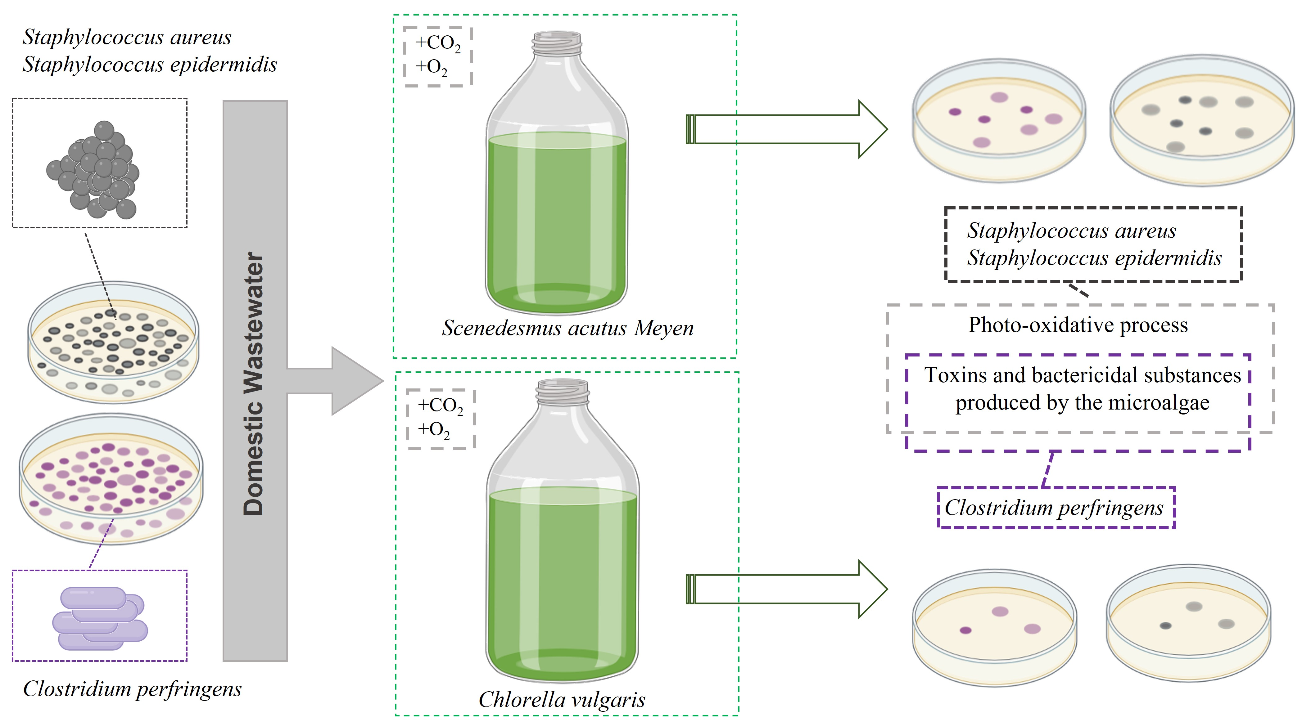
2.1. Algal-Bacterial Inoculum

Cultures of Scenedesmus acutus Meyen (≈97%) and Chlorella vulgaris (≈97%) were obtained from a pilot outdoor HRAP treating domestic wastewater at the Federal University of Mato Grosso do Sul (Campo Grande-MS, Brazil) after different experiments were performed [7,24]. In this case, the culture was collected and stored in a refrigerator at 4 °C before inoculation into the reactor. Aerobic nitrifying–denitrifying activated sludge was obtained from a WWTP in the same city. The initial TSS concentrations of the Scenedesmus acutus Meyen, Chlorella vulgaris, and activated sludge suspensions were 0.11, 0.15, and 0.21 g TSS L−1, respectively.

2.2. Gas and Domestic Wastewater

The CO2 source was a synthetic gas mixture (30% CO2/70% N2) (White Martins, Brazil), while the O2 source was atmospheric air (≈20% O2, ≈360 ppm CO2) (Brimblecombe 1996). Domestic wastewater was collected from a local WWTP, with a COD of 188.3 ± 20.8 mg L−1, 23.4 ± 0.1 mg L−1 ammonium (NH4+), (1.09 ± 1.29) × 105 (5.04 ± 5.11 log) CFU 100 mL−1 C. perfringens, (1.03 ± 1.21) × 106 (6.01 ± 6.08 log) CFU 100 mL−1 Staphylococcus spp., and a pH of 7.1 ± 0.2. The domestic wastewater was not autoclaved or diluted. The number of replicates performed to characterize the domestic wastewater was n = 8 for chemical oxygen demand (COD), ammonium (NH4+), C. perfringens, Staphylococcus spp., and pH value.

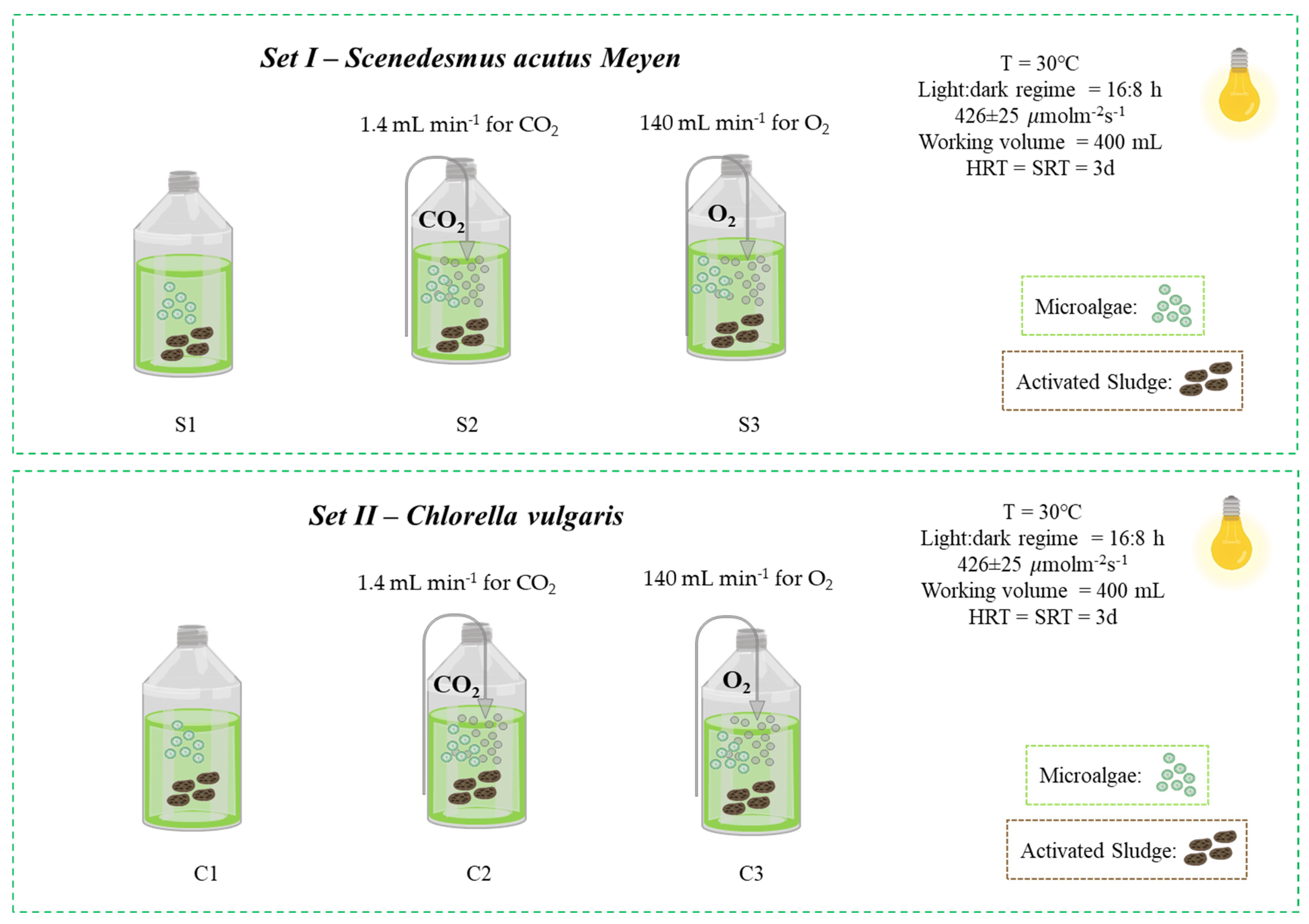
2.3. Experimental Setup and Sampling Procedure

To determine the influence of inoculum composition, CO2 addition, and O2 addition on the removal of C. perfringens and Staphylococcus spp., experiments were performed in two batch sets using Scenedesmus acutus Meyen inoculum (S, Set I) and Chlorella vulgaris inoculum (C, Set II). Three conditions were tested in both sets: only microalgae–bacteria inoculum (S1 and C1), microalgae–bacteria inoculum with CO2 addition (S2 and C2), and microalgae–bacteria inoculum with O2 addition (S3 and C3) (Table 1 and Figure 1). The two sets of experiments (I and II) were performed at different times to avoid contamination, and each condition tested (S1, S2, S3, C1, C2 and C3) was performed in duplicate. Both Sets (I and II) were run for 9 days, but the maximum removals and maximum algal growth occurred on the third day (3rd d) of the test, which is considered the end of the experiment. The reactors were fed with domestic wastewater only at the beginning of the test. The HRT and SRT of all reactors were equal to 3 days.
The experiments were conducted in laboratory-scale batch reactors using 500 mL sterilized glass bottles (400 mL working volume, with 5% microalgae inoculum and 5% activated sludge inoculum) closed with cotton wool stoppers. All photobioreactors were exposed to a light:dark regime of 16:8 h at a light intensity of 426 ± 25 µmol m−2 s−1 using LED lamps (100 W—light spectrum 400 to 700 nm) and constantly shaken at 200 rpm. All conditions were performed in duplicate and maintained at a constant temperature of 30 ± 1 °C. The gas supply rates were 1.4 mL min−1 for CO2 (Watson Marlon pump, Falmouth USA) and 140 mL min−1 for O2 (Salor Better pump, São Caetano do Sul, Brazil); CO2 addition rates were based on the systems of Posadas et. al. [5] and Ruas et. al. [6]. To prevent evaporation losses, CO2 and O2 were passed through scrubbing flasks filled with water before passing through the reactors. The ambient and cultivation temperatures, light intensity, pH, and DO concentration were measured daily at 10 a.m. Liquid samples (40 mL) were drawn every day to monitor the concentration of soluble (0.20 µm nylon filters) COD and N-NH4+ and insoluble (total concentration) TSS and C. perfringens and Staphylococcus spp. The determination of the CFU 100 mL−1 of C. perfringens and Staphylococcus spp. were performed in triplicate.

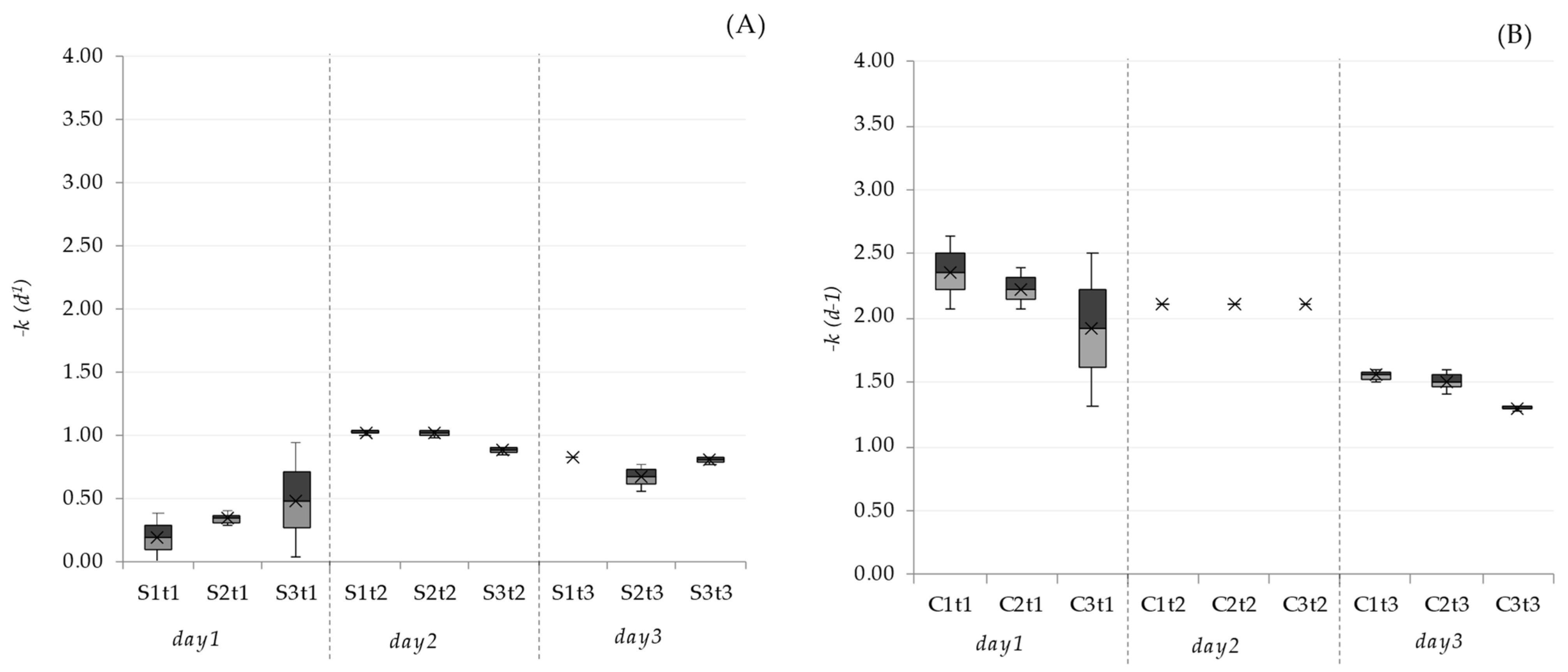
2.4. Inactivation Rates

The inactivation rates of each pathogen were estimated by calculating the first-order rate constant (k) as follows [25]:
l n ( C t C 0 ) = k t
where C0 is the initial concentration (CFU 100 mL−1) and Ct is the concentration (CFU 100 mL−1) at time t (d). The initial (C0) and final (Ct) concentrations were obtained from the average values (mean ± standard deviation) of the triplicates performed (n = 3).

2.5. Analytical Procedures

Temperature and DO were measured using a Jenway 9500 DO2 oximeter (Jenway-Cole-Parmer, Cambridgeshire, UK), light intensity was recorded with a Quantum MQ-200 photosynthetically active radiation meter (Apogee Instruments, Logan, USA), and pH and N-NH4+ were measured using pH and ammonia electrodes with an Orion Five Star multiparameter analyzer (Thermo Scientific, Bleiswijk, The Netherlands). C. perfringens and Staphylococcus spp. (Staphylococcus aureus and Staphylococcus epidermidis) levels were determined using membrane filtration with an M-PA Agar Base-M1354 and Baird Parker Agar Base M043 (HIMEDIA, Mumbai, India). The effect of a viable but nonculturable state was not considered in Staphylococcus spp. determination, and this effect was not detected in C. perfringens culture techniques [26]. In this study, the inactivation and removal of pathogenic bacteria refers to not detecting any cultivable colonies. All analyses were carried out according to the Standard Methods for the Examination of Water and Wastewater [27]. Microalgae were identified daily by microscopic examination (Olympus BX41, Miami, FL, USA) following Sournia [28].

2.6. Statistical Analysis

The pathogen removal under the different test conditions performed was evaluated using an analysis of variance (ANOVA) with Fisher’s least significant difference (LSD) test using a 95% confidence level. The differences in the environmental parameters (ambient temperature, light intensity, pH and DO) between the two Sets (I and II) were evaluated using a t test for independent samples. Spearman’s correlation was used to determine the behavior of Clostridium perfringens and Staphylococcus spp. removal in relation to pH, DO, temperature and TSS. The analyses were performed using Microsoft Excel® 2021 with Real Statistics Resource Pack software (Release 7.6) [29] and R® (R Foundation for Statistical Computing) [30].

3. Results and Discussion

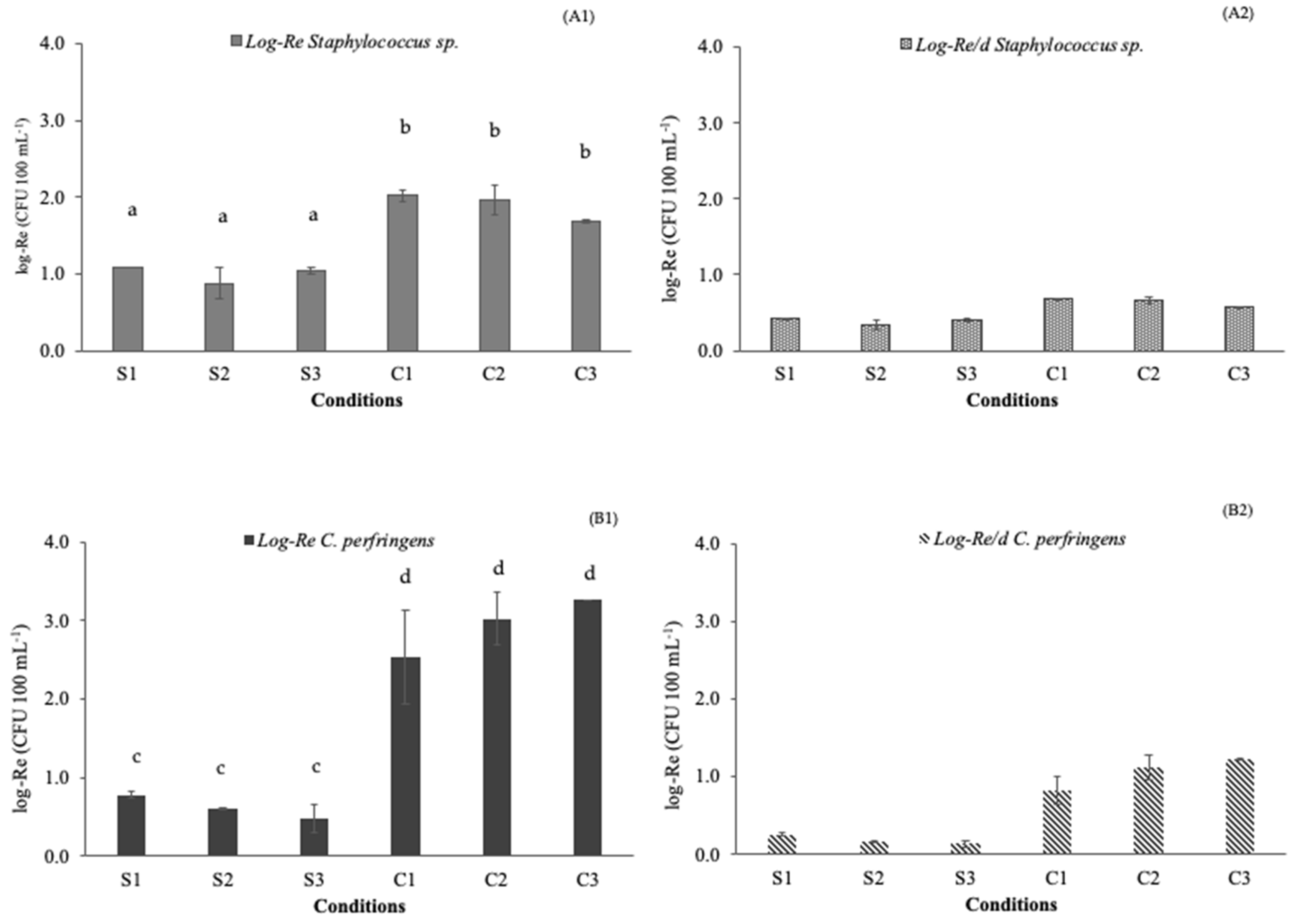
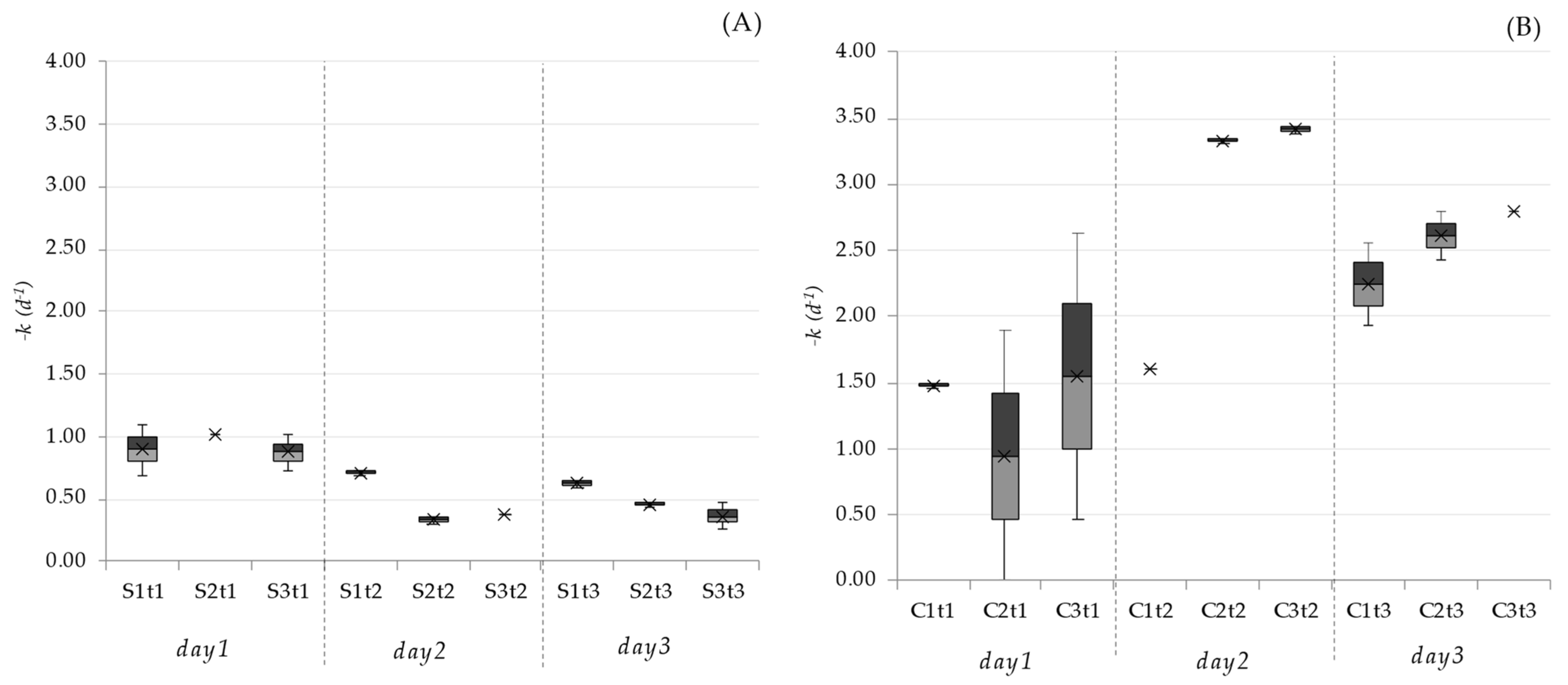
3.1. Pathogen Removal Efficiencies

The removal efficiency of C. perfringens was similar throughout Set I (Scenedesmus acutus Meyen), with log-Re removals of 0.78 ± 0.03 (S1), 0.60 ± 0.01 (S2), and 0.48 ± 0.19 (S3) (Figure 2). In Set II, using Chlorella vulgaris, the C. perfringens removal was also similar (no significant difference, p > 0.05), with log-Re removals of 2.54 ± 0.60 (C1), 3.03 ± 0.34 (C2), and 3.26 ± 0.00 (C3). However, the log-Re values were significantly different between Sets I and II. There were no significant differences in DO (p > 0.05), but pH values were different between Sets I and II (t(46) = 2,90; p < 0,001) (Table 2). Therefore, the different interactions and metabolic rates of each microalgae inoculum, Chlorella vulgaris and Scenedesmus acutus Meyen, impacted the physicochemical conditions of the medium, especially pH, which may have influenced the removal/inactivation rates of the pathogens studied. In addition to DO, light intensity and room temperature were similar between Sets I and II (Table 2).
The number of replicates performed in Set I and Set II was n = 20 for ambient temperature; n = 60 for light intensity; and n = 24 for pH and DO. DO: Dissolved Oxygen.
pH as a predominant factor in removal mechanisms is well established for E. coli (Davies-Colley et al., 1999) but not for Clostridium perfringens and Staphylococcus spp. As the highest removals were observed in Set II, with the highest pH values and the presence of the Chlorella vulgaris inoculum, these two factors are indicated as the main factors in the removal of these microorganisms. However, further studies evaluating the effects of environmental factors, especially DO and pH, and their interactions with sunlight should be evaluated in detail.
Different C. perfringens removals have been reported in HRAPs. García et al. [21] found maximum removals of 0.3 log units when operating a 0.32 m2 HRAP with a 6 d hydraulic retention time (HRT), while Ruas et al. [7] found maximum removals of 2.6 log units in a 0.13 m2 HRAP with a 5 d HRT and a Scenedesmus sp. microalgal population. This significant difference can be explained by the impact of the microalgal population on the removal of this bacteria (Figure 2(A1)). The role of microalgae in the removal of pathogens through toxin production has been pointed out as a disinfection mechanism in ponds [31]. Microalgae can produce compounds with bactericidal, fungicidal, antiviral, neuroprotective, and immunomodulatory properties [32], and different genera produce different specific biocomponents with different bactericidal properties that target pathogenic microorganisms differently.
Several studies have noted that these algal toxins are important factors in disinfection [33,34], while Oufdou et al. [34] observed that the blue–green algae Pseudanabaena sp. reduced the survival of E. coli, Salmonella sp., S. aureus, and Candida albicans but did not affect Candida tropicalis and even stimulated the growth of Vibrio cholerae, whereas Mezrioui et al. [33] observed that Chlorella sp. enhanced V. cholerae inactivation.
Krustok et al. [35] tested the influence of two sources of microalgal and bacterial inoculation (a local lake and the effluent itself) on the operating efficiency of a laboratory-scale batch photobioreactor with a 16-day HRT. The resulting microalgal communities were completely different: in the lake water reactor, >80 species from the phylum Chlorophyta were recorded, whereas in the other reactor, only 20 such species were present. Although both had similar nutrient removal efficiencies, the removal of pathogens such as C. perfringens and S. aureus was approximately twice as large in the lake water reactor, demonstrating the influence of the microalgal community on pathogen removal efficiency in a pond system. The efficiency of C. perfringens removal in activated sludge, anaerobic reactors, and maturation ponds (conventional wastewater systems) has been reported to be 0.5 ± 1.4, 0.3 ± 0.7, and 1.11 ± 1.26 log units, respectively [21,36], lower than the values found in our study.
In Set I, the Staphylococcus spp. removals were 1.08 ± 0.00 (S1), 0.87 ± 0.20 (S2), and 1.04 ± 0.05 (S3), while in Set II, they were 2.02 ± 0.08 (C1), 1.96 ± 0.18 (C2), and 1.68 ± 0.02 (C3) (Figure 2(A2)). The addition of CO2 and O2 had no significant effect on Staphylococcus spp. log-Re in either Set (p > 0.05). The same result was found by Ruas et al. [7] when studying the effect of CO2 addition on pathogenic bacteria removal in an HRAP. Alkalinity is an important factor for the mass transfer of CO2 to the culture medium; the higher the alkalinity available, the greater the availability of CO2 for microalgal metabolism [37], so the addition of CO2 to a medium with high alkalinity concentrations can have significant effects on pathogen removal, a process that has not yet been explored.
Additionally, the microalgal species significantly impacted Staphylococcus spp. removal, where Chlorella vulgaris also promoted higher removal efficiency. The effect of DO was discarded because there was no significant difference in this parameter between the Sets, as discussed above (Item 3.1). Staphylococcus spp. inactivation in microalgal–bacterial systems ranges from 1.2 to 1.7 log [7,21], while removals of 1.8 and 2.4 log units were achieved in pond systems with a 50 d HRT and with a soil filter, respectively [38,39].
Some extracts of phytoplankton [40], macroalgae [41], and microalgae [42] have shown a bactericidal effect against S. aureus, mainly through the inhibition of dehydrogenase, an important enzyme in energy processes [43]. Al-Gheethi et al. [44] observed S. aureus removal of more than four (4) log units in 6 d using a phycoremediation test with a Scenedesmus sp. inoculum, but highlighted the greater survival of this pathogen with lower concentrations of Scenedesmus sp. and the removal of four log units in the control test, showing the importance of photooxidative processes for the inactivation of this microorganism.
Photooxidation is an important disinfection mechanism in pond systems, in which photosensitizers absorb light and transfer this energy to other molecules, leading to the formation of reactive oxygen species that react with the membrane or DNA of the microorganisms, causing damage [45]. Exogenous photosensitizers are present in the aquatic environment, while endogenous photosensitizers are found inside the microorganisms, with the former having the most significant contribution. The main exogenous photosensitizers are humic substances, photosynthetic pigments, and dissolved organic matter, which are found in large quantities in microalgal–bacterial treatment systems. Different exogenous photosensitizers absorb varying amounts of energy and therefore induce different degrees of photooxidation [45]. Some substances produced by algae, such as phycobiliproteins, chlorophylls, and carotenoids, can act as photosensitizers absorbing different light spectra [46,47] and are already being used for the degradation of dyes and inactivation of microorganisms [48,49]. Therefore, cultures with different microalgae may present distinct concentrations of photosensitizers, resulting in varied inactivation of pathogenic microorganisms, as was observed in our study. Photooxidation is an important process in the inactivation of Staphylococcus sp., which has already shown a strong inactivation rate in the presence of photosensitizers [50]. Therefore, the microalgal population can be an important factor in HRAP disinfection by producing photosensitizers, a relevant theme for future research. Predation, as a mechanism for pathogen removal in domestic sewage, was not considered in this experiment, as it requires a different methodological approach than the one outlined.
In both sets, the inactivation rate (k) (Table 3, Figure 3 and Figure 4) of C. perfringens (0.35–2.47 d−1) and Staphylococcus sp. (0.66–1.53 d−1) was higher than that found by Al-Tameemi and Kadhim [11] (k maximum of 0.01 d−1), who tested the effect of pH, DO, and temperature in microalgal systems (Nostochopsis sp. And Mougeotia sp.) on the inactivation of C. perfringens in secondary domestic wastewater. The inactivation rate shown by Al-Tameemi and Kadhim [11] may have been lower due to the lower light intensity used in the study (≈50 µmol m−2 s−1), the lower initial concentration in the domestic effluent (3.7 log CFU 100mL−1) and the use of different algae genera.
The main parameters and culture conditions that significantly (p < 0.05) influenced the removal of the pathogens studied are described in Figure 5. The pH and algal growth parameters (TSS and NTU) showed statistically significant correlations.

3.2. COD and N-NH4+ Removal Efficiency, Productivity, and Specific Growth Rate (µ)

The COD removal efficiency (COD-Re) was significantly different between the two sets, with averages ranging from 38.1 to 79.0% (Set I) and from 82.4 to 94.9% (Set II) (Table 4). In Set I, removal under Condition S3 was significantly lower than under Conditions S1 and S2, although productivity was higher (≈201 mg TSS L−1 d−1). The continuous addition of CO2 at 140 mL min−1 may have caused stripping of O2 from the growth medium, decreasing the rate of COD removal by heterotrophic bacteria while favoring an increase in algal biomass by inserting CO2 from atmospheric air and maintaining more favorable DO concentrations for microalgae (high DO concentrations may inhibit photosynthesis) [51]. However, in Set II, COD removal was statistically equal in all three conditions, meaning that CO2 and O2 addition had no significant effect on COD removal (p > 0.05).
The productivity in Set I was significantly different in each of the studied conditions, indicating that the addition of CO2 and O2 improved biomass production in Scenedesmus acutus Meyen cultures. This result was expected because CO2 addition improves the photosynthetic capacity and carbon fixation rate of this microalgal species [52]. Ruas et al. [7] observed an increase in biomass productivity from 2.0 to 3.2 g m−2 d−1 in a reactor with CO2 addition, treating domestic wastewater with Scenedesmus sp. as the dominant microalgae. In batch experiments, where the effect of adding food wastewater and flue gas CO2 on the productivity and treatment efficiency of domestic wastewater was tested (Scenedesmus obliquus mixotrophic culture), both CO2 and food wastewater favored the growth of biomass, reaching a productivity of ≈75 mg L−1 d−1 [53]. However, in Set II, the biomass productivity was higher in C3 and statistically similar in C1 and C2, so the addition of O2 significantly influenced the biomass production of Chlorella vulgaris.
One of the main microalgae used for the treatment of domestic and agro-industrial wastewaters is Chlorella sp. because it tolerates adverse environmental conditions and has a great capacity to remove carbon and nutrients while achieving high biomass productivity [54,55]. The use of this microalgae tends to show better results in biomass production, COD removal, and lipid accumulation than the use of Scenedesmus sp. [56], as also observed in our study. The greater production of biomass in tests with Chlorella vulgaris inoculum may have amplified the effect that this alga has on pathogens, since greater biomass production would increase the production of toxins and bactericidal compounds.
The N-NH4+ removal (Table 4) was statistically equal at the 5% significance level (81.7–99.4%), except in S1 (62.8 ± 9.5%). In all conditions, the final pH was >9, which may have contributed to the removal of ammonia by volatilization [5], but considering the algal biomass composition of 7–9% N, the high removal rate of N-NH4+ (>62%), the productivity, the moderate temperature, and the availability of DO [5], assimilation was a relevant process in nitrogen removal in all tested conditions.

4. Conclusions

The removal of C. perfringens and Staphylococcus spp. was higher in tests using Chlorella sp. inoculum, demonstrating that different algae act in different ways in the removal of pathogens in microalgal–bacterial systems. The addition of CO2 and O2 did not have a significant effect on the removal of pathogenic bacteria. The use of different microalgae in the treatment of domestic wastewater can lead to the removal of pathogens, depending on the microorganism to be removed and the required degree of target removal. The biomass and COD-Re were enhanced in the culture with Chlorella sp., but in terms of ammonia removal, the microalgal inoculum did not influence the efficiency of the process. Therefore, the use of Chlorella sp. is more advantageous for removing carbon and nitrogen, as well as for inactivating pathogenic microorganisms such as C. perfringens and Staphylococcus spp.

Author Contributions

G.R.: Conceptualization, methodology, formal analysis, investigation, data curation, writing—original draft. S.L.F.: Investigation, writing—review and editing. B.A.B.d.R.: Investigation. M.L.S.: Conceptualization, validation, supervision, writing—review and editing. G.H.R.d.S.: Resources, validation, supervision, writing—review and editing. M.Á.B.: Resources, validation, supervision, funding acquisition, writing—review and editing. All authors have read and agreed to the published version of the manuscript.

Funding

This research was funded by the Conselho Nacional de Desenvolvimento Científico e Tecnológico—CNPq (Project number 429567/2016-2 and 308663/2021-7) and the Coordenação de Aperfeiçoamento de Pessoal de Nível Superior (CAPES) for the PhD grant awarded to Graziele Ruas (88882.458517/2019–01) and sandwich period (88881.190564/2018–01).

Data Availability Statement

The datasets generated and/or analyzed during the current study are available from the corresponding author upon reasonable request.

Acknowledgments

The authors would like to acknowledge the support obtained from the following Brazilian institutions: the Conselho Nacional de Desenvolvimento Científico e Tecnológico—CNPq (Project number 429567/2016-2 and 308663/2021-7); the Coordenação de Aperfeiçoamento de Pessoal de Nível Superior (CAPES) for the PhD grant awarded to Graziele Ruas (88882.458517/2019–01) and sandwich period (88881.190564/2018–01); the Fundação de Amparo à Pesquisa do Estado de Minas Gerais—FAPEMIG; and the Instituto Nacional de Ciência e Tecnologia em Estações Sustentáveis de Tratamento de Esgoto—INCT ETEs Sustentáveis (INCT Sustainable Sewage Treatment Plants).

Conflicts of Interest

The authors report no commercial or proprietary interest in any product or concept discussed in this article.

References

  1. WHO. Guidelines on Sanitation and Health; WHO: Geneva, Switzerland, 2018; ISBN 9789241514705. Available online: https://apps.who.int/iris/bitstream/handle/10665/274939/9789241514705-eng.pdf (accessed on 17 November 2022).
  2. UNICEF and WHO. Progress on Household Drinking Water, Sanitation and Hygiene 2000–2017: Special Focus on Inequalities. 2019; ISBN 978-92-806-5036-5. Available online: https://washdata.org/sites/default/files/documents/reports/2019-07/jmp-2019-wash-households.pdf (accessed on 17 November 2022).
  3. Sharafi, K.; Moradi, M.; Karami, A.; Khosravi, T. Comparison of the Efficiency of Extended Aeration Activated Sludge System and Stabilization Ponds in Real Scale in the Removal of Protozoan Cysts and Parasite Ova from Domestic Wastewater Using Bailenger Method: A Case Study, Kermanshah, Iran. Desalin. Water Treat. 2015, 55, 1135–1141. [Google Scholar] [CrossRef]
  4. Nirmalakhandan, N.; Selvaratnam, T.; Henkanatte-Gedera, S.M.; Tchinda, D.; Abeysiriwardana-Arachchige, I.S.A.; Delanka-Pedige, H.M.K.; Munasinghe-Arachchige, S.P.; Zhang, Y.; Holguin, F.O.; Lammers, P.J. Algal Wastewater Treatment: Photoautotrophic vs. Mixotrophic Processes. Algal Res. 2019, 41, 101569. [Google Scholar] [CrossRef]
  5. Posadas, E.; del Mar Morales, M.; Gomez, C.; Acién, F.G.; Muñoz, R. Influence of PH and CO2 Source on the Performance of Microalgae-Based Secondary Domestic Wastewater Treatment in Outdoors Pilot Raceways. Chem. Eng. J. 2015, 265, 239–248. [Google Scholar] [CrossRef] [Green Version]
  6. Ruas, G.; Serejo, M.L.; Paulo, P.L.; Boncz, M.Á. Evaluation of Domestic Wastewater Treatment Using Microalgal-Bacterial Processes: Effect of CO2 Addition on Pathogen Removal. J. Appl. Phycol. 2018, 30, 921–929. [Google Scholar] [CrossRef]
  7. Ruas, G.; Farias, S.L.; Scarcelli, P.G.; Serejo, M.L.; Boncz, M.A. The Effect of CO2 Addition and Hydraulic Retention Time on Pathogens Removal in HRAPs. Water Sci. Technol. 2020, 1–9. [Google Scholar] [CrossRef]
  8. Venczel, L.V.; Arrowood, M.; Hurd, M.; Sobsey, M. Inactation of Cryptosporidium Parvum Oocyts and Clostridium Perfringens Spores by a Mixed-Oxidant Disinfectant and by Free Chlorine. Appl. Environ. Microbiol. 1997, 63, 1598–1601. [Google Scholar] [CrossRef] [Green Version]
  9. Lanao, M.; Ormad, M.P.; Goñi, P.; Miguel, N.; Mosteo, R.; Ovelleiro, J.L. Inactivation of Clostridium Perfringens Spores and Vegetative Cells by Photolysis and TiO2 Photocatalysis with H2O2. Sol. Energy 2010, 84, 703–709. [Google Scholar] [CrossRef]
  10. Abreu-Acosta, N.; Vera, L. Occurrence and Removal of Parasites, Enteric Bacteria and Faecal Contamination Indicators in Wastewater Natural Reclamation Systems in Tenerife-Canary Islands, Spain. Ecol. Eng. 2011, 37, 496–503. [Google Scholar] [CrossRef]
  11. Al-Tameemi, F.A.H.; Kadhim, T.A. Significance of Algae in Eliminating and Deactivating Pathological Index Arganisms in Wastewater Stabilization Pond Systems. E3S Web Conf. 2019, 96, 4–10. [Google Scholar] [CrossRef] [Green Version]
  12. Payment, P.; Franco, E. Clostridium Perfringens and Somatic Coliphages as Indicators of the Efficiency of Drinking Water Treatment for Viruses and Protozoan Cysts. Appl. Environ. Microbiol. 1993, 59, 2418–2424. [Google Scholar] [CrossRef]
  13. Bitton, G. Wastewater Microbiology, 3rd ed.; John Wiley & Sons, Inc.: Hoboken, NJ, USA, 2005; Volume 53, ISBN 978-0-471-71791-1. [Google Scholar]
  14. WHO. Evaluating Household Water Treatment Options: Health-Based Targets and Microbiological Performance Specifications; WHO: Geneva, Switzerland, 2011; Available online: https://apps.who.int/iris/bitstream/handle/10665/44693/9789241548229_eng.pdf (accessed on 17 November 2022).
  15. European Commisssion Council. Directive 98/83/EC of 3 November 1998 on the Quality of Water Intended for Human Consumption; 1998. Available online: https://agris.fao.org/agris-search/search.do?recordID=CS2005000008 (accessed on 17 November 2022).
  16. Shoults, D.C.; Ashbolt, N.J. Decreased Efficacy of UV Inactivation of Staphylococcus Aureus after Multiple Exposure and Growth Cycles. Int. J. Hyg. Environ. Health 2019, 222, 111–116. [Google Scholar] [CrossRef] [PubMed]
  17. Bassetti, M.; Nicco, E.; Mikulska, M. Why Is Community-Associated MRSA Spreading across the World and How Will It Change Clinical Practice? Int. J. Antimicrob. Agents 2009, 34, S15–S19. [Google Scholar] [CrossRef] [PubMed]
  18. Duckworth, G. Revised Guidelines for the Control of Epidemic Methicillin-Resistant Staphylococcus Aureus. J. Hosp. Infect. 1990, 16, 351–377. [Google Scholar] [CrossRef]
  19. Börjesson, S.; Matussek, A.; Melin, S.; Löfgren, S.; Lindgren, P.E. Methicillin-Resistant Staphylococcus Aureus (MRSA) in Municipal Wastewater: An Uncharted Threat? J. Appl. Microbiol. 2010, 108, 1244–1251. [Google Scholar] [CrossRef] [PubMed]
  20. Rosenberg Goldstein, R.E.; Micallef, S.A.; Gibbs, S.G.; Davis, J.A.; He, X.; George, A.; Kleinfelter, L.M.; Schreiber, N.A.; Mukherjee, S.; Sapkota, A. Methicillin-Resistant Staphylococcus Aureus (MRSA) Detected at Four, U.S. Wastewater Treatment Plants. Environ. Health Perspect. 2012, 120, 1551–1558. [Google Scholar] [CrossRef] [PubMed] [Green Version]
  21. García, M.; Soto, F.; González, J.M.; Bécares, E. A Comparison of Bacterial Removal Efficiencies in Constructed Wetlands and Algae-Based Systems. Ecol. Eng. 2008, 32, 238–243. [Google Scholar] [CrossRef]
  22. Sutherland, D.L.; Howard-Williams, C.; Turnbull, M.H.; Broady, P.A.; Craggs, R.J. The Effects of CO2 Addition along a PH Gradient on Wastewater Microalgal Photo-Physiology, Biomass Production and Nutrient Removal. Water Res. 2014, 70C, 9–26. [Google Scholar] [CrossRef]
  23. Garcia-Gonzalez, L.; Geeraerd, A.H.; Spilimbergo, S.; Elst, K.; Van Ginneken, L.; Debevere, J.; Van Impe, J.F.; Devlieghere, F. High Pressure Carbon Dioxide Inactivation of Microorganisms in Foods: The Past, the Present and the Future. Int. J. Food Microbiol. 2007, 117, 1–28. [Google Scholar] [CrossRef]
  24. Serejo, M.L.; Farias, S.L.; Ruas, G.; Paulo, P.L.; Boncz, M.A. Surfactant Removal and Biomass Production in a Microalgal-Bacterial Process: Effect of Feeding Regime. Water Sci. Technol. 2020, 2, 1–10. [Google Scholar] [CrossRef] [PubMed]
  25. Liu, L.; Hall, G.; Champagne, P. The Role of Algae in the Removal and Inactivation of Pathogenic Indicator Organisms in Wastewater Stabilization Pond Systems. Algal Res. 2020, 46, 101777. [Google Scholar] [CrossRef]
  26. Colwell, R.R.; Grimes, D.J. Nonculturable Microorganisms in the Environment; Colweel, R.R., Grimes, D.J., Eds.; American Society for Microbiology: Washington, DC, USA, 2000; ISBN 9781475702736. [Google Scholar]
  27. APHA. Standard Methods for the Examination of Water, 22nd ed.; American Public Health Association, American Water Works Association, Water Environment Federation: New York, NY, USA, 2012; ISBN 9780875530130. [Google Scholar]
  28. Sournia, A. Phytoplankton Manual; UNESCO, Ed.; Muséum National d’Histoire Naturelle: Paris, France, 1978; ISBN 92-3-101572-9. [Google Scholar]
  29. Zaiontz, C. The Data Analysis for This Paper Was Generated Using the Real Statistics Resource Pack Software (Release 7.6). 2020. [Google Scholar]
  30. R Core Team R: A Language and Environment for Statistical Computing. Available online: https://www.r-project.org (accessed on 17 November 2022).
  31. Davies-Colley, R.J.; Craggs, R.J.; Park, J.; Sukias, J.P.S.; Nagels, J.W.; Stott, R. Virus Removal in a Pilot-Scale “advanced” Pond System as Indicated by Somatic and F-RNA Bacteriophages. Water Sci. Technol. 2005, 51, 107–110. [Google Scholar] [CrossRef] [PubMed]
  32. Maghembe, R.; Damian, D.; Makaranga, A.; Nyandoro, S.S.; Lyantagaye, S.L.; Kusari, S.; Hatti-Kaul, R. Omics for Bioprospecting and Drug Discovery from Bacteria and Microalgae. Antibiotics 2020, 9, 229. [Google Scholar] [CrossRef] [PubMed]
  33. Mezrioui, N.; Oudra, B.; Oufdou, K.; Hassani, L.; Loudiki, M.; Darley, J. Effect of Microalgae Growing on Wastewater Batch Culture on Escherichia Coli and Vibrio Cholerae Survival. Water Sci. Technol. 1994, 30, 295–302. [Google Scholar] [CrossRef]
  34. Oufdou, K.; Mezrioui, N.; Oudra, B.; Loudiki, M.; Barakate, M.; Sbiyyâa, B. Bioactive Compounds from Pseudanabaena Species (Cyanobacteria). Microbios 2001, 106, 21–29. [Google Scholar]
  35. Krustok, I.; Truu, J.; Odlare, M.; Truu, M.; Ligi, T.; Tiirik, K.; Nehrenheim, E. Effect of Lake Water on Algal Biomass and Microbial Community Structure in Municipal Wastewater-Based Lab-Scale Photobioreactors. Appl. Microbiol. Biotechnol. 2015, 99, 6537–6549. [Google Scholar] [CrossRef]
  36. Lucena, F.; Duran, A.E.; Morón, A.; Calderón, E.; Campos, C.; Gantzer, C.; Skraber, S.; Jofre, J. Reduction of Bacterial Indicators and Bacteriophages Infecting Faecal Bacteria in Primary and Secondary Wastewater Treatments. J. Appl. Microbiol. 2004, 97, 1069–1076. [Google Scholar] [CrossRef]
  37. del Rosario Rodero, M.; Posadas, E.; Toledo-Cervantes, A.; Lebrero, R.; Muñoz, R. Influence of Alkalinity and Temperature on Photosynthetic Biogas Upgrading Efficiency in High Rate Algal Ponds. Algal Res. 2018, 33, 284–290. [Google Scholar] [CrossRef]
  38. Mosteo, R.; Ormad, M.P.; Goñi, P.; Rodríguez-Chueca, J.; García, A.; Clavel, A. Identification of Pathogen Bacteria and Protozoa in Treated Urban Wastewaters Discharged in the Ebro River (Spain): Water Reuse Possibilities. Water Sci. Technol. 2013, 68, 575–583. [Google Scholar] [CrossRef]
  39. Scheurer, M.; Heß, S.; Lüddeke, F.; Sacher, F.; Güde, H.; Löffler, H.; Gallert, C. Removal of Micropollutants, Facultative Pathogenic and Antibiotic Resistant Bacteria in a Full-Scale Retention Soil Filter Receiving Combined Sewer Overflow. Environ. Sci. Process. Impacts 2015, 17, 186–196. [Google Scholar] [CrossRef] [Green Version]
  40. Schwartz, R.E.; Hirsch, C.F.; Sesin, D.F.; Flor, J.E.; Chartrain, M.; Fromtling, R.E.; Harris, G.H.; Salvatore, M.J.; Liesch, J.M.; Yudin, K. Pharmaceuticals from Cultured Algae. J. Ind. Microbiol. 1990, 5, 113–123. [Google Scholar] [CrossRef]
  41. Sánchez-Saavedra, M.D.P.; Licea-Navarro, A.; Bernáldez-Sarabia, J. Evaluation of the Antibacterial Activity of Different Species of Phytoplankton. Rev. Biol. Mar. Oceanogr. 2010, 45, 531–536. [Google Scholar] [CrossRef]
  42. Syed, S.; Arasu, A.; Ponnuswamy, I. The Uses of Chlorella Vulgaris as Antimicrobial Agent and as a Diet: The Presence of Bio-Active Compounds Which Caters the Vitamins, Minerals in General. Int. J. Bio-Sci. Bio-Technol. 2015, 7, 185–190. [Google Scholar] [CrossRef]
  43. Evangelista, V.; Barsanti, L.; Coltelli, P.; Frassanito, A.M.; Passarelli, V.; Vesentini, N.; Gualtieri, P. Algal Toxins: Nature, Occurrence, Effect and Detection; Springer Science & Business Media: Pisa, Italy, 2007; ISBN 978-1-4020-8480-5. [Google Scholar]
  44. Al-Gheethi, A.A.; Mohamed, R.M.; Jais, N.M.; Efaq, A.N.; Halid, A.A.; Wurochekke, A.A.; Amir-Hashim, M.K. Influence of Pathogenic Bacterial Activity on Growth of Scenedesmus Sp. and Removal of Nutrients from Public Market Wastewater. J. Water Health 2017, 15, 741–756. [Google Scholar] [CrossRef] [PubMed] [Green Version]
  45. Curtis, T.P.; Mara, D.D.; Silva, S.A. The Effect of Sunlight on Faecal Coliforms in Ponds: Implications for Research and Design. Water Sci. Technol. 1992, 26, 1729–1738. [Google Scholar] [CrossRef]
  46. Bermejo, R.; Talavera, E.M.; Alvarez-Pez, J.M.; Orte, J.C. Chromatographic Purification of Biliproteins from Spirulina Platensis High-Performance Liquid Chromatographic Separation of Their α and β Subunits. J. Chromatogr. A 1997, 778, 441–450. [Google Scholar] [CrossRef]
  47. Nurachman, Z.; Hartini, H.; Rahmaniyah, W.R.; Kurnia, D.; Hidayat, R.; Prijamboedi, B.; Suendo, V.; Ratnaningsih, E.; Panggabean, L.M.G.; Nurbaiti, S. Tropical Marine Chlorella Sp. PP1 as a Source of Photosynthetic Pigments for Dye-Sensitized Solar Cells. Algal Res. 2015, 10, 25–32. [Google Scholar] [CrossRef]
  48. Sinton, L.W.; Davies-Colley, R.J.; Bell, R.G. Inactivation of Enterococci and Fecal Coliforms from Sewage and Meatworks Effluents in Seawater Chambers. Appl. Environ. Microbiol. 1994, 60, 2040–2048. [Google Scholar] [CrossRef] [Green Version]
  49. Sharmila, J.; Saravanan, P.; Sivasankar, S.; Chamundeeswari, M. A Novel and an Eco-Friendly Approach for Organic Dyes Degradation Using Spirulina Platensis Cultivated Water. Catal. Today 2018, 340, 245–252. [Google Scholar] [CrossRef]
  50. Maraccini, P.A.; Wenk, J.; Boehm, A.B. Photoinactivation of Eight Health-Relevant Bacterial Species: Determining the Importance of the Exogenous Indirect Mechanism. Environ. Sci. Technol. 2016, 50, 5050–5059. [Google Scholar] [CrossRef] [Green Version]
  51. Cheah, W.Y.; Show, P.L.; Chang, J.; Ling, T.C.; Juan, J.C. Biosequestration of Atmospheric CO2 and Flue Gas-Containing CO2 by Microalgae. Bioresour. Technol. 2015, 184, 190–201. [Google Scholar] [CrossRef] [PubMed]
  52. Msanne, J.; Polle, J.; Starkenburg, S. An Assessment of Heterotrophy and Mixotrophy in Scenedesmus and Its Utilization in Wastewater Treatment. Algal Res. 2020, 48, 101911. [Google Scholar] [CrossRef]
  53. Ji, M.K.; Yun, H.S.; Park, Y.T.; Kabra, A.N.; Oh, I.H.; Choi, J. Mixotrophic Cultivation of a Microalga Scenedesmus Obliquus in Municipal Wastewater Supplemented with Food Wastewater and Flue Gas CO2 for Biomass Production. J. Environ. Manag. 2015, 159, 115–120. [Google Scholar] [CrossRef] [PubMed]
  54. Muñoz, R.; Guieysse, B. Algal-Bacterial Processes for the Treatment of Hazardous Contaminants: A Review. Water Res. 2006, 40, 2799–2815. [Google Scholar] [CrossRef] [PubMed]
  55. Wang, L.; Min, M.; Li, Y.; Chen, P.; Chen, Y.; Liu, Y.; Wang, Y.; Ruan, R. Cultivation of Green Algae Chlorella Sp. in Different Wastewaters from Municipal Wastewater Treatment Plant. Appl. Biochem. Biotechnol. 2010, 162, 1174–1186. [Google Scholar] [CrossRef]
  56. Li, Y.; Zhou, W.; Hu, B.; Min, M.; Chen, P.; Ruan, R.R. Integration of Algae Cultivation as Biodiesel Production Feedstock with Municipal Wastewater Treatment: Strains Screening and Significance Evaluation of Environmental Factors. Bioresour. Technol. 2011, 102, 10861–10867. [Google Scholar] [CrossRef]
Figure 1. Experimental set-up of the two Sets performed. In Set I, the conditions S1, S2, and S3 were tested, while in Set II, the conditions C1, C2, and C3 where tested. In both trials, light intensity, hydraulic retention time (HRT), solid retention time (SRT), light intensity, and ambient temperature were constant. S1: Scenedesmus spp. inoculum; S2: Scenedesmus spp. inoculum + addition of CO2; S3: Scenedesmus spp. inoculum + addition of O2. C1: Chlorella vulgaris. inoculum; C2: Chlorella vulgaris. inoculum + addition of CO2; C3: Chlorella vulgaris. inoculum + addition of O2.
Water 15 00005 g001
Figure 2. Removal efficiency in logarithmic units (log) and logarithmic units per day (log d−1) for C. perfringens ((A1,A2), respectively) and Staphylococcus sp. ((B1,B2), respectively) on average ± standard deviation. Values with the same letter (a, b, c, d) are statistically equal (α = 0.05). S1: Scenedesmus spp. inoculum; S2: Scenedesmus spp. inoculum + addition of CO2; S3: Scenedesmus spp. inoculum + addition of O2. C1: Chlorella vulgaris. inoculum; C2: Chlorella vulgaris. inoculum + addition of CO2; C3: Chlorella vulgaris. inoculum + addition of O2.
Water 15 00005 g002
Figure 3. Daily inactivation rates of Staphylococcus sp. in Set I (A) and Set II (B). S1: Scenedesmus spp. inoculum; S2: Scenedesmus spp. inoculum + addition of CO2; S3: Scenedesmus spp. inoculum + addition of O2. C1: Chlorella vulgaris. inoculum; C2: Chlorella vulgaris. inoculum + addition of CO2; C3: Chlorella vulgaris. inoculum + addition of O2; t1, t2, and t3 indicate day 1, day 2, and day 3, respectively.
Water 15 00005 g003
Figure 4. Daily inactivation rates of Clostridium perfringens. in Set I (A) and Set II (B). S1: Scenedesmus spp. inoculum; S2: Scenedesmus spp. inoculum + addition of CO2; S3: Scenedesmus spp. inoculum + addition of O2. C1: Chlorella vulgaris. inoculum; C2: Chlorella vulgaris. inoculum + addition of CO2; C3: Chlorella vulgaris. inoculum + addition of O2; t1, t2, and t3 indicate day 1, day 2, and day 3, respectively.
Water 15 00005 g004
Figure 5. Spearman’s correlation coefficients obtained between inoculum (Scenedesmus acutus Meyen and Chlorella vulgaris), CO2 supplementation, aeration (O2), TSS concentration, NTU, pH, and DO. TSS: Total Suspended Solid; NTU: Nephelometric Turbidity Unit; DO: Dissolved Oxygen; pH: potential of hydrogen. Only the values without markings (X) are statistically significant (p > 0.05).
Water 15 00005 g005
Table 1. Experimental design testing different combinations of inoculum, CO2 addition, and O2 addition for Scenedesmus acutus Meyen inoculum (S) and Chlorella vulgaris inoculum (C).
Set I II
ConditionsS1S2S3C1C2C3
Scenedesmus Acutus Meyen+++---
Chlorella vulgaris---+++
CO2-+--+-
O2--+--+
Table 2. Environmental parameters monitored during the experiment: ambient temperature, light intensity, pH, and dissolved oxygen (DO).
ParametersUnitSet ISet II
Ambient temperature°C30 ± 130 ± 1
Light Intensityµmol m−2 s−1406 ± 19446 ± 32
pH-7.87 ± 0.928.68 ± 1.02
DOmg O2 L−15.42 ± 2.427.42 ± 4.59
Table 3. Inactivation rates (k) of Staphylococcus spp. and C. perfringens in Sets I and II.
Set I II
ConditionS1S2S3C1C2C3
C. perfringens day−10.600.460.351.792.242.47
Staphylococcus spp. day−10.840.660.811.531.471.27
Note: S1: Scenedesmus spp. inoculum; S2: Scenedesmus spp. inoculum + addition of CO2; S3: Scenedesmus spp. inoculum + addition of O2. C1: Chlorella vulgaris. inoculum; C2: Chlorella vulgaris. inoculum + addition of CO2; C3: Chlorella vulgaris. inoculum + addition of O2.
Table 4. COD and N-NH4+ removal efficiencies, mean and final pH and DO, productivity, and specific growth rate (µ) in all conditions (average ± standard deviation).
Set I II
ConditionS1S2S3C1C2C3
COD-Re (%)57.64 ± 0.78 a,b79.00 ± 7.81 c38.12 ± 1.56 c,d,e,f82.47 ± 8.55 c,d94.96 ± 5.04 a,e92.97 ± 3.05 b,f
N-NH4+-Re (%)62.82 ± 9.598.64 ± 1.3599.41 ± 0.5181.66 ± 18.386.66 ± 13.399.36 ± 0.63
pH (mean)8.02 ± 1.007.67 ± 1.117.91 ± 0.948.80 ± 1.128.53 ± 1.168.71 ± 1.17
pH final9.22 ± 0.029.27 ± 0.019.20 ± 0.019.49 ± 0.059.73 ± 0.049.59 ± 0.07
DO (mean)
(mg O2 L−1)
5.81 ± 3.365.90 ± 2.704.54 ± 1.479.48 ± 4.958.88 ± 5.083.89 ± 1.49
DO final
(mg O2 L−1)
5.15 ± 0.076.15 ± 0.494.95 ± 0.078.98 ± 0.837.04 ± 2.575.43 ± 0.64
Productivity
(mg TSS L−1 d−1)
114.6 ± 4.2 a,b,c,d153.0 ± 9.8 e201.7 ± 21.6 a,f216.0 ± 5.6 b,g273 ± 15.2 c,e,h384 ± 14.6 d,e,f,g,h
µ (d−1)0.300.400.490.490.560.68
Note: The pH, DO, TSS, and N-NH4+-Re values showed no statistical difference between the treatments (p > 0.05). Values marked with the same letters (a,b,c,d,e,f,g,h) have statistically significant differences. S1: Scenedesmus spp. inoculum; S2: Scenedesmus spp. inoculum + addition of CO2; S3: Scenedesmus spp. inoculum + addition of O2. C1: Chlorella vulgaris. inoculum; C2: Chlorella vulgaris. inoculum + addition of CO2; C3: Chlorella vulgaris. inoculum + addition of O2.
Disclaimer/Publisher’s Note: The statements, opinions and data contained in all publications are solely those of the individual author(s) and contributor(s) and not of MDPI and/or the editor(s). MDPI and/or the editor(s) disclaim responsibility for any injury to people or property resulting from any ideas, methods, instructions or products referred to in the content.

