Article

Arsenite Methyltransferase Is an Important Mediator of Hematotoxicity Induced by Arsenic in Drinking Water

1
Department of Pharmaceutical Sciences, The University of New Mexico College of Pharmacy, Albuquerque, NM 87131, USA
2
Department of Biology, New Mexico Highlands University, Las Vegas, NM 87701, USA
3
Department of Nutrition, Gillings School of Global Public Health, University of North Carolina at Chapel Hill, Chapel Hill, NC 27599, USA
4
Department of Pathology, Stony Brook University, Stony Brook, NY 11794, USA
*
Author to whom correspondence should be addressed.
Water 2023, 15(3), 448; https://doi.org/10.3390/w15030448
Submission received: 31 October 2022 / Revised: 10 January 2023 / Accepted: 19 January 2023 / Published: 22 January 2023
(This article belongs to the Special Issue Arsenic in Drinking Water and Human Health)

Abstract

:
Chronic arsenic exposures via the consumption of contaminated drinking water are clearly associated with many deleterious health outcomes, including anemia. Following exposure, trivalent inorganic arsenic (AsIII) is methylated through a series of arsenic (+III oxidation state) methyltransferase (As3MT)-dependent reactions, resulting in the production of several intermediates with greater toxicity than the parent inorganic arsenicals. The extent to which inorganic vs. methylated arsenicals contribute to AsIII-induced hematotoxicity remains unknown. In this study, the contribution of As3MT-dependent biotransformation to the development of anemia was evaluated in male As3mt-knockout (KO) and wild-type, C57BL/6J, mice following 60-day drinking water exposures to 1 mg/L (ppm) AsIII. The evaluation of hematological indicators of anemia revealed significant reductions in red blood cell counts, hemoglobin levels, and hematocrit in AsIII-exposed wild-type mice as compared to unexposed controls. No such changes in the blood of As3mt-KO mice were detected. Compared with unexposed controls, the percentages of mature RBCs in the bone marrow and spleen (measured by flow cytometry) were significantly reduced in the bone marrow of AsIII-exposed wild-type, but not As3mt-KO mice. This was accompanied by increased levels of mature RBCS in the spleen and elevated levels of circulating erythropoietin in the serum of AsIII-exposed wild-type, but not As3mt-KO mice. Taken together, the findings from the present study suggest that As3MT-dependent biotransformation has an essential role in mediating the hematotoxicity of AsIII following drinking water exposures.

1. Introduction

Exposure to inorganic arsenic through the consumption of contaminated drinking water is a global public health issue [1]. It is estimated that greater than 200 million people worldwide are exposed to arsenic in their drinking water at levels known to be associated with many deleterious health outcomes [1,2,3,4,5,6]. Many regions impacted by arsenic contamination are also disproportionately affected by anemia [7,8,9]. Anemia is a widespread hematological disorder that adversely impacts the health of more than a billion people worldwide [10,11,12]. Anemia is identified based on alterations to several hematological parameters, including red blood cell counts (RBCs), hemoglobin levels, and RBC volumes (Kassebaum et al., 2014; WHO, 2015). Studies in human populations with chronic arsenic exposures report strong associations between arsenic and anemia [3,7,8,9,11,13,14]; however, the underlying mechanistic basis of these interactions remains to be fully understood.
In drinking water, arsenic occurs primarily as arsenate (AsV) or arsenite (AsIII) [15,16]. AsIII is generally regarded as the most toxic inorganic arsenical [17], and once ingested, is biotransformed in the liver, kidneys, lungs, and testes through a series of oxidative methylation and reduction reactions catalyzed by the arsenic (+3 oxidation state) methyltransferase (As3MT) enzyme [18,19]. During this multistep process, AsIII is converted into monomethylarsonic acid (MMAV), monomethylarsonous acid (MMAIII), dimethylarsinic acid (DMAV), and dimethylarsinous acid (DMAIII) [18,19].
The biotransformation of arsenic is dependent on the activity of As3MT [20,21]. Arsenic biotransformation has an essential role in detoxification but also produces intermediates with greater in vitro toxicity than the parent inorganic arsenicals [19,22]. Variations in As3mt produced by single-nucleotide polymorphisms result in interindividual differences in arsenic biotransformation and are associated with differential sensitivities to arsenic toxicity [14,23,24,25].
RBCs develop primarily in the bone marrow through the process of erythropoiesis [26,27]. Erythropoiesis is tightly regulated and orchestrated dominantly by the actions of the hormone erythropoietin (EPO) [26,27]. In response to a variety of intrinsic and extrinsic cues, erythromegakaryocytic progenitor cells in the bone marrow differentiate toward the RBC lineage [26,28,29]. The first stage of committed RBC differentiation is burst-forming unit-erythroid cells (BFU-E), which in response to EPO, further differentiate into colony-forming unit-erythroid cells (CFU-E), proerythroblasts, basophilic erythroblasts, polychromatophilic erythroblasts, and orthochromatophilic erythroblasts, ultimately producing mature RBCs that exit the bone marrow and enter systemic circulation [26,27].
Previous findings from our laboratory show that in vivo drinking water exposures of mice to AsIII suppress the differentiation of erythroid progenitor cells in the bone marrow, resulting in the development of anemia [30,31]. We have also shown in vitro that the impairment of erythropoiesis is likely mediated by AsIII-induced disruption of GATA-1 and GATA-2 function [31,32]. However, the extent that AsIII directly mediates the suppression of erythropoiesis in vivo remains unknown. In the present study, we evaluated the contribution of the As3MT-dependent biotransformation to the arsenic-induced hematotoxicity identified in our previous experiments [14,30,31,32,33,34]. This study provides novel information regarding the relative contribution of inorganic AsIII vs. methylated metabolites to the development of AsIII-induced anemia. This information is critical for understanding how polymorphisms in As3mt that reduce AsIII methylation capacity may influence the risk of anemia via the impairment of RBC production in the bone marrow.

2. Materials and Methods

2.1. Reagents

Sodium meta-AsIII (NaAsO2, ≥99% purity, CAS 7784-46-5, Cat. No. S7400), Iscove’s Modified Dulbecco’s Medium, and cell-culture-grade water were purchased from Sigma-Aldrich (St. Louis, MO, USA). Dulbecco’s phosphate-buffered saline without Ca+2 or Mg+2 (DPBS-) and fetal bovine serum were purchased from Atlanta Biologicals (Flowery Branch, GA, USA). L-Glutamine (200 mM) and penicillin/streptomycin (10,000 (mg/mL)/10,000 (U/mL)) were purchased from Life Technologies (Grand Island, NY, USA). Serum-free, M3436 MethocultTM methylcellulose medium containing erythropoietin for mouse erythroid progenitor cells (Cat. No. 03436) was purchased from STEMCELL Technologies (Cambridge, MA, USA). The EPO Quantikine ELISA kit for mice (Cat. No. MEP00B) was purchased from R&D Systems (Minneapolis, MN, USA). Acridine orange/propidium iodide (AO/PI) (Cat. No. CS2-0106) was purchased from Nexcelom Bioscience (Manchester, UK). Flow cytometry antibodies, rat anti-mouse PE CD71 (clone R17217; Cat. No. 113803), rat anti-mouse Alexa647 TER119 (clone TER-119; Cat. No. 116218), and the Zombie Aqua Fixable Viability Stain (Cat. No. 423102) were purchased from Biolegend (San Diego, CA, USA).

2.2. Mice

All experiments involving mice were performed at the University of New Mexico (UNM) Health Science Center (HSC) in accordance with the protocols approved by the UNM Office of Animal Care Compliance Committee (Albuquerque, NM, USA). Wild-type, C57BL/6J, male mice (7 weeks of age) were purchased from Jackson Laboratory (Bar Harbor, MA, USA). Male As3mt-knockout (KO) mice on a C57BL/6 background were generously provided by Dr. Miroslav Styblo from the University of North Carolina at Chapel Hill. As3mt-KO mice were originally developed by the Environmental Protection Agency, and their production is described in detail by Dobna et al., 2009 [35]. As3mt-KO mice were bred and maintained at the UNM HSC and utilized for experiments at 7 weeks of age. Male mice were exclusively utilized for this study to reduce the complications associated with gender differences and to facilitate comparisons with our previous findings from in vivo and in vitro studies using male mice [30,31,32,33].

2.3. Drinking Water Exposures

Wild-type and As3mt-KO mice were housed 2 or 3 per cage and exposed via their drinking water to the control (tap water from the UNM HSC in Albuquerque, NM, which was reported between 2015 and 2020 to contain approximately 6–8 μg/L (ppb) total arsenic; see https://ehs.unm.edu/environmental-affairs/drinking-water-quality.html) or 1 mg/L (ppm) AsIII for 60 days (n = 5 mice/group). The duration of exposure was selected based on the circulating half-life of mature RBCs, which in mice is approximately 45 days; i.e., the total circulating RBC pool will be entirely replenished by newly produced cells after 45 days [26,27].
Stock solutions of AsIII were prepared using cell-culture-grade water and diluted weekly into mouse drinking water pouches to yield the desired dosing concentration of 1 ppm AsIII. The arsenic concentrations in the stock solutions and dosing water (i.e., water collected from the mouse cages post-dosing) were verified by inductively coupled plasma-mass spectrometry [36]. Drinking water intake was monitored by weighing the water pouches at the beginning and end of each week, using the change in weight as an estimation of water consumption (1 mL ≈ 1 g H2O). The dose of AsIII utilized in the present study was selected, taking into account that mice metabolize inorganic arsenic more efficiently than humans [37], resulting in the need to use higher doses in laboratory mouse studies. Although the dose of AsIII used in this study exceeds the United States Environmental Protection Agency and World Health Organization drinking water standard of 10 ppb, it is well within the range of environmentally relevant arsenic levels experienced by many human populations around the world in their drinking water [3,5,7,9,11,38].

2.4. Collection of Blood, Preparation of Serum, and Hematology Analysis

The blood was collected and processed for hematology analysis or used for the preparation of the serum, as described by Medina et al., 2017 [30]. Whole blood was collected from each mouse at the time of euthanasia using cardiac puncture directly into either EDTA-coated 250 μL tubes (Cat. No. 2-03003, Fisher Scientific, Pittsburgh, PA, USA) or 1.5 mL microcentrifuge tubes for hematology analysis or serum preparation, respectively. An Abaxis VetScan HM5 hematology analyzer (Abaxis, Union City, CA, USA) was used for the analysis of the hematology parameters (i.e., complete blood count and differential). The serum was prepared by clotting the blood at room temperature for 2 h followed by centrifugation at 2000× g for 30 min. The serum was collected and stored at −80 °C prior to analysis.

2.5. Isolation of Primary Mouse Bone Marrow Cells

Bone marrow cells were isolated from the femur and tibia sets of each mouse, as described previously [39]. In brief, the cells were flushed from the femur and tibia bones and resuspended in Isocove’s Modified Dulbecco’s Medium (IMDM) supplemented with 2% heat-inactivated fetal bovine serum (HI FBS), 2 mM L-glutamine, 100 mg/mL streptomycin, and 100 units/mL penicillin. Viability and cell counts were measured using acridine orange/propidium iodide (AO/PI) and a Nexcelom Cellometer Auto 2000.

2.6. Isolation of Primary Mouse Spleen Cells

The spleens were collected and weighed prior to the isolation of the cells, as previously described [40]. Briefly, in a 65 mm dish containing cold RPMI 1640 with 10% HI FBS, 2 mM L-glutamine, and 100 mg/mL penicillin/streptomycin, single-cell suspensions were prepared through the homogenization of each spleen between the frosted ends of two glass microscope slides (Fisher Scientific, Pittsburgh, PA, USA). The cell counts and viabilities were assessed using AO/PI and a Nexcelom Cellometer 2000.

2.7. Bone Marrow BFU-E

BFU-E assays were set up in accordance with version 3.4.0 of STEMCELL Technologies Technical Manual for Mouse Colony-Forming Unit Assays using MethoCult™ and as described previously [30,31,32]. The bone marrow cells from the femurs and tibias were suspended at 0.5 × 10⁶ cells/mL, and 300 μL of this solution (1.5 × 10⁵ cells) was added to 3 mL MethoCult M3436 (STEMCELL Technologies). The samples were thoroughly mixed by vortexing, and 1.1 mL (5.5 × 10⁴ cells) of each suspension was plated in duplicate in specially coated 35 mm culture dishes designed for methylcellulose assays (STEMCELL Technologies). Duplicate sample dishes were placed with one uncovered 35 mm dish containing cell-culture-grade water into a closed 100 mm dish and incubated for 14 days in a humidified incubator at 37 °C with 5% CO2. Following incubation, BFU-E colonies containing ≥30 cells were identified based on morphological characteristics and counted using an inverted microscope. BFU-E data are reported as the number of colonies per total viable cells recovered from the femur and tibia sets of each mouse, as described previously by Ezeh et al., 2016 [39].

2.8. Flow Cytometry

Erythroid progenitor cell subsets in the bone marrow and spleen were evaluated using flow cytometry based on CD71 and TER119 surface marker expression [14,30,31,32,33]. The bone marrow and spleen cells (1 × 10⁶ cells) were stained with 0.5 μL of Zombie Aqua Fixable Viability Stain in 100 μL DPBS- for 15 min at room temperature in the dark. The cells were then washed with flow wash/stain buffer (DPBS−with 2% heat-inactivated FBS and 0.09% sodium azide) and stained with 0.5 μg of CD71-PE and Ter119-Alexa647 monoclonal antibodies for 30 min at room temperature in the dark. The samples were resuspended in 0.5 mL flow wash/stain buffer and analyzed using an Invitrogen Attune NxT Flow Cytometer (Carlsbad, CA, USA). Flow cytometry data were analyzed using FlowJo version 10 (FlowJo LLC, Ashland, OR, USA).

2.9. Mouse EPO ELISA

The EPO levels were measured in the serum using the Mouse Quantikine ELISA Kit for EPO (R&D Systems), as described previously [30]. The serum from each mouse and EPO standards (0, 47, 94, 188, 375, 750, and 1500 pg/mL) were assayed in triplicate, according to the directions provided with the kit. Monoclonal antibody conjugated to horse radish peroxidase was utilized for the detection of EPO in the samples. Colorimetric reactions were stopped by adding 0.25 N hydrochloric acid to each well, and absorbance was measured immediately at 450 nm and 540 nm using a SpectraMax® 340PC microplate reader (Molecular Devices, Sunnyvale, CA, USA). The data were adjusted for optical imperfections by subtracting measurements at 540 nm from those at 450 nm. The absolute EPO levels were extrapolated using a four-parameter logistic standard curve.

3. Arsenic Speciation by Hydride Generation-Cryotrapping-Inductively Coupled Plasma-Mass Spectrometry

Arsenic species were measured in the bone marrow cells, spleen cells, and the plasma using hydride generation-cryotrapping-inductively coupled plasma-mass spectrometry (HG-CT-ICP-MS) according to previously established methodologies [1,2,3]. The bone marrow and spleen cells (5 × 10⁶ total cells) were lysed using ice-cold deionized water. The plasma samples were simply diluted with deionized water. Each sample was treated with 2% cysteine prior to analysis to reduce all arsenic species to their trivalent oxidation state. Pentavalent arsenic standards (>98% purity) treated with cysteine were used to generate calibration curves [4] to quantify total inorganic arsenic (AsV and AsIII), total methyl arsenic (MMAV and MMAIII), and total dimethyl arsenic (DMAV and DMAIII) The HG-CT-ICP-MS level of detection for methylated arsenic species was 0.04 pg, and for inorganic arsenic species, it was 2.0 pg.

Statistics

The data were analyzed with Excel 16.65 and GraphPad Prism 9.4.0. FlowJo version 10 (FlowJo LLC) was utilized to process all flow cytometry data. Five mice from each genotype (n = 5/group) were placed into control (tap water) or exposure groups and were utilized for statistical analyses. The differences between the control and AsIII exposure groups for either wild-type or As3mt-KO mice were evaluated using a one-way ANOVA followed by Tukey’s post hoc test at a significance level of p < 0.05. The results from all assays include two-three technical replicates and are representative of two independent experiments, with comparable results attained.

4. Results

4.1. AsIII Exposure Does Not Significantly Modify Mouse Body Weights, Drinking Water Consumption, Bone Marrow or Spleen Cell Recoveries, or Spleen Weights in Wild-Type and As3mt-KO Mice

The objective of this study was to evaluate whether AsIII exposure alone, in the absence of As3MT-mediated biotransformation, results in the development of anemia through the impairment of RBC differentiation in the bone marrow. Male wild-type and As3mt-KO mice were exposed continuously to 1 ppm AsIII via their drinking water for 60 days. Throughout the duration of the 60-day study, no signs of overt toxicity were observed for either genotype, including no significant changes to body weight, weekly drinking water consumption, and spleen weights (Table 1). In addition, no significant differences in the numbers of total viable cells recovered from the bone marrow or spleens of each mouse were detected for either wild-type or As3mt-KO mice following exposure to AsIII for 60 days (Table 1).

4.2. Hematological Indicators of Anemia Are Reduced Following AsIII Exposure in Wild-Type, but Not As3mt-KO Mice

Following the 60-day AsIII exposure, blood was collected from each mouse using cardiac puncture and utilized for the analysis of hematological indicators of anemia [10,30,38,41], including RBC counts, hematocrit (Hct), hemoglobin (Hgb) levels, mean corpuscular hemoglobin (MCH) levels, and mean corpuscular volumes (MCV) were measured using an Abaxis VetScan hematology analyzer (Table 2). Wild-type mice exposed to 1 ppm AsIII had significantly lower RBC counts (p < 0.05), Hct (p < 0.01), and Hgb levels (p < 0.05) compared to the wild-type controls, but no such differences were identified in the As3mt-KO mice (Table 2). No statistically significant differences in RBC counts, Hct, Hgb levels, or MCVs were detected in the As3mt-KO mice following AsIII exposure for 60 days (Table 2). These findings suggest that arsenic biotransformation, catalyzed by the As3MT enzyme, has an important role in mediating the hematotoxicity of AsIII, as evidenced by the reduced hematological indicators of anemia in wild-type but not As3mt-KO mice.

4.3. Abundance and Distribution of Arsenic Species in the Bone Marrow, Spleen, and Plasma of AsIII Exposed Wild-Type and As3mt-KO Mice

The abundance and distribution of arsenic and intracellular arsenic species were measured in the bone marrow, spleen, and plasma of the control and AsIII-exposed wild-type and As3mt-KO mice using HG-CT-ICP-MS (Table 3). AsIII exposure substantially increased total intracellular arsenic and inorganic arsenicals (iAs) in the bone marrow, spleen, and plasma collected from the wild-type or As3mt-KO mice (Table 3). Relative to the untreated controls, statistically significant increases (p < 0.05 or lower) in the levels of methylated (MAs) and dimethylated arsenicals (DMAs) were detected in all three tissues (bone marrow, spleen, and plasma) from wild-type mice (Table 3). Arsenic measured in the tissues of the control mice is likely contributed via small amounts of arsenic present in chow and drinking water (~6–8 ppb). As expected, iAs were most abundant across all three tissues (bone marrow, spleen, and plasma) in AsIII-exposed As3mt-KO mice (Table 3). Intriguingly, methylated and dimethylated arsenicals (MAs and DMAs) were the major forms of arsenic detected in the bone marrow and plasma of wild-type mice, whereas iAs were the dominant forms detected in the spleen (Table 3).

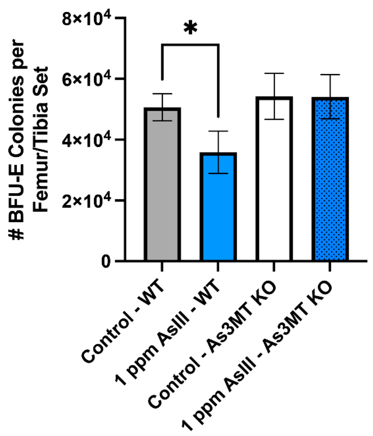
4.4. Attenuation of BFU-E Colony Formation in Wild-Type, but Not As3mt-KO Mice Following 60-Day AsIII Exposure

To determine whether the anemia identified via hematology analysis was significantly contributed to by the loss of RBC progenitor differentiation, the colony-forming ability of BFU-E cells in the bone marrow was evaluated following the exposure of wild-type and As3mt-KO mice to 1 ppm AsIII for 60 days (Figure 1). AsIII exposure was found to significantly attenuate BFU-E colony formation in wild-type (p < 0.05) but not As3mt-KO mice (Figure 1). These findings suggest that As3MT-mediated biotransformation is important for the toxicity of AsIII to early developing RBCs.

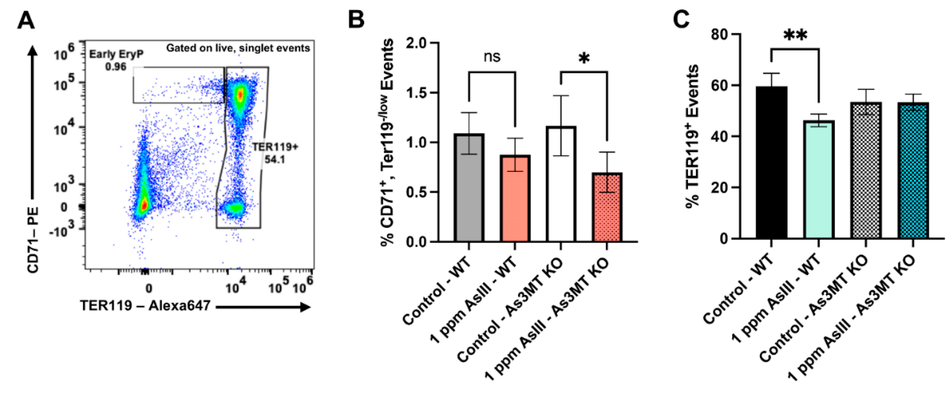
4.5. Early and Late-Stage Erythroblast Subsets Are Altered in the Bone Marrow of Wild-Type and As3mt-KO Mice Following Exposure to AsIII

To further understand the ramifications of impaired arsenic biotransformation on the differentiation of RBCs, the proportion of early- and late-stage erythroid progenitor cells were evaluated in the bone marrow based on CD71 and TER119 surface marker expression using flow cytometry [14,30,31,32,33] (Figure 2A–C). The percentages of early-stage RBC progenitors showed a trend of reduction in wild-type mice and were significantly reduced in As3mt-KO mice (p < 0.05) following the 60-day AsIII exposure (Figure 2B). AsIII was also found to significantly decrease (p < 0.01) the percentage of late-stage, TER119+, RBC progenitor cells in the bone marrow of wild-type but not As3mt-KO mice (Figure 2C). This suggests that the suppressive effects of AsIII on early RBCs are dependent on the As3MT-mediated biotransformation of AsIII.

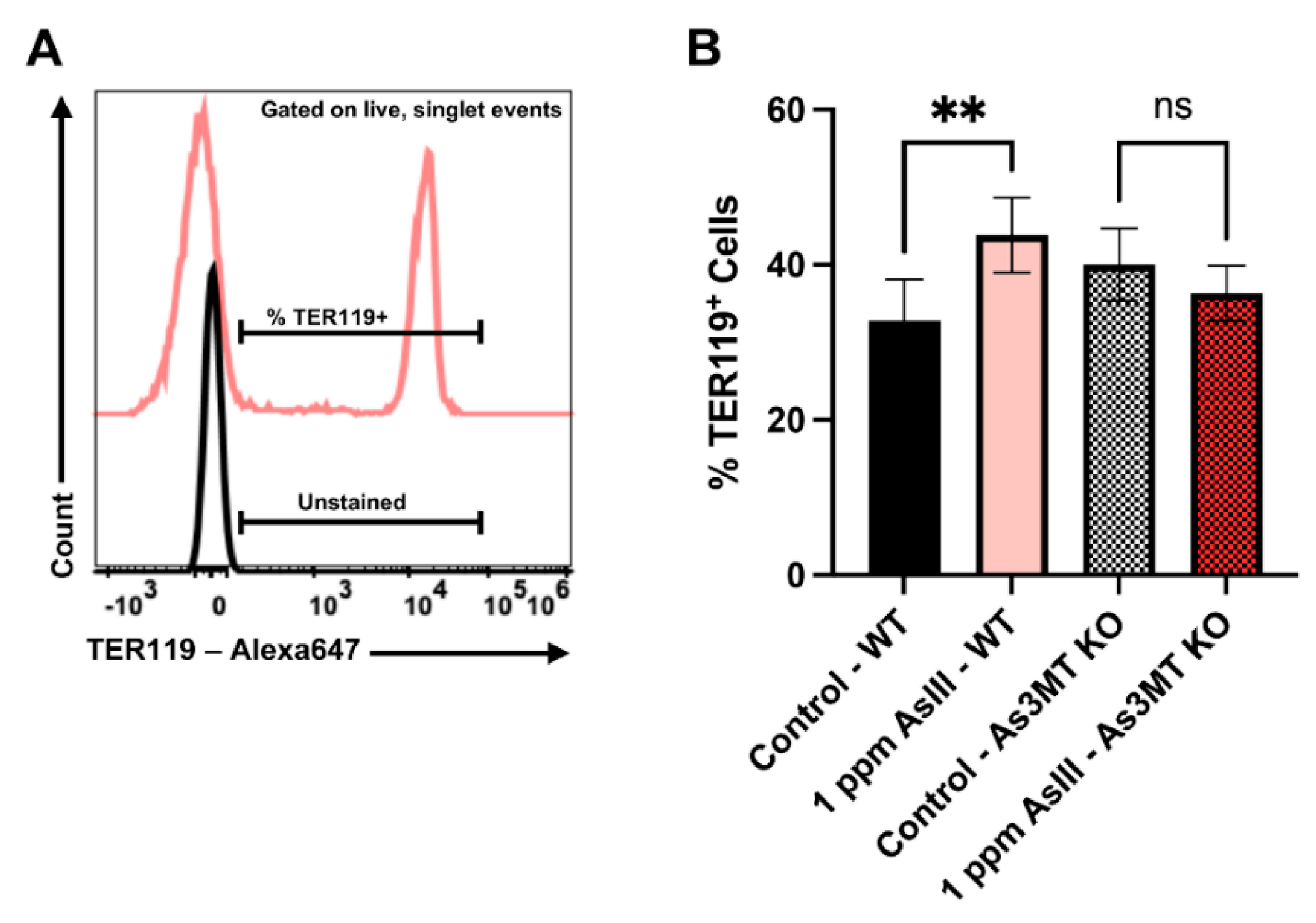
4.6. AsIII Exposure Increases Late-Stage, TER119+ Erythroblasts in the Spleens of Wild-Type, but Not As3mt-KO Mice

The proportions of late-stage RBC progenitor cells in the spleens of wild-type and As3mt-KO mice were evaluated following exposure to AsIII for 60 days based on TER119 surface marker expression using flow cytometry [14,30,31,32,33]. There was a significant increase (p < 0.01) in the percentage of late-stage TER119+ RBCs in the spleen of wild-type mice (Figure 3A,B). No significant alterations in the proportion of TER119+ RBCs were found in the spleens of As3mt-KO mice (Figure 3A,B). Taken together, these results suggest that suppressed RBC output from the bone marrow of wild-type mice induces erythropoietic stress, causing a compensatory increase in RBC production from the spleen. This further underscores the role of biotransformation, mediated by the As3MT enzyme, to the impaired differentiation of RBCs.

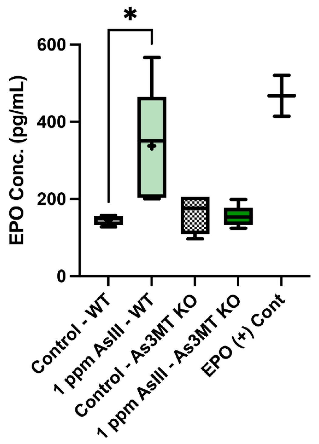
4.7. Levels of Circulating EPO Are Increased Following AsIII Exposure in Wild-Type, but Not As3mt-KO Mice

During instances of hypoxic stress, such as those produced by decreased RBC production from the bone marrow, the release of EPO from the kidney is increased. Increased EPO production promotes the survival and rapid expansion of the early erythroid progenitor pool in the bone marrow [26]. To further determine if the reduced hematological indicators of anemia and compromised production of mature RBCs from the bone marrow were accompanied by an increase in serum EPO levels, the concentration of EPO in the serum of wild-type and As3mt-KO mice was measured using the Mouse EPO ELISA Kit (R&D Systems). A significant increase (p < 0.05) in circulating EPO concentrations in the serum of 1 ppm AsIII-exposed wild-type mice was detected (Figure 4). Circulating EPO levels were not significantly altered in As3mt-KO mice following AsIII exposure (Figure 4). Collectively, these findings suggest that the biotransformation of AsIII produces erythropoietic stress, as indicated by suppressed RBC production in the bone marrow, increased production of mature RBCs from the spleen, and elevated levels of circulating EPO in wild-type but not As3mt-KO mice.

5. Discussion

Millions of people worldwide are exposed to arsenic in their drinking water at levels known to produce numerous detrimental health outcomes, including cancer, cardiovascular disease, immune suppression, and anemia [1,2,3,5,14,15,16]. Despite our growing understanding of the mechanistic basis of arsenic-induced diseases, a significant knowledge gap remains in our understanding of the relative contribution of inorganic vs. organic arsenical species to the development of such ailments. To our knowledge, this is the first study to evaluate the effects of arsenic biotransformation on RBC progenitor cell development and anemia. Our findings provide novel evidence demonstrating a role for biotransformation in arsenic-induced anemia. We found that AsIII exposure resulted in the development of anemia in an As3MT-dependent manner. In addition, the suppressive effects of AsIII on the differentiation of RBC progenitors in the bone marrow were also found to be associated with the As3mt genotype.
The findings from our group have established that exposure to environmentally relevant levels of AsIII results in the development of anemia via the suppression of erythropoiesis in the bone marrow [14,30,31,32,33,34]. In the present study, we found that the As3mt genotype has a critical role in mediating the hematotoxicity of arsenic. Intriguingly, the absence of the As3MT enzyme substantially reduced the effects of AsIII on RBCs in the bone marrow, suggesting the process of biotransformation of inorganic arsenic (i.e., the production of methylated arsenical intermediates) is an important driver of hematotoxicity in vivo. Our lab and others report that MMAIII is more toxic than AsIII in vitro [17,42,43,44] and produces stronger suppressive effects to early developing RBCs in vitro [32]. This is significant because, in the present study, we found the predominant arsenic species in the bone marrow of AsIII-exposed wild-type mice were methylated arsenicals. This is consistent with our previous results showing that MMAIII is the major arsenical present in the bone marrow of mice exposed to AsIII in their drinking water [40]. Dosimetry studies in As3mt-KO mice following drinking water exposure to AsIII report similar arsenic metabolite profiles as those observed in this study, with the predominant arsenicals present in As3mt-KO mice being inorganic [45].
These findings suggest that arsenic metabolism may play a significant role in impairing erythropoiesis through the generation of MMAIII, which in the absence of the As3MT enzyme, largely does not occur [45]. Numerous epidemiological studies in populations chronically exposed to arsenic have shown that arsenic methylation capacity is associated with elevated disease risk [46,47,48,49,50,51,52]. The proportions of excreted arsenic metabolites are indicative of arsenic methylation efficiency [24,37,53]. Higher levels of DMA relative to MMA in urine are indicative of more efficient arsenic metabolism and are associated with decreased toxicity [19,54]. MMAIII exposure may therefore be a significant contributing factor to anemia in people chronically exposed to arsenic. Consistent with this hypothesis, our previous studies revealed an inverse association between urinary MMA and RBC counts in a cohort of men from rural Bangladesh [38]. Additionally, findings from the present study identified significant impairments to erythropoiesis that were associated with the As3mt-genotype.
Our previous results show that AsIII exposure results in the development of anemia via the suppression of RBC differentiation in the bone marrow of male C57BL/6J mice [30]. Consistent with these findings, the formation of BFU-E colonies and percentages of late-stage erythroblasts were reduced in wild-type mice. Whereas our previous studies focused on the role of arsenic exposure in the suppression of erythropoiesis and the development of anemia, the present study extends beyond these works by evaluating the role of As3MT-mediated biotransformation in the hematotoxicity of AsIII. Although BFU-E colony formation was not significantly altered in As3mt-KO mice, the percentages of early erythroid progenitor cells were significantly reduced. These findings suggest that different stages of early RBC differentiation may exhibit varying sensitivities to different arsenical species. The extent to which specific inorganic or organic arsenicals contribute to this phenomenon in vivo remains unclear but will be the focus of our ongoing investigations. The current study only indicates the critical role of arsenic methylation in AsIII-induced anemia. Whether methylation plays a similar role in other arsenic-mediated adverse health effects remains unknown and warrants further study.
Anemic conditions, such as those caused by impairment of bone marrow erythropoiesis, induce a physiological response mechanism known as stress erythropoiesis to cope with the loss of RBCs [55,56]. An important distinction between mice and humans is that the primary site of stress erythropoiesis in mice is the spleen, whereas, in humans, it occurs predominately in the bone marrow [55,56,57]. Consistent with the hypothesis, the wild-type mice in the present study demonstrated distinguishing factors of stress erythropoiesis, including elevated percentages of mature erythroblasts in the spleen and increased circulating EPO levels. These findings suggest that the AsIII-induced suppression of bone marrow erythropoiesis resulted in the activation of stress erythropoiesis in the spleen.
Taken together, the findings from the present study provide novel support for the role of biotransformation in arsenic-induced hematotoxicity and anemia. These results emphasize the importance of considering arsenic biotransformation as a mediator of toxicity in humans. This information is essential for understanding the molecular mechanisms by which arsenic exposure produces adverse health effects and provides a critical framework for understanding how biotransformation influences the risk of arsenic-associated anemias, which is critical for the development of potential preventative and therapeutic strategies in the future.

Author Contributions

Funding acquisition: S.M. and K.J.L.; conceptualization: S.M., M.S. and K.J.L.; formal analysis, investigation, and data curation: S.M., H.Z., L.V.S.-M., Z.A.Y., K.J.M., G.W., A.M.B., X.Z., M.S. and K.J.L.; writing—original draft preparation: S.M.; writing—review and editing: S.M., H.Z., L.V.S.-M., Z.A.Y., K.J.M., G.W., A.M.B., X.Z., M.S. and K.J.L. All authors have read and agreed to the published version of the manuscript.

Funding

This work was funded by National Institutes of Environmental Health Sciences (NIEHS) Grant Number RO1 ES029369, R01 ES029369-03S1; National Institute of General Medical Sciences (NIGMS) of the National Institutes of Health (NIH) Grant Number 1R16 GM146669-01, NIH NIGMS UNM Center for Metals in Biology and Medicine Grant Number P20 GM130422, Institutional Development Award (IDeA) P20 GM103451; NIEHS Superfund Research Program Grant Number P42 ES025589; and the National Science Foundation Louis Stokes Alliance for Minority Participation program.

Institutional Review Board Statement

The study was conducted in accordance with the UNM Office of Animal Care Compliance Committee (Albuquerque, NM, USA) at the UNM HSC according to protocol number 21-201104-HSC, approval date 5 May 2022.

Informed Consent Statement

Not applicable.

Data Availability Statement

Data generated during the present study are available from the corresponding author (Sebastian Medina; [email protected]) upon reasonable request.

Acknowledgments

The authors would like to thank Fredine T. Lauer and Tamara A. Anderson of the UNM College of Pharmacy for their technical assistance with sample collection and assay setup.

Conflicts of Interest

The authors have no conflict of interest to declare.

References

  1. World Health Organization. Arsenic in Drinking-Water. 2019. Available online: https://www.who.int/publications/i/item/arsenic-in-drinking-water-background-document-for-development-of-who-guidelines-for-drinking-water-quality (accessed on 1 October 2022).
  2. Ferrario, D.; Gribaldo, L.; Hartung, T. Arsenic Exposure and Immunotoxicity: A Review Including the Possible Influence of Age and Sex. Curr. Environ. Health Rep. 2016, 3, 1–12. [Google Scholar] [CrossRef] [PubMed]
  3. Heck, J.E.; Chen, Y.; Grann, V.R.; Slavkovich, V.; Parvez, F.; Ahsan, H. Arsenic exposure and anemia in Bangladesh: A population-based study. J. Occup. Environ. Med. 2008, 50, 80–87. [Google Scholar] [CrossRef] [PubMed]
  4. Hughes, M.F. Arsenic toxicity and potential mechanisms of action. Toxicol. Lett. 2002, 133, 1–16. [Google Scholar] [CrossRef] [Green Version]
  5. Naujokas, M.F.; Anderson, B.; Ahsan, H.; Aposhian, H.V.; Graziano, J.H.; Thompson, C.; Suk, W.A. The broad scope of health effects from chronic arsenic exposure: Update on a worldwide public health problem. Environ. Health Perspect. 2013, 121, 295–302. [Google Scholar] [CrossRef]
  6. Tyler, C.R.; Allan, A.M. The Effects of Arsenic Exposure on Neurological and Cognitive Dysfunction in Human and Rodent Studies: A Review. Curr. Environ. Health Rep. 2014, 1, 132–147. [Google Scholar] [CrossRef] [Green Version]
  7. Kile, M.L.; Faraj, J.M.; Ronnenberg, A.G.; Quamruzzaman, Q.; Rahman, M.; Mostofa, G.; Afroz, S.; Christiani, D.C. A cross sectional study of anemia and iron deficiency as risk factors for arsenic-induced skin lesions in Bangladeshi women. BMC Public Health 2016, 16, 158. [Google Scholar] [CrossRef] [Green Version]
  8. Majumdar, K.K.; Guha Mazumder, D.N.; Ghose, N.; Ghose, A.; Lahiri, S. Systemic manifestations in chronic arsenic toxicity in absence of skin lesions in West Bengal. Indian J. Med. Res. 2009, 129, 75–82. [Google Scholar] [PubMed]
  9. Surdu, S.; Bloom, M.S.; Neamtiu, I.A.; Pop, C.; Anastasiu, D.; Fitzgerald, E.F.; Gurzau, E.S. Consumption of arsenic-contaminated drinking water and anemia among pregnant and non-pregnant women in northwestern Romania. Environ. Res. 2015, 140, 657–660. [Google Scholar] [CrossRef] [Green Version]
  10. World Health Organization. The Global Prevalence of Anaemia in 2011. 2015. Available online: https://apps.who.int/iris/handle/10665/177094 (accessed on 1 October 2022).
  11. Hopenhayn, C.; Bush, H.M.; Bingcang, A.; Hertz-Picciotto, I. Association between arsenic exposure from drinking water and anemia during pregnancy. J. Occup. Environ. Med. 2006, 48, 635–643. [Google Scholar] [CrossRef]
  12. Kassebaum, N.J.; Jasrasaria, R.; Naghavi, M.; Wulf, S.K.; Johns, N.; Lozano, R.; Regan, M.; Weatherall, D.; Chou, D.P.; Eisele, T.P.; et al. A systematic analysis of global anemia burden from 1990 to 2010. Blood 2014, 123, 615–624. [Google Scholar] [CrossRef]
  13. Breton, C.V.; Houseman, E.A.; Kile, M.L.; Quamruzzaman, Q.; Rahman, M.; Mahiuddin, G.; Christiani, D.C. Gender-specific protective effect of hemoglobin on arsenic-induced skin lesions. Cancer Epidemiol. Biomark. Prev. 2006, 15, 902–907. [Google Scholar] [CrossRef] [PubMed] [Green Version]
  14. United States Environmental Protection Agency. 2012 Edition of the Drinking Water Standards and Health Advisories Table; United States Environmental Protection Agency: Washington, DC, USA, 2012.
  15. Agency for Toxic Substances and Disease Registry. Toxicological Profile for Arsenic; Agency for Toxic Substances and Disease Registry: Atlanta, GA, USA, 2007.
  16. Agency for Toxic Substances and Disease Registry. Addendum to the Toxicological Profile for Arsenic; Agency for Toxic Substances and Disease Registry: Atlanta, GA, USA, 2016.
  17. Styblo, M.; Del Razo, L.M.; Vega, L.; Germolec, D.R.; LeCluyse, E.L.; Hamilton, G.A.; Reed, W.; Wang, C.; Cullen, W.R.; Thomas, D.J. Comparative toxicity of trivalent and pentavalent inorganic and methylated arsenicals in rat and human cells. Arch. Toxicol. 2000, 74, 289–299. [Google Scholar] [CrossRef] [PubMed]
  18. Thomas, D.J.; Styblo, M.; Lin, S. The cellular metabolism and systemic toxicity of arsenic. Toxicol. Appl. Pharmacol. 2001, 176, 127–144. [Google Scholar] [CrossRef] [PubMed]
  19. Vahter, M. Mechanisms of arsenic biotransformation. Toxicology 2002, 181–182, 211–217. [Google Scholar] [CrossRef]
  20. Lin, S.; Shi, Q.; Nix, F.B.; Styblo, M.; Beck, M.A.; Herbin-Davis, K.M.; Hall, L.L.; Simeonsson, J.B.; Thomas, D.J. A novel S-adenosyl-L-methionine:arsenic(III) methyltransferase from rat liver cytosol. J. Biol. Chem. 2002, 277, 10795–10803. [Google Scholar] [CrossRef] [Green Version]
  21. Thomas, D.J.; Li, J.; Waters, S.B.; Xing, W.; Adair, B.M.; Drobna, Z.; Devesa, V.; Styblo, M. Arsenic (+3 oxidation state) methyltransferase and the methylation of arsenicals. Exp. Biol. Med. 2007, 232, 3–13. [Google Scholar]
  22. Douillet, C.; Huang, M.C.; Saunders, R.J.; Dover, E.N.; Zhang, C.; Styblo, M. Knockout of arsenic (+3 oxidation state) methyltransferase is associated with adverse metabolic phenotype in mice: The role of sex and arsenic exposure. Arch. Toxicol. 2017, 91, 2617–2627. [Google Scholar] [CrossRef]
  23. Engstrom, K.; Vahter, M.; Mlakar, S.J.; Concha, G.; Nermell, B.; Raqib, R.; Cardozo, A.; Broberg, K. Polymorphisms in arsenic(+III oxidation state) methyltransferase (AS3MT) predict gene expression of AS3MT as well as arsenic metabolism. Environ. Health Perspect. 2011, 119, 182–188. [Google Scholar] [CrossRef] [Green Version]
  24. Vahter, M. Variation in human metabolism of arsenic. In Arsenic Exposure and Health Effects; Chappell, W.R., Abernathy, C.O., Calderon, R.L., Eds.; Elsevier Science Ltd.: Amsterdam, The Netherlands, 1999; pp. 267–279. [Google Scholar]
  25. Vahter, M.; Concha, G.; Nermell, B.; Nilsson, R.; Dulout, F.; Natarajan, A.T. A unique metabolism of inorganic arsenic in native Andean women. Eur. J. Pharmacol. 1995, 293, 455–462. [Google Scholar] [CrossRef]
  26. Dzierzak, E.; Philipsen, S. Erythropoiesis: Development and differentiation. Cold Spring Harb. Perspect. Med. 2013, 3, a011601. [Google Scholar] [CrossRef]
  27. Palis, J. Primitive and definitive erythropoiesis in mammals. Front. Physiol. 2014, 5, 3. [Google Scholar] [CrossRef] [PubMed]
  28. Orkin, S.H.; Zon, L.I. Hematopoiesis: An evolving paradigm for stem cell biology. Cell 2008, 132, 631–644. [Google Scholar] [CrossRef] [PubMed] [Green Version]
  29. Rieger, M.A.; Schroeder, T. Hematopoiesis. Cold Spring Harb. Perspect. Biol. 2012, 4, a008250. [Google Scholar] [CrossRef] [PubMed]
  30. Medina, S.; Xu, H.; Wang, S.C.; Lauer, F.T.; Liu, K.J.; Burchiel, S.W. Low level arsenite exposures suppress the development of bone marrow erythroid progenitors and result in anemia in adult male mice. Toxicol. Lett. 2017, 273, 106–111. [Google Scholar] [CrossRef] [PubMed]
  31. Zhou, X.; Medina, S.; Bolt, A.M.; Zhang, H.; Wan, G.; Xu, H.; Lauer, F.T.; Wang, S.C.; Burchiel, S.W.; Liu, K.J. Inhibition of red blood cell development by arsenic-induced disruption of GATA-1. Sci. Rep. 2020, 10, 19055. [Google Scholar] [CrossRef]
  32. Medina, S.; Bolt, A.M.; Zhou, X.; Wan, G.; Xu, H.; Lauer, F.T.; Liu, K.J.; Burchiel, S.W. Arsenite and monomethylarsonous acid disrupt erythropoiesis through combined effects on differentiation and survival pathways in early erythroid progenitors. Toxicol. Lett. 2021, 350, 111–120. [Google Scholar] [CrossRef]
  33. Medina, S.; Zhang, H.; Santos-Medina, L.V.; Wan, G.; Bolt, A.M.; Zhou, X.; Burchiel, S.W.; Liu, K.J. Arsenic impairs the lineage commitment of hematopoietic progenitor cells through the attenuation of GATA-2 DNA binding activity. Toxicol. Appl. Pharmacol. 2022, 452, 116193. [Google Scholar] [CrossRef]
  34. Wan, G.; Medina, S.; Zhang, H.; Pan, R.; Zhou, X.; Bolt, A.M.; Luo, L.; Burchiel, S.W.; Liu, K.J. Arsenite exposure inhibits the erythroid differentiation of human hematopoietic progenitor CD34(+) cells and causes decreased levels of hemoglobin. Sci. Rep. 2021, 11, 22121. [Google Scholar] [CrossRef]
  35. Drobna, Z.; Naranmandura, H.; Kubachka, K.M.; Edwards, B.C.; Herbin-Davis, K.; Styblo, M.; Le, X.C.; Creed, J.T.; Maeda, N.; Hughes, M.F.; et al. Disruption of the arsenic (+3 oxidation state) methyltransferase gene in the mouse alters the phenotype for methylation of arsenic and affects distribution and retention of orally administered arsenate. Chem. Res. Toxicol. 2009, 22, 1713–1720. [Google Scholar] [CrossRef] [Green Version]
  36. Medina, S.; Lauer, F.T.; Castillo, E.F.; Bolt, A.M.; Ali, A.S.; Liu, K.J.; Burchiel, S.W. Exposures to uranium and arsenic alter intraepithelial and innate immune cells in the small intestine of male and female mice. Toxicol. Appl. Pharmacol. 2020, 403, 115155. [Google Scholar] [CrossRef]
  37. Vahter, M. Methylation of inorganic arsenic in different mammalian species and population groups. Sci. Prog. 1999, 82 Pt 1, 69–88. [Google Scholar] [CrossRef] [PubMed]
  38. Parvez, F.; Medina, S.; Santella, R.M.; Islam, T.; Lauer, F.T.; Alam, N.; Eunus, M.; Rahman, M.; Factor-Litvak, P.; Ahsan, H.; et al. Arsenic exposures alter clinical indicators of anemia in a male population of smokers and non-smokers in Bangladesh. Toxicol. Appl. Pharmacol. 2017, 331, 62–68. [Google Scholar] [CrossRef] [PubMed]
  39. Ezeh, P.C.; Xu, H.; Wang, S.C.; Medina, S.; Burchiel, S.W. Evaluation of Toxicity in Mouse Bone Marrow Progenitor Cells. Curr. Protoc. Toxicol. 2016, 67, 18.9.1.–18.9.2. [Google Scholar] [CrossRef] [PubMed] [Green Version]
  40. Xu, H.; McClain, S.; Medina, S.; Lauer, F.T.; Douillet, C.; Liu, K.J.; Hudson, L.G.; Styblo, M.; Burchiel, S.W. Differential sensitivities of bone marrow, spleen and thymus to genotoxicity induced by environmentally relevant concentrations of arsenite. Toxicol. Lett. 2016, 262, 55–61. [Google Scholar] [CrossRef] [PubMed] [Green Version]
  41. Balarajan, Y.; Ramakrishnan, U.; Ozaltin, E.; Shankar, A.H.; Subramanian, S.V. Anaemia in low-income and middle-income countries. Lancet 2011, 378, 2123–2135. [Google Scholar] [CrossRef]
  42. Ezeh, P.C.; Xu, H.; Lauer, F.T.; Liu, K.J.; Hudson, L.G.; Burchiel, S.W. Monomethylarsonous acid (MMA+3) Inhibits IL-7 Signaling in Mouse Pre-B Cells. Toxicol. Sci. 2016, 149, 289–299. [Google Scholar] [CrossRef] [Green Version]
  43. Petrick, J.S.; Jagadish, B.; Mash, E.A.; Aposhian, H.V. Monomethylarsonous acid (MMA(III)) and arsenite: LD(50) in hamsters and in vitro inhibition of pyruvate dehydrogenase. Chem. Res. Toxicol. 2001, 14, 651–656. [Google Scholar] [CrossRef] [PubMed]
  44. Xu, H.; Medina, S.; Lauer, F.T.; Douillet, C.; Liu, K.J.; Styblo, M.; Burchiel, S.W. Genotoxicity induced by monomethylarsonous acid (MMA(+3)) in mouse thymic developing T cells. Toxicol. Lett. 2017, 279, 60–66. [Google Scholar] [CrossRef]
  45. Chen, B.; Arnold, L.L.; Cohen, S.M.; Thomas, D.J.; Le, X.C. Mouse arsenic (+3 oxidation state) methyltransferase genotype affects metabolism and tissue dosimetry of arsenicals after arsenite administration in drinking water. Toxicol. Sci. 2011, 124, 320–326. [Google Scholar] [CrossRef] [Green Version]
  46. Ahsan, H.; Chen, Y.; Kibriya, M.G.; Slavkovich, V.; Parvez, F.; Jasmine, F.; Gamble, M.V.; Graziano, J.H. Arsenic metabolism, genetic susceptibility, and risk of premalignant skin lesions in Bangladesh. Cancer Epidemiol. Biomark. Prev. 2007, 16, 1270–1278. [Google Scholar] [CrossRef] [Green Version]
  47. Chen, Y.; Parvez, F.; Gamble, M.; Islam, T.; Ahmed, A.; Argos, M.; Graziano, J.H.; Ahsan, H. Arsenic exposure at low-to-moderate levels and skin lesions, arsenic metabolism, neurological functions, and biomarkers for respiratory and cardiovascular diseases: Review of recent findings from the Health Effects of Arsenic Longitudinal Study (HEALS) in Bangladesh. Toxicol. Appl. Pharmacol. 2009, 239, 184–192. [Google Scholar] [CrossRef] [PubMed]
  48. Chen, Y.C.; Guo, Y.L.; Su, H.J.; Hsueh, Y.M.; Smith, T.J.; Ryan, L.M.; Lee, M.S.; Chao, S.C.; Lee, J.Y.; Christiani, D.C. Arsenic methylation and skin cancer risk in southwestern Taiwan. J. Occup. Environ. Med. 2003, 45, 241–248. [Google Scholar] [CrossRef] [PubMed]
  49. Huang, Y.K.; Pu, Y.S.; Chung, C.J.; Shiue, H.S.; Yang, M.H.; Chen, C.J.; Hsueh, Y.M. Plasma folate level, urinary arsenic methylation profiles, and urothelial carcinoma susceptibility. Food Chem. Toxicol. 2008, 46, 929–938. [Google Scholar] [CrossRef] [PubMed]
  50. Lin, Y.C.; Chen, W.J.; Huang, C.Y.; Shiue, H.S.; Su, C.T.; Ao, P.L.; Pu, Y.S.; Hsueh, Y.M. Polymorphisms of Arsenic (+3 Oxidation State) Methyltransferase and Arsenic Methylation Capacity Affect the Risk of Bladder Cancer. Toxicol. Sci. 2018, 164, 328–338. [Google Scholar] [CrossRef] [Green Version]
  51. Luo, L.; Li, Y.; Gao, Y.; Zhao, L.; Feng, H.; Wei, W.; Qiu, C.; He, Q.; Zhang, Y.; Fu, S.; et al. Association between arsenic metabolism gene polymorphisms and arsenic-induced skin lesions in individuals exposed to high-dose inorganic arsenic in northwest China. Sci. Rep. 2018, 8, 413. [Google Scholar] [CrossRef] [Green Version]
  52. Yu, R.C.; Hsu, K.H.; Chen, C.J.; Froines, J.R. Arsenic methylation capacity and skin cancer. Cancer Epidemiol. Biomark. Prev. 2000, 9, 1259–1262. [Google Scholar]
  53. Gamble, M.V.; Liu, X.; Ahsan, H.; Pilsner, R.; Ilievski, V.; Slavkovich, V.; Parvez, F.; Levy, D.; Factor-Litvak, P.; Graziano, J.H. Folate, homocysteine, and arsenic metabolism in arsenic-exposed individuals in Bangladesh. Environ. Health Perspect. 2005, 113, 1683–1688. [Google Scholar] [CrossRef] [Green Version]
  54. Vahter, M.; Concha, G. Role of metabolism in arsenic toxicity. Pharmacol. Toxicol. 2001, 89, 1–5. [Google Scholar] [CrossRef]
  55. Paulson, R.F.; Shi, L.; Wu, D.C. Stress erythropoiesis: New signals and new stress progenitor cells. Curr. Opin. Hematol. 2011, 18, 139–145. [Google Scholar] [CrossRef] [Green Version]
  56. Socolovsky, M. Molecular insights into stress erythropoiesis. Curr. Opin. Hematol. 2007, 14, 215–224. [Google Scholar] [CrossRef]
  57. Kim, T.S.; Hanak, M.; Trampont, P.C.; Braciale, T.J. Stress-associated erythropoiesis initiation is regulated by type 1 conventional dendritic cells. J. Clin. Investig. 2015, 125, 3965–3980. [Google Scholar] [CrossRef] [PubMed]
Figure 1. Drinking water exposure to AsIII suppresses the formation of BFU-E colonies in wild-type, but not As3mt-KO mice following exposure to control (untreated, tap water) or 1 ppm AsIII in their drinking water for 60 days. Number of BFU-E colonies (expressed relative to the number of viable cells recovered per femur and tibia set of each mouse) formed in EPO containing serum-free methylcellulose-based medium (i.e., Methocult M3436; STEMCELL Technologies) for 14 days. Data are expressed as mean ± SD of duplicate cultures per mouse. Statistically significant differences compared to untreated control in one-way ANOVA followed by Tukey’s post hoc test; n = 5 mice/group, * p < 0.05.
Water 15 00448 g001
Figure 2. Percentages of early and late-stage erythroblasts were altered in the bone marrow of wild-type and As3mt-KO mice following 60-day drinking water exposure to 1 ppm AsIII. (A) Representative flow cytometry dot plot showing the gating strategy used to define early erythroid progenitors (Early EryP; CD71+, TER119-/low events) or late-stage erythroblasts (CD71+,−, TER119+ events) in the bone marrow. Percentages of (B) early and (C) late-stage erythroblasts were measured by flow cytometry in bone marrow collected from wild-type and As3mt-KO mice following 60-day exposure to control (untreated, tap water) or 1 ppm AsIII. Data are expressed as mean ± SD. Statistically significant difference compared to control in one-way ANOVA followed by Tukey’s post hoc test; n = 5 mice/group, * p < 0.05, ** p < 0.01, non-statistically significant.
Water 15 00448 g002
Figure 3. AsIII exposure increases the percentage of mature TER119+ erythroblasts in the spleens of wild-type mice. (A) Representative flow cytometry histograms showing an unstained sample and the gating strategy used to enumerate the percentage of TER119+ erythroblasts in the spleen. (B) Percentages of TER119+ erythroblasts in the spleen of wild-type and As3mt-KO mice following 60-day drinking water exposures control (untreated, tap water) or 1 ppm AsIII. Data are expressed as mean ± SD. Statistically significant difference compared to control in one-way ANOVA followed by Tukey’s post hoc test; n = 5 mice/group, ** p < 0.01, ns = non-statistically significant.
Water 15 00448 g003
Figure 4. Elevated serum EPO levels in wild-type mice following drinking water exposure to AsIII. EPO concentrations were measured using the Mouse EPO Quantikine ELISA Kit (R&D Systems) in serum collected from wild-type and As3mt-KO mice following exposure to control (untreated, tap water) or 1 ppm AsIII for 60 days. EPO positive (+) control was a standard prepared at a known concentration of 500 pg/mL. In the box plot, crosses indicate the mean and black horizontal lines indicate the median. Data are expressed as mean ± SD. Statistically significant difference compared to control in one-way ANOVA followed by Tukey’s post hoc test; n = 5 mice/group, * p < 0.05.
Water 15 00448 g004
Table 1. AsIII exposure does not significantly alter body weight, weekly drinking water consumption, spleen weights, or bone marrow and spleen cell recoveries in male wild-type or As3mt-KO mice exposed via drinking water to control (tap water) or 1 ppm AsIII for 60 days a.
Wild-TypeAs3mt-KO
ParameterControl1 ppm AsIIIControl1 ppm AsIII
Body Wt. (g)32.1 ± 3.1831.38 ± 1.1229.50 ± 1.7829.99 ± 3.23
Water Consumption (mL/mouse/wk)21.00 ± 1.5021.00 ± 4.0017.00 ± 0.9819.00 ± 2.00
Bone Marrow Cell Recovery (Total viable cells per femur/tibia set, ×106)57.90 ± 6.3851.14 ± 11.7566.92 ± 8.2463.26 ± 11.06
Spleen Wt. (g)80.80 ± 8.2099.80 ± 18.4979.4 ± 4.8976.4 ± 7.30
Spleen Cell Recovery (Total viable cells, ×106)153 ± 15.3150 ± 15.398.66 ± 15.30109.24 ± 18.97
a Data are expressed as mean ± SD, n = 5.
Table 2. Alterations to hematological indicators of anemia (i.e., RBC counts, Hct, Hgb levels, MCH levels, and MCVs) measured in whole blood collected from male wild-type and As3mt-KO mice following drinking water exposure to control (tap water) or 1 ppm AsIII for 60 days a.
Wild-TypeAs3mt-KO
ParameterControl1 ppm AsIIIControl1 ppm AsIII
RBC Count (×1012/L)12.17 ± 0.1311.21 ± 0.22 ***11.66 ± 0.2511.47 ± 0.75
Hct (%)52.08 ± 0.5149.1 ± 0.38 ***48.56 ± 1.2648.12 ± 3.47
Hgb (g/dL)16.7 ± 0.4716.12 ± 0.30 *14.96 ± 2.2115.66 ± 1.21
MCH (pg)13.98 ± 0.5314.38 ± 0.4613.58 ± 0.2913.66 ± 0.30
MCV (fL)43 ± 0.7143.8 ± 0.8441.8 ± 0.4541.8 ± 0.45
a Data are expressed as mean ± SD, n = 5, * p < 0.05, *** p < 0.0001, in one-way ANOVA followed by Tukey’s multiple-comparison post hoc test.
Table 3. Arsenic species measured using HG-CT-ICP-MS in bone marrow cells, spleen cells, and plasma collected from male wild-type and As3mt-KO mice following exposure to control (tap water) or 1 ppm AsIII for 60 days a,b.
TissuesGenotypeExposure As Amount by Species (pg)
iAsMAsDMAsTotal As
Bone MarrowWild-typeControl1.50 ± 0.820.22 ± 0.090.75 ± 0.152.46 ± 0.89
1 ppm AsIII4.80 ± 2.432.66 ± 2.14 *23.23 ± 9.71 ****30.69 ± 13.78
As3mt-KOControl 11.63 ± 1.020.37 ± 0.120.25 ± 0.1212.25 ± 1.06
1 ppm AsIII134.27 ± 28.71 ****1.55 ± 0.240.23 ± 0.09136.05 ± 28.50 ****
SpleenWild-typeControl6.68 ± 4.820.19 ± 0.130.26 ± 0.147.14 ± 4.97
1 ppm AsIII10.04 ± 3.320.75 ± 0.38 **5.22 ± 1.61 ****16.00 ± 2.65
As3mt-KOControl 30.14 ± 11.020.33 ± 0.130.21 ± 0.0830.68 ± 11.18
1 ppm AsIII89.33 ± 17.79 ****0.98 ± 0.20 **0.14 ± 0.0590.45 ± 17.90 ****
PlasmaWild-typeControl12.83 ± 6.472.29 ± 0.91218.98 ± 54.90234.11 ± 61.23
1 ppm AsIII92.32 ± 39.29117.6 ± 3.56 ****6956.65 ± 1495.13 ****7166.57 ± 1533.08 ****
As3mt-KOControl 243.74 ± 142.482.44 ± 1.321.49 ± 0.62247.67 ± 144.19
1 ppm AsIII1537.93 ± 257.47 ****26.33 ± 2.14 ****8.81 ± 4.671573.07 ± 254.08
a Data are expressed as mean ± SD; n = 5; * p < 0.05, ** p < 0.01, **** p < 0.0001 in one-way ANOVA followed by Tukey’s multiple-comparison post hoc test. b Inorganic arsenicals (iAs; AsV and AsIII); methylated As (MAs; MMAV and MMAIII); demethylated As (DMAs; DMAV and DMAIII); Total As (iAs + MAs + DMAs).
Disclaimer/Publisher’s Note: The statements, opinions and data contained in all publications are solely those of the individual author(s) and contributor(s) and not of MDPI and/or the editor(s). MDPI and/or the editor(s) disclaim responsibility for any injury to people or property resulting from any ideas, methods, instructions or products referred to in the content.

