Article

The Mutual Effect of Microparticles and Antidepressants on the Protozoan Spirostomum ambiguum (Müller, 1786) Ehrenberg, 1835

Department of Toxicology and Food Sciences, Medical University of Warsaw, Banacha 1 str., 02-097 Warszawa, Poland
*
Authors to whom correspondence should be addressed.
Water 2023, 15(3), 552; https://doi.org/10.3390/w15030552
Submission received: 29 December 2022 / Revised: 23 January 2023 / Accepted: 26 January 2023 / Published: 31 January 2023
(This article belongs to the Section Water Quality and Contamination)

Abstract

:
Antidepressants, especially selective serotonin re-uptake inhibitors, which are among the most commonly used pharmaceuticals, are ubiquitous in effluents and freshwaters. Microparticles, including microplastics, show sorption properties to different compounds, thus becoming a potential vector of toxic substances. This study aimed to evaluate the effects of four antidepressants on the protozoan Spirostomum ambiguum in the presence of four types of microplastics and baker’s yeast. The Spirotox, measuring the acute toxicity, and food uptake inhibition assay were applied. The microparticles did not influence the toxicity of the tested antidepressants in the acute toxicity assay. Moreover, they did not adsorb the drugs during a seven-day incubation in dark. However, sublethal levels of sertraline and duloxetine decreased the number of food vacuoles formed by the protozoa. The highest effect was observed in the case of the suspension of edible particles of baker’s yeast, where a significant decrease in the number of food vacuoles was observed in the sertraline concentration as low as 0.025 mg L−1. A lower but statistically significant effect was observed when wettable microparticles of phenolic resin were used as the artificial food source. These results indicate that serotonin re-uptake inhibitors can interfere with the feeding processes of ciliates.

1. Introduction

Antidepressants are one of the most commonly used drug groups. Fluoxetine (FLX, methyl({3-phenyl-3-[4-(trifluoromethyl)phenoxy]propyl})amine), sertraline (SER, (1S,4S)-4-(3,4-dichlorophenyl)-N-methyl-1,2,3,4-tetrahydronaphthalen-1-amine), and paroxetine (PAR, (3S,4R)-3-[(2H-1,3-benzodioxol-5-yloxy)methyl]-4-(4-fluorophenyl) piperidine) are selective serotonin re-uptake inhibitors (SSRIs, ATC classification: N06AB) approved for clinical use in the late 1980s and early 1990s. Duloxetine (DLX, (+)-(S)-N-methyl-3-(naphthalen-1-yloxy)-3-(thiophen-2-yl)propan-1-amine) is a dual serotonin and norepinephrine re-uptake inhibitor (SNRI, Anatomical Therapeutic Chemical (ATC) Classification System: N06AX) approved in 2004. They are among the 80 most prescribed pharmaceuticals in the USA with 38 (SER), 27 (FLX), 21 (DLX), and 10 (PAR) million prescriptions in 2019 [1]. Pharmaceutically active compounds end up in the environment primarily with municipal sewage after use. Antidepressants have been detected in rivers in several countries, e.g., Canada [2], the USA [3,4], Egypt [5], Poland [6], and the Czech Republic [7]. SER was detected in effluents at a concentration of up to 1 µg L−1 [8]. SER and FLX were considered potential bioaccumulative compounds [7,9,10]. In our previous study, SER was found to be the most bioaccumulative antidepressant in the protozoan Spirostomum ambiguum with the bioaccumulation factor of 34,092 L/kg [11]. Antidepressants, especially SSRI, are considered one of the most ecotoxic groups of pharmaceuticals, with effective concentration (EC50) values for algae as low as 12 and 24 µg L−1 for SER and FLX, respectively [12]. Besides investigational pharmaceuticals, a strong focus has recently been placed on new emerging contaminants—microplastics.
Evaluating the presence, existence, and toxic effects of micro- and nanoplastics in aquatic environments is currently a huge challenge for scientists [13]. Microplastics end up in the environment along with sewage as a result of the degradation of macroplastics. They have been detected not only in marine environments but also in freshwater environments [13]. They are persistent in the environment, and as their specific density is close to the density of water, microparticles can be suspended in the water column for a long time [14,15,16]. The acute toxicity of chemically inert, polymeric microparticles in environmentally realistic concentrations is unlikely [17,18]. However, as plastics have high surface hydrophobicity, they may adsorb hydrophobic organic pollutants and influence their environmental fate [19,20]. Microparticles may act as a vector facilitating the transport of toxic, hydrophobic substances and metals over long distances [21,22]. Research has reported the sorption of hydrophilic chemicals, including pharmaceuticals [23]; however, studies on the adsorption of antidepressants on plastic microparticles are limited [13]. The majority of the studies on microplastics have been carried out using standardized purchased microbeads made of pure polymers [22,23,24,25,26,27]. In the present study, microplastics prepared from household products were used. They may contain additives such as plasticizers, pigments, fillers, and flame retardants that could affect the organisms present in the environment [16]. The use of microparticles prepared from everyday materials better reflects the real-world scenario of environmental pollution.
Protozoa are ubiquitous in aquatic environments, playing a crucial role in freshwaters and wastewater treatment plants as primary consumers. Ciliated protozoa feed primarily through phagocytosis. As they cannot distinguish between digestible and inert particles [28], they may be affected by the toxic compounds adsorbed on the surface of the microparticles and/or additives leached from the plastics. Furthermore, the sorption of toxicants on the microparticles may decrease their bioavailability by the test organisms. S. ambiguum is a sensitive ciliated protozoan that has been used in ecotoxicology for more than 35 years [29]. Its large size enables us to observe both morphological changes in the cell and the formation of food vacuoles. In our previous study, high sensitivity of S. ambiguum to antidepressants was observed [11]. Analysis of the uptake of microplastics made of various materials showed that vacuoles can be formed from a suspension of both edible and inedible particles [30]. Another study showed that serotonin receptors detected in the protozoan Tetrahymena cells have a structure similar to that of the receptors of higher animals, including humans [31]. Serotonin is involved in various physiological processes of protozoa, including the stimulation of phagocytosis [32]. These findings show that SSRI and SNRI may have an indirect effect on the phagocytosis process in protozoa.
The primary aim of this research was to evaluate the acute toxicity of four antidepressants — SER, FLX, PAR, and DLX—toward S. ambiguum in the absence and presence of four types of microplastics and baker’s yeast, which served as a natural food source. We can expect both higher toxicity of the mixture due to the synergistic effect of both components, and lower toxicity due to the lower bioavailability of drugs adsorbed on MP. In addition, to determine the sorption of the studied pharmaceuticals on the microparticles, the concentration of the pharmaceuticals in the water phase of the suspensions was determined using HPLC-PDA. In particular, this study aimed to evaluate the influence of sublethal levels of the antidepressants on the ingestion of edible (baker’s yeast), inedible wettable (phenolic resin, PhR), and inedible hydrophobic (polystyrene, PS) microparticles by S. ambiguum. The tested microplastics were prepared from everyday household products. Colored plastics were used to increase the visibility of the microparticles both in aquatic suspensions and inside the protozoa.

2. Materials and Methods
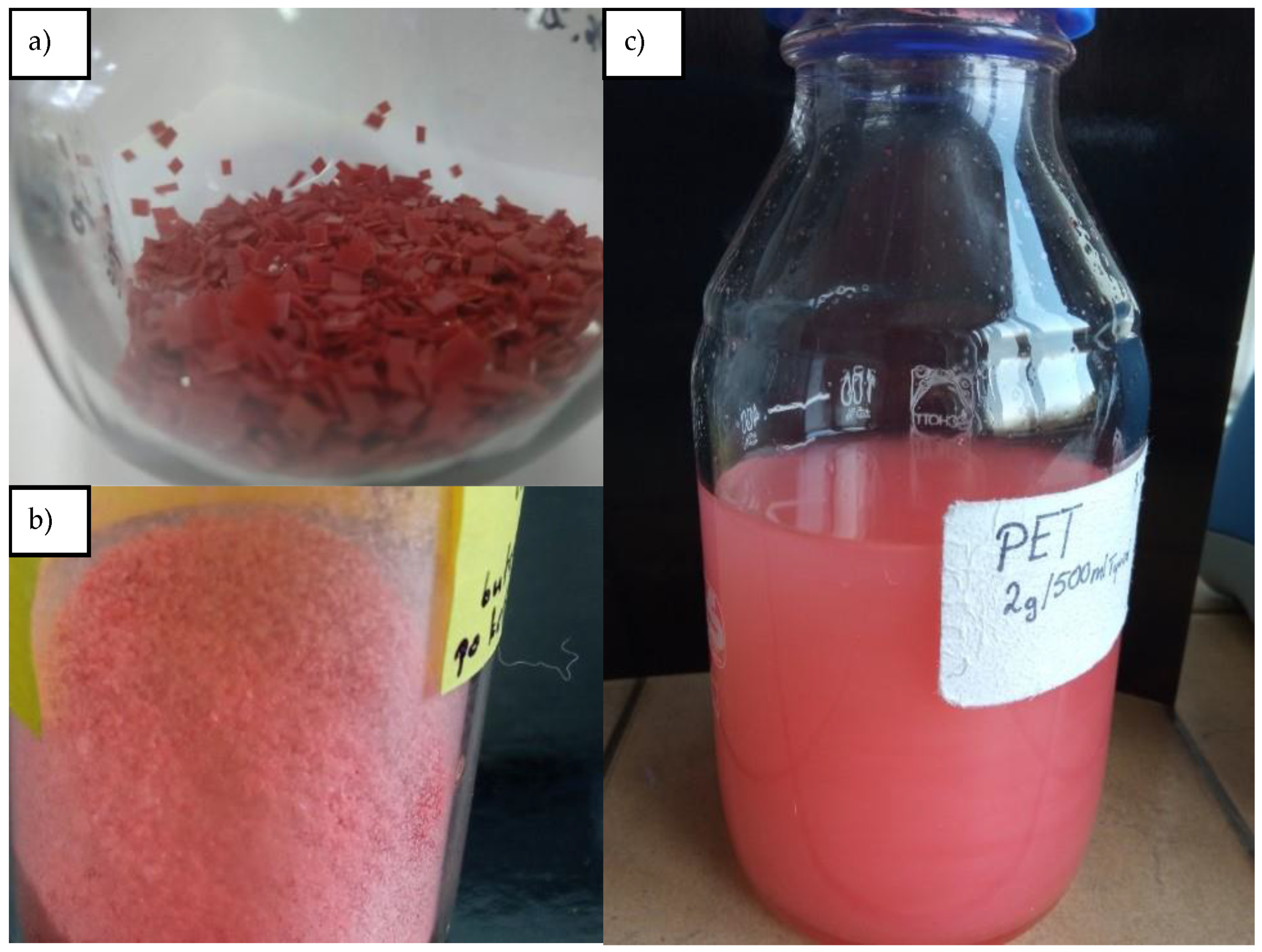
2.1. Microparticles

Four types of colored plastics were used in this study: black polystyrene from CD packaging (PS), red polyethylene terephthalate from ketchup bottles (PET), white polyvinyl chloride from tap water pipes (PVC), and gray phenolic resin from laboratory worktops (PhR). These plastics were cleaned, cut into small pieces and then ground in liquid nitrogen using a cryomill, type 6770, SPEX (Metuchen, NJ, USA). In this grinding process, larger particles were broken down into smaller fragments without altering their physical and chemical properties [33]. After the grinding process, the particles were sieved through a 100-µm steel mesh. Suspensions of stock microplastics were prepared by mixing 2 g of the microparticles smaller than 100 µm with 500 mL of the Tyrode’s medium in glass bottles. The suspensions were sonicated in an ultrasonic bath, type Sonic 6, Polsonic (Warsaw, Poland) for 20 min at 25 °C and stored in dark at 25 °C (Figure 1). The concentration of the microparticles (MPs) in the stock suspensions was measured using a Bürker counting chamber, following a previously described procedure [30]. Particle size distribution of the tested suspensions followed that of a previous study [30]. In our previous study, these microparticles were found to be not toxic to S. ambiguum even at concentrations of up to 107 particles mL−1 [30]. Working microplastic suspensions were prepared just before each analysis step. For this purpose, the stock suspension was shaken and dispersed in the ultrasonic bath (10 min at 25 °C), and an appropriate amount of the suspension was immediately transferred to the Tyrode’s medium. Baker’s yeast (Saccharomyces cerevisiae, BY) was used as a source of edible natural food. The stock suspension of BY was prepared by mixing 10 mg of freeze-dried baker’s yeast with 10 mL of the Tyrode’s medium. In this study, all synthetic microplastics and cells of BY were referred to as MPs.

2.2. Antidepressants

The following antidepressants were tested: standards of DLX hydrochloride (CAS No. 136434-34-9), SER hydrochloride (CAS No. 79559-97-0), FLX hydrochloride (CAS No. 56296-78-7), and PAR hydrochloride (CAS No. 110429-35-1). All four antidepressants were obtained from Toronto Research Chemicals (Toronto, Canada). They were of high purity grade and stored at −20 °C in glass bottles. The standard stock solutions of the compounds (1 mg mL−1) were prepared in a mixture of methanol/water (50:50 ratio, v:v) and stored in dark glass bottles at −20 °C. The working solutions were prepared just before each analysis step by diluting the stock solutions with the Tyrode’s medium.
Deionized water was prepared using the Milli-Q® Direct water purification system from Merck (Darmstadt, Germany). HPLC-grade acetonitrile, methanol, and trifluoroacetic acid (TFA) were purchased from Merck (Darmstadt, Germany).

2.3. Liquid Chromatography Analyses (HPLC-PDA)

The concentration of the pharmaceuticals was measured using a Shimadzu HPLC instrument equipped with LC-10ATvp pumps and an SPD-M10Avp diode-array detector (PDA). Separation was carried out on a Purosphere STAR RP-18, LiChroCART 50 × 4 mm column from Merck (Darmstadt, Germany). The injection volume was 20 µL, and the flow rate of the mobile phase was maintained at 1 mL min−1. The gradient program was as follows: 0.05% TFA in water/0.05% TFA in acetonitrile 75/25–10/90 in 6 min. The wavelength range of the PDA was set to be 200–350 nm. Quantitative analysis was carried out at 217, 225, 225, and 294 nm for DLX, SER, FLX, and PAR, respectively. HPLC-PDA analyses were carried out to evaluate the sorption of the pharmaceuticals on MPs and their degradation during toxicity experiments. Subsamples (0.5 mL) from the microplates were transferred directly to Eppendorf tubes, diluted two-fold with deionized water, centrifuged (10,000× g, 5 min), and analyzed within 2 h.

2.4. Microscopic Imaging

An ultrahigh-accuracy digital Keyence VHX 7000 microscope from Keyence International (Mechelen, Belgium) was used for both analysis of MPs and observation of structures inside the protozoa. Its dedicated image analysis system enables the observation in both 2D and 3D and the measurement of a wide range of microparticles. The number of MPs and their size and shape were analyzed using the software from Keyence International VHX-7000_970F communication and analyzer, version 1.3.11.2.

2.5. Preparation of S. ambiguum

The ciliated protozoan S. ambiguum has been cultured in our laboratory for more than 35 years, as described previously [34]. Prior to the tests, to remove residues from the S. ambiguum cultures, the protozoa were rinsed thrice with the Tyrode’s medium and incubated in a fresh medium for at least 1 h.

2.6. Standard Toxicity Measurements

The influence of the MPs on the acute toxicity of the tested pharmaceuticals was determined using the Spirotox assay with S. ambiguum, according to the standard operational protocol [34] with some modifications. In brief, two replicates of eleven 1.5-fold dilution series of the sample were prepared directly in 24-well polystyrene microplates. Two control samples without the tested pharmaceuticals were placed in each microplate. Then, 10 organisms of S. ambiguum were transferred to each well containing 1 mL of the sample. The suspension of the MPs (106 particles mL−1) in Tyrode’s medium was used as the diluent and the control. The suspensions were prepared from the stock suspensions (2.1) immediately prior to analysis. According to the preliminary tests, the MPs used in the present study were not acutely toxic to S. ambiguum up to a concentration of 107 particles mL−1. The Tyrode’s medium was used to prepare the reference assay for each toxicity series. The pH of Tyrode’s medium was 7.4 ± 0.2. The following concentrations were tested: 2–2000 µg L−1 (SER and FLX) and 5–5000 µg L−1 (DLX and PAR). Four replicates were performed for each treatment. The endpoints, morphological deformations, and lethality were observed using a dissection microscope after 24 h, 48 h, and 7 d of incubation of plates at 25 °C in dark. The protozoa were not fed during the analysis to prevent the introduction of additional factors that could interfere with the results. As reported in a previous study, S. ambiguum can survive in a nonnutritive inorganic medium for 8 d [30]. Based on all observed endpoints, EC50 and EC20 values were calculated using the graphical interpolation method. Based on the initial concentrations of the tested pharmaceuticals, the toxicity values were expressed in mg L−1.

2.7. Ingestion Studies

To evaluate the influence of antidepressants on the formation of food vacuoles by S. ambiguum, the organisms were incubated in the MP suspensions both in the presence and in the absence of sublethal levels of the tested pharmaceuticals. Three drug concentrations were applied: high level, medium level, and low level, corresponding to the EC20 values of 1.00, 0.33, and 0.10, respectively, obtained using the Spirotox experiment. The experiment was carried out in triplicate in a 6-well polystyrene plate with 10 mL of the sample and 100 organisms per well. The controls contained the same suspension of MPs without the pharmaceuticals. After 24 h of incubation, 10–15 protozoa were collected from each well and immobilized with 0.4 mM Ni(NO3)2 solution. The number of vacuoles inside each protozoan was counted using the Keyence VHX 7000. The tests were carried out in duplicate.

2.8. Calculations and Statistics

Preliminary statistical analysis was conducted using the Microsoft Office Excel data analysis tool pack. Dixon’s Q and/or Grubbs test was used for the identification and rejection of outliers. Student’s t test was used for the comparison of results, with a significance level of 0.05.

3. Results

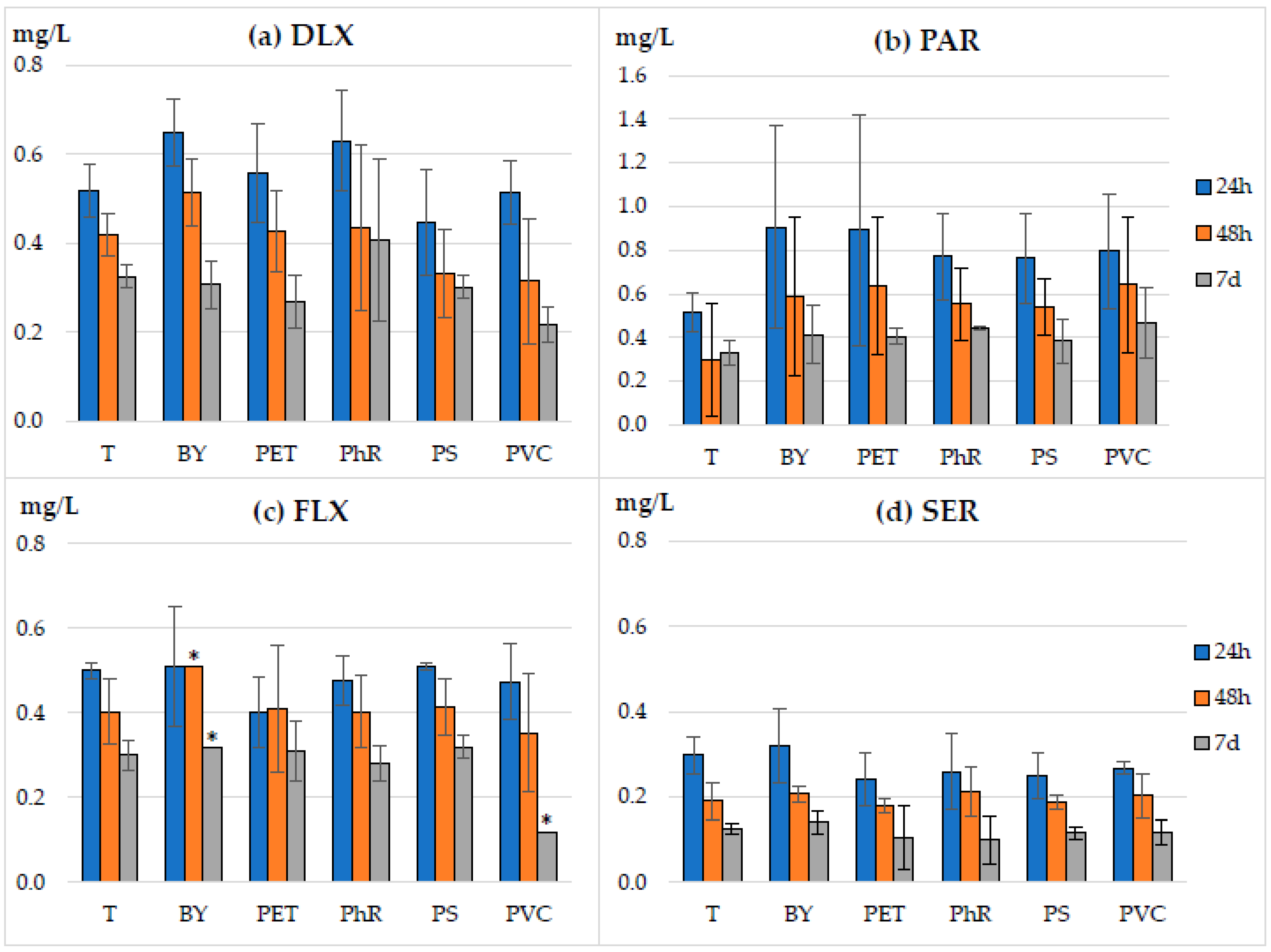
3.1. Sorption of Antidepressants on Microparticles

To evaluate the sorption of the antidepressants on the microplastics (PET, PhR, PS, and PVC) and BY, the MP suspensions were added to the drug solutions prepared in the Tyrode’s medium up to the concentration of 106 particles mL−1. The initial concentration of the drugs was 5 mg L−1. The suspensions were incubated for 7 d in dark. In the majority of the cases, the concentration of the tested antidepressants in water analyzed using HPLC after 1 and 7 d of incubation did not change by more than 20% (Figure 2).
The only exceptions were the PhR suspension samples with PAR and SER, and the FLX suspension sample with BY, in which the drug concentrations decreased by 37, 20, and 31%, respectively. Moreover, in PhR suspensions, the decrease in SER and PAR concentration was observed after 24 h, and these levels remained constant for the next 6 d. In the control sample (Tyrode’s medium without MP suspensions), the concentration of the tested pharmaceuticals decreased only by 20%, indicating that the tested pharmaceuticals were not degraded under the test conditions.

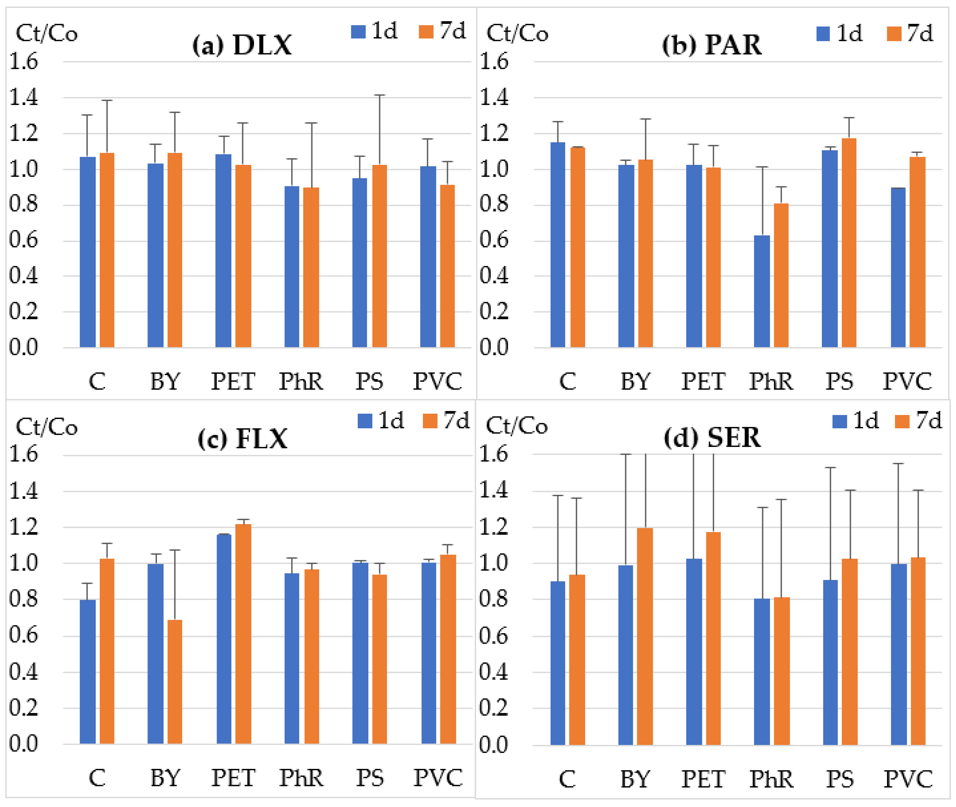
3.2. Influence of Microparticles on the Toxicity of Antidepressants Evaluated in the Spirotox Assay

This is the first study to report the influence of various microplastics on the toxicity of antidepressants toward protozoa. Figure 3 and Figure 4 summarize the results of the first part of the experiment—toxicity assessment using standard effects, i.e., deformations of the cells and lethality. No toxic effect above the 20% threshold was observed in the control samples, i.e., MP suspensions without the tested pharmaceuticals. The median EC50 values were used to reflect the toxic effect of the tested compounds (Figure 3). SER was the most toxic antidepressant, with a 24 h-EC50 of 0.3–0.5 mg L−1. The other antidepressants were two-fold less toxic than SER. The threshold toxicity values indicating the threat to the protozoan population (EC20) were up to two-fold lower than EC50 values (Figure 4). This indicates that the low effects of the concentration quickly became severe, affecting the entire population of the protozoa.
After 24 h of incubation, the toxicity of the drug–microparticle mixtures was slightly lower than that of the drugs alone, especially for DLX and PAR. However, the EC50 values of DLX–PVC and FLX–PVC samples showed a slight increase in toxicity after 7 d. In both cases, these differences did not exceed 30% and were statistically insignificant. A similar trend was observed for EC20 values (Figure 4).

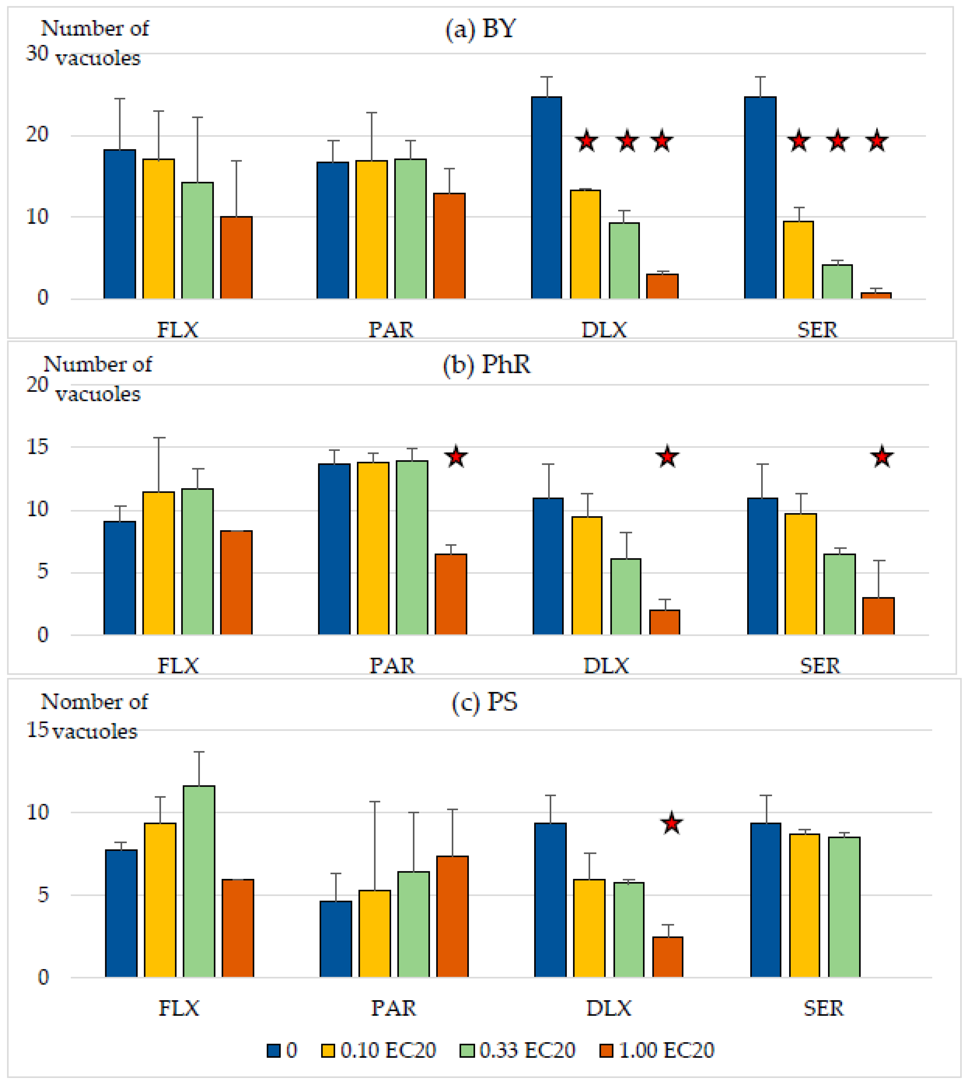
3.3. Influence of Low Levels of Antidepressants on the Formation of Vacuoles by S. ambiguum

The 24 h EC20 values recorded in the Spirotox test with the Tyrode’s medium were used in the second part of the experiment (ingestion study) (Table 1). Three antidepressant levels were tested, with concentrations equal to EC20 values of 0.10, 0.33, and 1.00. The protozoa were incubated for 24 h in the MP–antidepressant mixtures that were taken up by the cells. Three suspensions were used: BY as natural food, PhR as wettable inedible plastic microparticles, and PS as hydrophobic inedible microplastics. The number of food vacuoles was counted using the Keyence VHX 700 microscope, and the results are presented in Figure 5.
The number of food vacuoles formed by S. ambiguum depended on the type of the particle and the type and concentration of the pharmaceutical. As expected, yeast cells were the most frequently ingested particles by the protozoa. In the control samples without the drugs, S. ambiguum formed an average of 16.7–21.3 food vacuoles containing numerous yeast cells (Figure 6a).
The wettable (PhR) and hydrophobic (PS) particles were ingested to a much lower extent, with the number of food vacuoles ranging from 10.0 to 13.7 and from 4.6 to 9.3, respectively. DLX and SER inhibited the formation of vacuoles to the highest extent. For SER and DLX, the number of vacuoles was more than twofold lower at concentrations equal to 0.1 EC20: 0.025 and 0.063 mg L−1, respectively (Figure 5). Both of these drugs also decreased PhR uptake, but at concentrations equal to 0.3 EC20 and higher. However, in the case of PS, the addition of 0.25 mg L−1 of SER resulted in a toxic reaction of protozoa, cell deformation, and the complete absence of food vacuoles inside the cells. On the other hand, FLX and PAR did not affect the formation of food vacuoles. The only exception was the highest PAR concentration in the PhR suspension, where the number of vacuoles formed was two-fold lower than in the control (Figure 5b).
Each of the vacuoles contained several particles, which is best observed in the case of BY (Figure 6). PhR particles were poorly visible due to the poor contrast between them and other cell components (Figure 6d–f).

4. Discussion

The increasing consumption of drugs, especially antidepressants, increases their impact on aquatic organisms. Xenobiotics occur as complex mixtures in aquatic environments, including both water-soluble emerging pollutants and suspensions of plastic microparticles. The preliminary step in understanding the effects of these mixtures is to evaluate the effects of their high concentrations on organisms via short-term tests used in biotests. Ciliated protozoan S. ambiguum has been used in ecotoxicology for many years. Endpoints of both classic [29,34] and recently developed tests [30] enable the understanding of the influence of samples on important physiological processes. In the present study, higher than environmental concentrations of both antidepressants and microplastics were investigated. The OECD recommendations were followed, and the highest tested concentration resulted in the test effect, whereas the lowest did not.
During the 7-d incubation of DLX, PAR, FLX, and SER in the Tyrode’s medium in dark, their concentration decreased by a maximum of 20%. These findings confirm the previous observation that antidepressants are stable in water solutions in dark [8,35]. Many studies have reported the bioaccumulation of antidepressants in protozoa, invertebrates, and fish [11,12,36]. Due to their high lipophilicity, SER, PAR, and FLX were classified as potentially bioaccumulative compounds [9,12], and it is highly likely that they adsorb to organic matter, e.g., sediments and suspended microparticles. However, the sorption of antidepressants to abiotic material has rarely been tested in the literature. Kucharski et al. [37] detected venlafaxine, amitriptyline, and FLX in 80, 30, and 40% of sediment samples in the Odra River estuary, with average concentrations of 10, 9, and 8 ng g−1, respectively. In the previous decade, many papers on the sorption interactions of drugs with microplastics have been published [27,38]. The sorption of drugs on microplastics may be attributable to various interactions, depending on both the structure of the drug and the physicochemical properties of the sorbent [27,38]. Furthermore, the sorption properties of plastics can be modified by additives added to them, e.g., plasticizers. Therefore, to evaluate the real impact, microplastics obtained from everyday products were used in this work.
HPLC results indicated that the tested antidepressants were not adsorbed on PS, PET, and PVC microparticles. A little higher sorption of the drugs was observed in BY and PhR suspensions. This observation is in line with those of Frydkjær et al. [24], who reported that the sorption of lipophilic phenanthrene to microplastics was much lower than that to yeast, bacteria, and plankton. Taking into account the concentration of microplastics in freshwaters being several orders of magnitude lower than the concentrations of microorganisms, the sorption of organic compounds on microplastics and their subsequent transport over long distances are unlikely. This also applies to regions receiving wastewater effluents, where pharmaceutical concentrations are significantly higher and therefore the ecological impact is higher. However, as the behavior and the mechanism of the sorption of organic pollutants to microplastics depends on numerous factors [39], each mixture should be analyzed in a case-by-case basis.
To study the toxic effects of plastics on protozoa, plastics from commercial products were used, which may contain different types of additives such as plasticizers, fillers, flame retardants, and pigments [16,40]. The use of these materials better reflects the real situation because, in aquatic environments, secondary microplastics are prevalent, which are formed as a result of the degradation of plastics from households [16,41]. Levels of MPs detected in the environment are much lower than the concentrations used in the present study, which was 106 particles mL−1. Koelmans et al. [42] reported that the levels of MPs in effluents and freshwaters ranged from 10−2 to 108 particles m−3. However, it should be taken into account that the determination of the content of small MPs (below 50 µm) in environmental samples is challenging, and much larger particles are reported in most of the studies. Concentrations of small particles have been estimated only in effluents and freshwaters.
In the majority of the cases, the toxicity of MP–antidepressant mixtures did not increase more than two-fold when the incubation time was extended from 1 to 7 days (Figure 3 and Figure 4). The exceptions were all samples of SER, which showed a 2–2.7-fold increase in toxicity, and PVC and PET mixtures with DLX. The highest increase was observed for SER, which was probably due to the bioaccumulation of SER in the protozoa [11].
The presence of plastic microparticles did not affect the toxicity of the tested pharmaceuticals toward S. ambiguum. This is the first study to report the influence of various microplastics on the toxicity of antidepressants. The acute toxicity of antidepressants in the presence of MPs has not been previously reported for protozoa. However, mixtures of SER and different MPs have been shown to affect other organisms [25]. Wei et al. [25] found a synergistic immunotoxic effect of SER and nano-PS toward a bivalve Tegillarca granosa. However, no effects were observed in the case of larger micro-PS particles. Schmieg et al. [43] carried out a study of the influence of irregularly shaped polystyrene microplastics (<50 µm) on another antidepressant —amitriptyline — on the early life stages of brown trout. They conducted two experiments using eggs at different stages of development and summarized that amitriptyline exposure exerted a significant effect on fish development, causing increased acetylcholinesterase activity and inhibition of two carboxylesterases. However, in their study, microplastics alone neither influenced the development of fish nor changed the biochemical parameters and effects of amitriptyline. Zhang et al. [44] studied the effect of polystyrene microplastics on the distribution and bioaccumulation of roxithromycin in fish (Oreochromis niloticus). In addition, they applied a suite of biomarkers at the molecular level in red tilapia tissues to evaluate the interactive effects between the tested compounds. They observed acute toxicity in the tested organisms but suggested that the presence of PS could dramatically increase roxithromycin accumulation levels in various tissues of fish. Furthermore, co-exposure to the tested pharmaceutical and PS showed a complex biochemical response in red tilapia.
The results of this study did not support the hypothesis of possible interactions between antidepressants and plastic microparticles. MPs may, through drug absorption, either increase or decrease xenobiotic toxicity. The increase in toxicity could be due to the introduction of higher amounts of toxic substances into the cells during phagocytosis, whereas the decrease in toxicity could be due to the decrease in the bioavailability of the substance. Even unrealistically high levels of microplastics used in this study did not adsorb these drugs and did not change their toxicity. However, the opposite reaction was observed. The presence of low levels of antidepressants affected the food intake by protozoa. The structure of the oral apparatus and the process of formation of vacuoles in ciliates are well known [45,46]. However, only a few studies have used naturally occurring substances or xenobiotics that could modify this process. Some natural substances in food products can increase or decrease the attractiveness toward them, such as yeast protein hydrolyzate or amino acids. Buduma et al. [47] reported that Tetrahymena thermophila had a receptor linked to the phagocytosis process, which was activated by the antihistamine diphenhydramine. In the present study, for the first time, low concentrations of SER and DLX were shown to inhibit the uptake of a natural food source, i.e., yeast cells; higher concentrations were shown to affect the uptake of hydrophilic microparticles; and even the highest tested concentrations did not affect the uptake of hydrophobic particles. Protozoa cannot distinguish between edible particles and inedible ones [26,30]. However, Dürichen et al. [26], based on observations of the ingestion of surface-modified particles, suggested that T. thermophila had receptor systems to recognize food particles. Little is known about the molecules and receptors that determine phagocytosis in protozoa. Serotonin is an important neurotransmitter associated with various physiological mechanisms in vertebrates and invertebrates [48]. The SSRI FLX, SER, and fluvoxamine affect different aquatic organisms at low µg L−1 [12,48], and their mechanism of action on lower organisms is not elucidated. Serotonin is involved in numerous physiological processes of Tetrahymena, including movement, chemotaxis, cell growth, secretion, excretion, hormone production, and phagocytosis [31,32]. Tetrahymena serotonin receptors are extremely sensitive and selective [49]. Thus, SSRI and SNRI may have an indirect effect on phagocytosis in ciliated protozoa. This reaction of the tested antidepressants on the food intake by protozoa may have a significant impact on their populations, especially in wastewater, where total drug concentrations often exceed 1 µg L−1. As the growth of the population is limited in wastewater, wastewater treatment processes may slow down, and also, the balance between bacterial populations and ciliates, which play an important role, may be disturbed in the activated sludge ecosystem in sewage treatment plants.

5. Conclusions and Recommendations

The present study is a preliminary laboratory model study investigating the interaction and toxicity of antidepressants and MPs detected in aquatic environments. The protozoan S. ambiguum was used as a suitable bioindicator due to its size, ease of making in vivo observations of sublethal effects, including the formation of food vacuoles, and high sensitivity to antidepressants.
In this study, no effect of MPs on the toxicity of antidepressants was observed, and it was not confirmed whether these chemicals adsorb on the surface of plastic particles. Thus, there is a low probability that microplastics present in the environment at much lower concentrations have a detrimental effect on protozoa, and hence, no interactions between MPs and pharmaceuticals are anticipated. Furthermore, in this study, the additives contained in microplastics showed no toxic effects on toxicity or food uptake. In the future, such research should also be carried out on aged microplastics, which may have different sorption properties than pristine ones.
However, sublethal levels of SER and DLX were found to reduce the uptake of natural food—yeast cells—by the protozoa. This effect can be of great importance for the ciliate population and can also lead to ecosystem changes, disturbing the balance between bacteria and protozoa. Future studies should focus on determining whether the effect of antidepressants is additive, which significantly depends on the understanding of the mechanisms of toxicity. A mixture of active drug substances is released into the environment by means of municipal wastewater. Although each of the substances is present in extremely low concentrations, their combined effect can be noticeable in the population.
Another interesting issue is understanding other aspects of the effect of serotonin and drugs affecting its action on protozoa. Molecules such as serotonin affect numerous signaling pathways in multicellular organisms and, in protozoa, may enable interpopulation cooperation. However, behavioral research is needed to verify these hypotheses.

Author Contributions

Conceptualization, J.C., A.D. and G.N.-J.; methodology, J.C., A.D., M.W. and G.N.-J.; software, J.C. and A.D.; validation, J.C., A.D. and G.N.-J.; formal analysis, J.C., A.D. and G.N.-J.; investigation, J.C., A.D., W.L., J.M. and G.N.-J.; resources, J.C., A.D., W.L., J.M. and G.N.-J.; data curation, J.C., A.D. and G.N.-J.; writing—original draft preparation, J.C., A.D. and G.N.-J.; writing—review and editing, J.C., A.D., M.W. and G.N.-J.; visualization, J.C., A.D. and G.N.-J.; supervision, A.D. and G.N.-J.; project administration, G.N.-J.; funding acquisition, G.N.-J. All authors have read and agreed to the published version of the manuscript.

Funding

This research was funded by the National Science Center in Poland (grant number: UMO-2019/35/B/NZ8/01388).

Institutional Review Board Statement

Not applicable.

Informed Consent Statement

Not applicable.

Data Availability Statement

Not applicable.

Acknowledgments

Thank for Łukasz Pajchel and Magdalena Kubuj for technical support.

Conflicts of Interest

The authors declare no conflict of interest. The funders had no role in the design of the study; in the collection, analyses, or interpretation of the data; in the writing of the manuscript; or in the decision to publish the results.

References

  1. Available online: www.clincalc.com (accessed on 29 August 2022).
  2. Lajeunesse, A.; Gagnon, C.; Sauve, S. Determination of basic antidepressants and their N-desmethyl metabolites in raw sewage and wastewater using solid-phase extraction and liquid chromatography-tandem mass spectrometry. Anal. Chem. 2008, 80, 5325–5333. [Google Scholar] [CrossRef] [PubMed]
  3. Schultz, M.M.; Furlong, E.T.; Kolpin, D.W.; Werner, S.L.; Schoenfuss, H.L.; Barber, L.B.; Blazer, V.S.; Norris, D.O.; Vajda, A.M. Antidepressant pharmaceuticals in two U.S. effluent-impacted streams: Occurrence and fate in water and sediment, and selective uptake in fish neural tissue. Environ. Sci. Technol. 2010, 44, 1918–1925. [Google Scholar] [CrossRef] [PubMed]
  4. Scircle, A.; Cizdziel, J.V.; Tisinger, L.; Anumol, T.; Robey, D. Occurrence of Microplastic Pollution at Oyster Reefs and Other Coastal Sites in the Mississippi Sound, USA: Impacts of Freshwater Inflows from Flooding. Toxics 2020, 8, 35. [Google Scholar] [CrossRef] [PubMed]
  5. Khan, F.R.; Shashoua, Y.; Crawford, A.; Drury, A.; Sheppard, K.; Stewart, K.; Sculthorp, T. The Plastic Nile’: First Evidence of Microplastic Contamination in Fish from the Nile River (Cairo, Egypt). Toxics 2020, 8, 22. [Google Scholar] [CrossRef] [Green Version]
  6. Giebułtowicz, J.; Nałęcz-Jawecki, G. Occurrence of antidepressant residues in the sewage-impacted Vistula and Utrata rivers and in tap water in Warsaw (Poland). Ecotoxicol. Environ. Saf. 2014, 104, 103–109. [Google Scholar] [CrossRef]
  7. Grabicova, K.; Grabic, R.; Fedorova, G.; Fick, J.; Cerveny, D.; Kolarova, J.; Turek, J.; Zlabek, V.; Randak, T. Bioaccumulation of psychoactive pharmaceuticals in fish in an effluent dominated streams. Water Res. 2017, 124, 654–662. [Google Scholar] [CrossRef]
  8. Bojanowska-Czajka, A.; Pyszyńska, M.; Majkowska-Pilip, A.; Wawrowicz, K. Degradation of selected antidepressants sertraline and citalopram in ultrapure water and surface water using gamma radiation. Processes 2022, 10, 63. [Google Scholar] [CrossRef]
  9. Howard, P.H.; Muir, D.C.G. Identifying new persistent and bioaccumulative organics among chemicals in commerce II: Pharmaceuticals. Environ. Sci. Technol. 2011, 45, 6938–6946. [Google Scholar] [CrossRef]
  10. Puckowski, A.; Mioduszewska, K.; Łukaszewicz, P.; Borecka, M.; Caban, M.; Maszkowska, J.; Stepnowski, P. Bioaccumulation and analytics of pharmaceutical residues in the environment: A review. J. Pharm. Biomed. Anal. 2016, 127, 232–255. [Google Scholar] [CrossRef]
  11. Nałęcz-Jawecki, G.; Wawryniuk, M.; Giebułtowicz, J.; Olkowski, A.; Drobniewska, A. Influence of selected antidepressants on the ciliated protozoan Spirostomum ambiguum: Toxicity, bioaccumulation, and biotransformation products. Molecules 2020, 25, 1476. [Google Scholar] [CrossRef]
  12. Silva, L.J.G.; Pereira, A.; Meisel, L.M.; Lino, C.M.; Pena, A. Reviewing the serotonin reuptake inhibitors (SSRIs) footprint in the aquatic biota: Uptake, bioaccumulation and ecotoxicology. Environ. Pollut. 2015, 197, 127–143. [Google Scholar] [CrossRef] [PubMed]
  13. Akdogan, Z.; Guven, B. Microplastics in the environment: A critical review of current understanding and identification of future research needs. Environ. Pollut. 2019, 254, 113011. [Google Scholar] [CrossRef] [PubMed]
  14. De Hann, W.P.; Sanchez-Vidal, A.; Canalas, M. Floating the microplastics and aggregate formation in the Western Mediterranean Sea Marine Pollut. Bull. 2019, 140, 523–535. [Google Scholar] [CrossRef]
  15. Hidalgo-Ruz, V.; Gutow, L.; Thompson, R.C.; Thiel, M. Microplastics in the Marine Environment: A Review of the Methods Used for Identification and Quantification. Environ. Sci. Technol. 2012, 46, 3060–3075. [Google Scholar] [CrossRef] [PubMed]
  16. Abeynayaka, A.; Werellagama, I.; Ngoc-Bao, P.; Hengesbaugh, M.; Gajanayake, P.; Nallaperuma, B.; Karkour, S.; Bui, X.T.; Itsubo, N. Microplastics in Wastewater Treatment Plants. In Current Developments in Biotechnology and Bioengineering; Elsevier: Amsterdam, The Netherlands, 2022; pp. 311–337. [Google Scholar] [CrossRef]
  17. Rehse, S.; Kloas, W.; Zarfl, C. Short-term exposure with high concentrations of pristine microplastic particles leads to immobilisation of Daphnia magna. Chemosphere 2016, 153, 91–99. [Google Scholar] [CrossRef] [PubMed]
  18. Kaposi, K.L.; Mos, B.; Kelaher, B.P.; Dworjanyn, S.A. Ingestion of microplastic has limited impact on a marine larva. Environ. Sci. Technol. 2014, 48, 1636–1645. [Google Scholar] [CrossRef] [Green Version]
  19. Ma, Y.; Huang, A.; Cao, S.; Sun, F.; Wang, L.; Guo, H. Effects of nanoplastics and microplastics on toxicity, bioaccumulation, and environmental fate of phenanthrene in fresh water. Environ. Pollut. 2016, 219, 166–173. [Google Scholar] [CrossRef]
  20. Teuten, E.L.; Rowland, S.J.; Galloway, T.S.; Thompson, R.C. Potential for plastics to transport hydrophobic contaminants. Environ. Sci. Technol. 2007, 41, 7759–7764. [Google Scholar] [CrossRef]
  21. Hartmann, N.B.; Rist, S.; Bodin, J.; Jensen, L.H.S.; Schmidt, S.N.; Mayer, P.; Meibom, A.; Baun, A. Microplastics as vectors for environmental contaminants: Exploring sorption, desorption, and transfer to biota. Integr. Environ. Assess. Manag. 2017, 13, 488–493. [Google Scholar] [CrossRef]
  22. Ma, J.; Zhao, J.; Zhu, Z.; Li, L.; Yu, F. Effect of microplastic size on the adsorption behavior and mechanism of triclosan on polyvinyl chloride. Environ. Pollut. 2019, 254, 113104. [Google Scholar] [CrossRef]
  23. Liu, G.; Zhu, Z.; Yang, Y.; Sun, Y.; Yu, F.; Ma, J. Sorption behavior and mechanism of hydrophilic organic chemicals to virgin and aged microplastics in freshwater and seawater. Environ. Pollut. 2019, 246, 26–33. [Google Scholar] [CrossRef] [PubMed]
  24. Frydkjær, C.K.; Iversen, N.; Roslev, P. Ingestion and Egestion of Microplastics by the Cladoceran Daphnia magna: Effects of Regular and Irregular Shaped Plastic and Sorbed Phenanthrene. Bull. Environ. Contam. Toxicol. 2017, 99, 655–661. [Google Scholar] [CrossRef]
  25. Wei, S.; Yu, H.; Shuge, S.; Yu, T.; Weishang, Z.; Xueying, D.; Guangxu, L. Immunotoxicities of microplastics and sertraline, alone and in combination, to a bivalve species: Size-dependent interaction and potential toxication mechanism. J. Hazard. Mater. 2020, 396, 122603. [Google Scholar] [CrossRef]
  26. Dürichen, H.; Siegmund, L.; Burmester, A.; Fischer, M.S.; Wöstemeyer, J. Ingestion and digestion studies in Tetrahymena pyriformis based on chemically modified microparticles. Eur. J. Protistol. 2016, 52, 45–57. [Google Scholar] [CrossRef] [PubMed]
  27. Razanajatovo, R.M.; Ding, J.; Zhang, S.; Jiang, H.; Zou, H. Sorption and desorption of selected pharmaceuticals by polyethylene microplastics. Mar. Pollut. Bull. 2018, 136, 516–523. [Google Scholar] [CrossRef] [PubMed]
  28. Christaki, U.; Dolan, J.R.; Pelegri, S.; Rassoulzadegan, F. Consumption of picoplankton-size particles by marine ciliates: Effects of physiological state of the ciliate and particle quality. Limnol. Oceanogr. 2003, 43, 458–464. [Google Scholar] [CrossRef]
  29. Nałęcz-Jawecki, G. Spirotox—Spirostomum ambiguum acute toxicity test—10 years of experience. Environ. Toxicol. 2004, 19, 359–364. [Google Scholar] [CrossRef]
  30. Nałęcz-Jawecki, G.; Chojnacka, J.; Wawryniuk, M.; Drobniewska, A. Influence of nano- and small microplastics on ciliated protozoan Spirostomum ambiguum (Müller, 1786) Ehrenberg, 1835. Water 2021, 13, 2857. [Google Scholar] [CrossRef]
  31. Csaba, G. Presence in and effects of pineal indoleamines at very low level of phylogeny. Experientia 1993, 49, 627–634. [Google Scholar] [CrossRef]
  32. Csaba, G.; Kovacs, P.; Pallinger, E. How does the unicellular Tetrahymena utilize the hormones that it produces? Paying a visit to the realm of atto- and zeptomolar concentrations. Cell Tissue Res. 2007, 327, 199–203. [Google Scholar] [CrossRef]
  33. Ogonowski, M.; Schür, C.; Jarsen, A.; Gorokhova, E. The effects of natural and anthropogenetic microparticles on individual fitness in Daphnia magna. PLoS ONE 2016, 11, e01550632016. [Google Scholar] [CrossRef] [PubMed] [Green Version]
  34. Nałęcz-Jawecki, G.; Sawicki, J. Toxicity of inorganic compounds in the Spirotox test—A miniaturized version of the Spirostomum ambiguum test. Arch. Environ. Contam. Toxicol. 1998, 34, 1–5. [Google Scholar] [CrossRef]
  35. Trawiński, J.; Skibiński, R. Studies on photodegradation process of psychotropic drugs: A review. Environ. Sci. Pollut. Res. 2017, 24, 1152–1199. [Google Scholar] [CrossRef] [PubMed] [Green Version]
  36. Meredith-Williams, M.; Carter, L.J.; Fussell, R.; Raffaelli, D.; Ashauer, R.; Boxall, A.B. Uptake and depuration of pharmaceuticals in aquatic invertebrates. Environ. Pollut. 2012, 165, 250–258. [Google Scholar] [CrossRef] [PubMed]
  37. Kucharski, D.; Nałęcz-Jawecki, G.; Drzewicz, P.; Skowronek, A.; Mianowicz, K.; Strzelecka, A.; Giebułtowicz, J. The assessment of environmental risk related to the occurrence of pharmaceuticals in bottom sediments of the Odra River estuary (SW Baltic Sea). Sci. Total Environ. 2022, 828, 154446. [Google Scholar] [CrossRef]
  38. Puckowski, A.; Ćwięk, W.; Mioduszewska, K.; Stepnowski, P.; Białk-Bielińska, A. Sorption of pharmaceuticals on the surface of microplastics. Chemosphere 2021, 263, 127976. [Google Scholar] [CrossRef] [PubMed]
  39. Wang, F.; Zhang, M.; Sha, W.; Wang, Y.; Hao, H.; Dou, Y.; Li, Y. Sorption behavior and mechanisms of organic contaminants to nano and microplastics. Molecules 2020, 25, 1827. [Google Scholar] [CrossRef]
  40. Atugoda, T.; Vithanage, M.; Wijesekara, H.; Bolan, N.; Sarmah, A.K.; Bank, M.S.; You, S.; Ok, Y.S. Interactions between microplastics, pharmaceuticals and personal care products: Implications for vector transport. Environ. Int. 2021, 149, 106367. [Google Scholar] [CrossRef]
  41. Rhodes, C.J. Plastic pollution and potential solutions. Sci. Prog. 2018, 101, 207–226. [Google Scholar] [CrossRef]
  42. Koelmans, A.A.; Nor, N.H.M.; Hermsen, E.; Kooi, M.; Mintenig, S.M.; De France, J. Microplastics in Freshwaters and Drinking Water: Critical Review and Assessment of Data Quality. Water Res. 2019, 155, 410–422. [Google Scholar] [CrossRef] [PubMed]
  43. Schmieg, H.; Burmester, J.K.Y.; Krais, S.; Ruhl, A.S.; Tisler, S.; Zwiener, C.; Köhler, H.R.; Triebskorn, R. Interacting effects of polystyrene microplastics and the antidepressant amitriptyline on early life stages of brown trout (Salmo trutta f. fario). Water 2020, 12, 2361. [Google Scholar] [CrossRef]
  44. Zhang, S.; Ding, J.; Razanajatovo, R.M.; Jiang, H.; Zou, H.; Zhu, W. Interactive effects of polystyrene microplastics and roxithromycin on bioaccumulation and biochemical status in the freshwater fish red tilapia (Oreochromis niloticus). Sci. Total Environ. 2019, 648, 1431–1439. [Google Scholar] [CrossRef] [PubMed]
  45. Wloga, G.; Frankel, J. From molecules to morphology: Cellular organization of Tetrahymena thermophila. Methods Cell Biol. 2012, 109, 83–140. [Google Scholar] [CrossRef] [PubMed]
  46. Ishida, M.; Allen, R.D.; Fok, A.K. Phagosome formation in Paramecium: Roles of somatic and oral cilia and of solid particles as revealed by video microscopy. J. Eucaryot. Microbiol. 2001, 48, 640–646. [Google Scholar] [CrossRef] [PubMed]
  47. Buduma, N.; Balabanian, J.; Dalvi, P.; Chia, S.; Dhaliwal, A.; Eliya, D.; Boothby, J.; Bros-Seemann, S.; Kibler, R.; Khuri, S.; et al. Modulation of phagocytosis in Tetrahymena thermophila by histamine and the antihistamine diphenhydramine. Acta Protozool. 2013, 52, 317–323. [Google Scholar] [CrossRef]
  48. Santos, L.H.M.L.M.; Rodríguez-Mozaz, S.; Barceló, D. Microplastics as vectors of pharmaceuticals in aquatic organisms—An overview of their environmental implications. CSCEE 2021, 3, 100079. [Google Scholar] [CrossRef]
  49. Csaba, G. The hormonal system of the unicellular Tetrahymena: A review with evolutionary aspects. A Microbiol. Immunol. Hung. 2012, 59, 131–156. [Google Scholar] [CrossRef]
Figure 1. Prepared samples of microplastics. (a) cut plastic ketchup bottle made of PET; (b) milled and sieved microparticles; (c) stock suspension of the microplastics.
Water 15 00552 g001
Figure 2. The relative concentration of drugs (a) duloxetine, (b) paroxetine, (c) fluoxetine, and (d) sertraline after 1 and 7 d of incubation in microparticle suspensions in relation to the initial level (C0).
Water 15 00552 g002
Figure 3. Acute toxicity data in the Spirotox test (EC50) of the tested pharmaceuticals with and without microparticles, (a) duloxetine; (b) paroxetine; (c) fluoxetine; and (d) sertraline. T—Tyrode’s medium, BY—baker’s yeast, PhR—phenolic resin, PVC—polyvinyl chloride, PET—ethyl polyterephthalate, PS—polystyrene.
Water 15 00552 g003
Figure 4. Acute toxicity data in the Spirotox test (EC20) of the tested pharmaceuticals with and without microparticles, (a) duloxetine; (b) paroxetine; (c) fluoxetine; and (d) sertraline. T—Tyrode’s medium, BY—baker’s yeast, PhR—phenolic resin, PVC—polyvinyl chloride, PET—ethyl polyterephthalate, PS—polystyrene, *—the toxic effect in the control and the lowest concentration higher than 20%.
Water 15 00552 g004
Figure 5. The number of food vacuoles formed by S. ambiguum incubated in the tested antidepressant solutions after administration: (a) baker’s yeast; (b) phenolic resin microparticles; and (c) polystyrene microparticles. Asterisk indicates the statistical difference in comparison with the control (sample without pharmaceutical)—p < 0.05.
Water 15 00552 g005
Figure 6. Food vacuoles formed by Spirostomum ambiguum. (ac) baker’s, yeast; (d,e), and (f) phenolic resin microparticles; (gi) polystyrene microparticles. Water 15 00552 i001 
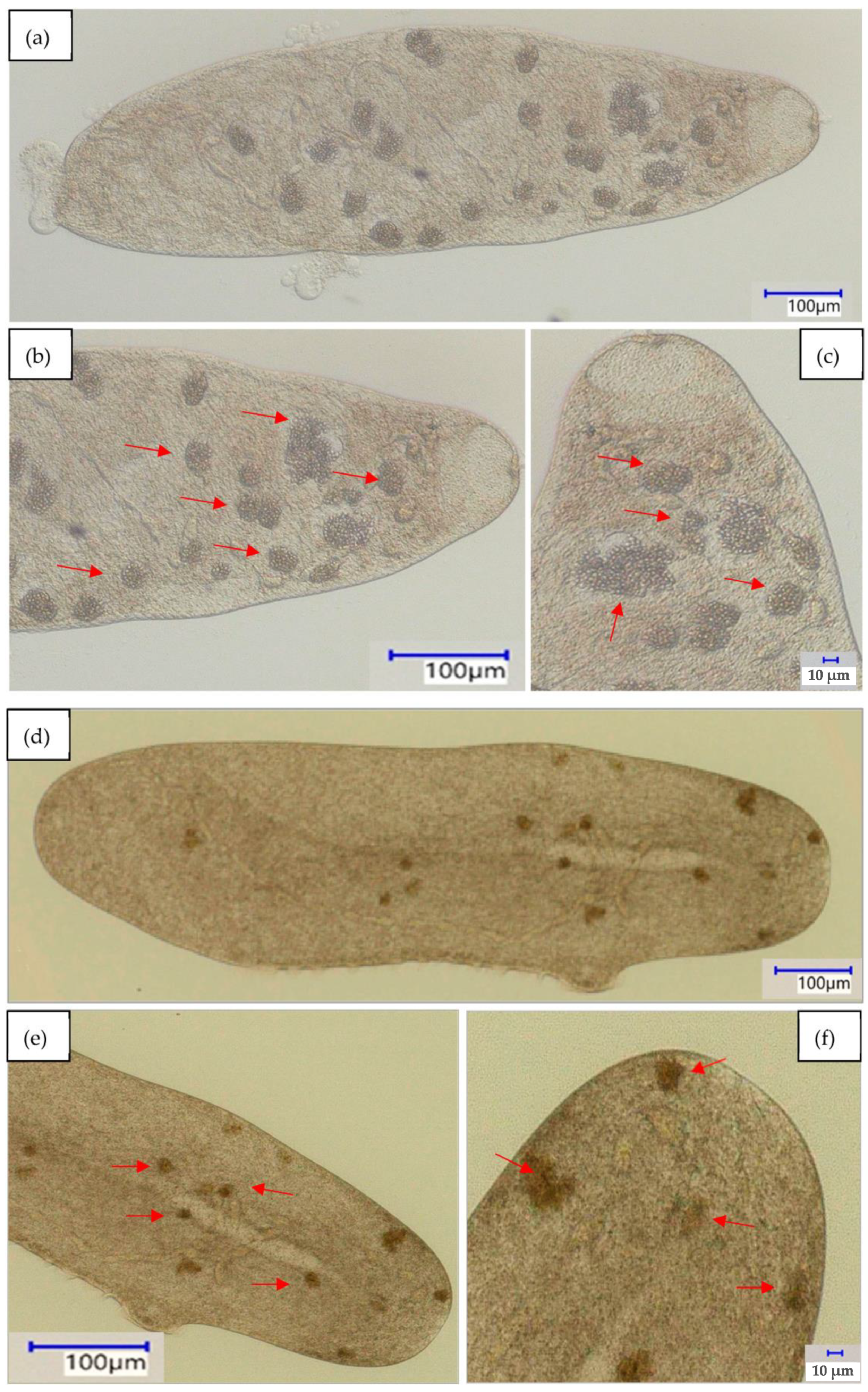
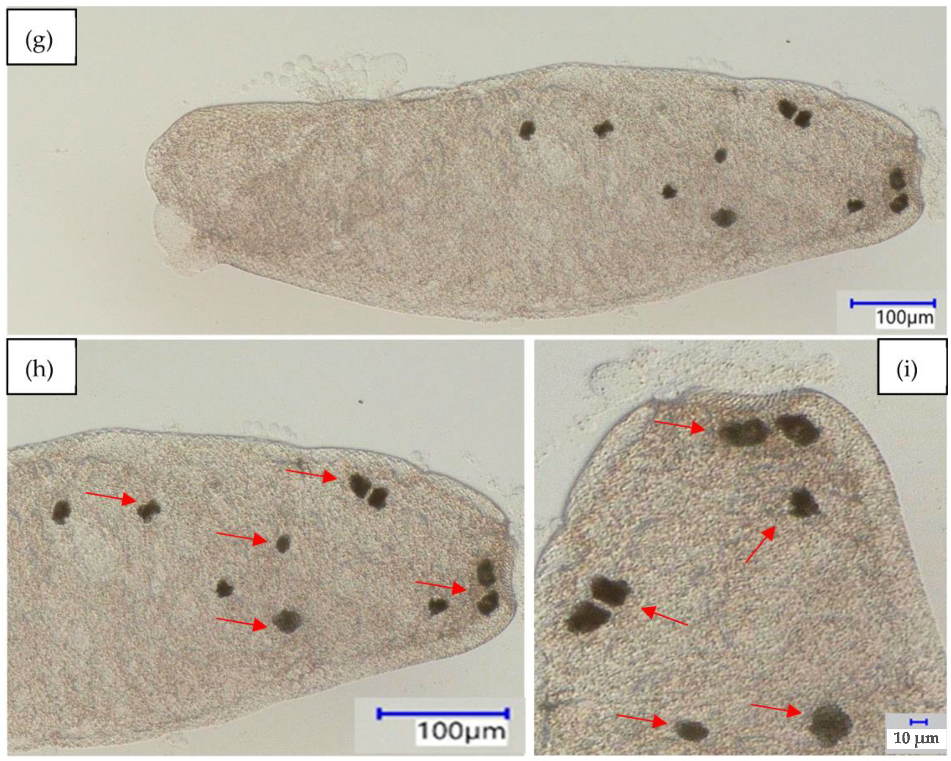
enlargement and mark food vacuoles (b,c) baker’s yeast; (e,f) phenolic resin microparticles; and (h,i) polystyrene microparticles.
Water 15 00552 g006aWater 15 00552 g006b
Table 1. The concentrations of the tested pharmaceuticals used in the ingestion assay.
Pharmaceutical1.00 EC20
[mg L−1]
0.33-EC20
[mg L−1]
0.10-EC20
[mg L−1]
Sertraline0.250.0760.025
Duloxetine0.630.190.063
Fluoxetine0.650.200.064
Paroxetine0.880.260.088
Disclaimer/Publisher’s Note: The statements, opinions and data contained in all publications are solely those of the individual author(s) and contributor(s) and not of MDPI and/or the editor(s). MDPI and/or the editor(s) disclaim responsibility for any injury to people or property resulting from any ideas, methods, instructions or products referred to in the content.

