
Comparison of Two Methods for SARS-CoV-2 Detection in Wastewater: A Case Study from Sofia, Bulgaria

1
Faculty of Biology, Sofia University “St. Kliment Ohridski”, 8 Dragan Tsankov Blvd., 1766 Sofia, Bulgaria
2
Center of Competence “Clean Technologies for Sustainable Environment—Water, Waste, Energy for Circular Economy”, Sofia University “St. Kliment Ohridski”, 15 Tsar Osvoboditel Blvd., 1000 Sofia, Bulgaria
3
Sofia Tech Park, 111, Tsarigradsko Shose Blvd., 1784 Sofia, Bulgaria
4
Sofiyska Voda AD, 4 Business Park Sofia, Building 4, 1766 Sofia, Bulgaria
*
Author to whom correspondence should be addressed.
Water 2023, 15(4), 658; https://doi.org/10.3390/w15040658
Submission received: 23 December 2022
/
Revised: 28 January 2023
/
Accepted: 4 February 2023
/
Published: 8 February 2023
(This article belongs to the Special Issue SARS-CoV-2 in Wastewater: Methods, Epidemiology and Future Goals)
Abstract
:Wastewater surveillance for monitoring the spread of SARS-CoV-2 remains important even in the current endemic stage of the COVID-19 outbreak. This approach has already demonstrated its value by providing early warnings of coronavirus spread in different communities. The aim of the present publication is to share relevant experience from the Center of Competence “Clean&Circle”, obtained in the development of an effective strategy for SARS-CoV-2 detection in the wastewater of Sofia, Bulgaria. Using four different RNA concentration/extraction methods, we revealed that the key hindering factor for successful viral detection was the presence of PCR inhibitors in the wastewater. The most efficient way to overcome their presence turned out to be the application of a specialized polymerase in the RT-PCR detection setup. Our data showed that using such an enzyme increases the detection efficiency from 1.9% to 70.5% in samples with a spiked control virus. We also evaluated the recovery rates of viral particles by using silica columns (71%), PEG precipitation (23%), ultrafiltration (15%), and MCE filtration (10%). These results support the international effort to unify and standardize the various techniques used for SARS-CoV-2 monitoring in wastewater.
1. Introduction
The COVID-19 pandemic caused by the virus SARS-CoV-2 challenged contemporary human society in terms of healthcare, social organization, and economic measures. This provoked an unprecedented development of the testing methodology and led to a remarkable increase in studies on the subject. One of the fields that marked a significant progress was water based epidemiology [1]. Since the first reports demonstrated the stability of the SARS-CoV-2 genetic material in wastewater [2,3,4], many investigations have been conducted to explore the reliability of the information obtained [5,6], the prospects for the application of wastewater-based monitoring of the infection level in communities [7,8], or the best methods for data collection [9,10,11]. Although wastewater-based monitoring of SARS-CoV-2 is already being implemented in practice, there is no unified and standardized method for detecting the virus RNA concentration [11]. The protocol for the determination of the number of virus copies in the samples consists of two main parts: (1) the concentration and extraction of the RNA [12]; and (2) quantification by RT-qPCR or RT-dPCR [13]. For the first part, any different methods have been tested: precipitation with PEG and AlCl3 [14], ultrafiltration [13], ultracentrifugation [15], skimmed milk flocculation [16], and filtration with electronegative and electropositive filters [10,17]. Studies tend to give priority to PEG (polyethylene glycol) precipitation, ultrafiltration, silica-based columns, and MCE (mixed cellulose esters) filtration [12,17,18]. The main difficulties in performing the concentration procedure relate to the cost of the materials, the unavailability of some consumables, and above all, the low reproducibility of the results. Deviations in the data often exceed half of the reported values [17]. PEG precipitation has a recovery efficiency of SARS-CoV-2 and various surrogate viruses ranging from 1% to 76%, according to some studied [17,19]. For ultrafiltration, the percentages indicated by researchers vary from 6% to 64% [20,21]. The MCE filtration has reported recovery rates from 4.8% to 66%, but it is worth mentioning that this method often gives false negative results [22,23]. Approaches based on the use of silica columns are widely used in practice, as Kordatou [24] mentions. However, they are not widely represented in studies that allow determining the specificities of the SARS-CoV-2 genetic material concentration and extraction in wastewater. Pecson et al. made a comparative analysis of 36 standard operating procedures, registering no significant differences in their results [25]. Dimitrakopoulos et al. reported high reproducibility and 38–39% viral RNA recovery efficiency for different concentrations and subsequent extraction methods using the Manual Enviro Wastewater TNA kit (Promega, United States) [13]. A similar method was preferred by Mondal [26], Kabdasli [27], and Billoud [28] for conducting epidemiological studies in different cities.
In addition to the numerous studies on identifying an optimal method for detecting the new coronavirus, there are some suggestions that the quantification of SARS-CoV-2 does not particularly depend on the experimental protocol that has been used [25] or on the PCR methodology that has been applied [13]. However, many authors indicate that the presence of inhibitors should be taken into account in wastewater studies [29,30]. No real experimental data from the detection of SARS-CoV-2 in wastewater supporting this assumption can be found in the literature. Moreover, such waters greatly vary in terms of composition and the presence of putative inhibitors. These substances lead to failed PCR reactions or low reproducibility of results. Because of the differences in the waters, it is difficult to predict the extent to which they affect the results. Thus, experimenters often experience difficulties in establishing the reasons for unsuccessful analyzes and in taking measures to eliminate the negative effects. A simple and effective solution is proposed in the present study—the use of polymerases adapted to inhibitors.
The present study was conducted as a joint activity of the Center of Competence “Clean technologies for a sustainable environment—water, waste, energy for a circular economy” (Clean&Circle) with leading organization SU “St. Kliment Ohridski” and Wastewater Treatment Plant (WWTP) “Kubratovo” (Sofiyska Voda AR, Veolia group). It was motivated by the need for the city of Sofia to select and apply an appropriate protocol for monitoring the COVID-19 epidemic based on the specific characteristics of the city’s wastewater. The aim of this research was to investigate the efficiency of viral RNA recovery, using four of the most common methods for concentrating viral particles from wastewater. Two RT-qPCR detection setups based on polymerases with and without increased tolerance to the presence of PCR inhibitors were used in the experiments.
2. Materials and Methods
For the purposes of the present study, samples were taken from the wastewater of Sofia at the entrance of the city’s treatment plant. Those were transported at 4 °C in clean, new containers (provided by the WWTP Sofia) and processed immediately upon arrival at the laboratory. The experiments were performed according to GLP in the microbiological laboratory. Sterile containers, tubes, pipette tips, extraction and PCR consumables, and other laboratory materials were used. The concentration and TNA extraction procedures were performed in separate, specially dedicated areas in the laboratories. PCR analysis was prepared in a PCR cabinet. Pasteurization of the row samples was performed to avoid any biological risk [31]. The suspended particles were then removed by centrifugation at 4000 g for 30 min at 4 °C using a swinging-bucket rotor. The supernatant was used for the subsequent nucleic acid concentration and extraction steps.
Control samples with spiked inactivated viral particles (105 copies/mL), provided by the National Center for Infectious and Parasitic Diseases, Bulgaria, were used to determine viral RNA recovery efficiency.
The research presented here includes three different methods for concentrating viral particles from wastewater:
- PEG precipitation: 10% of PEG 8000 was used in combination with 2.25% NaCl for the precipitation. To sediment the viral RNA in the pellet, the mixture was centrifuged in a swinging-bucket rotor at 12,000 g for 120 min at 4 °C without braking force. The pellet was resuspended in 200 µL of nuclease-free water.
- MCE filtration: In the concentration approach based on electronegative filtration, MCE filters (Merck Millipore Ltd., Ireland, Carrigtohill) were used. Before the procedure, the pH of the samples was measured and adjusted to neutral if needed. MgCl2 was added to the sample to a final concentration of 25 mM. 50 mL of the sample was filtered through an MCE filter with a pore size of 0.45 µm using a sterile filtration funnel. The filter was then cut into pieces and immediately placed in a 2 mL bead beating tube (Zymo Research, Irvine, CA, USA) with 200 µL of saline solution (0.9% NaCl). After that, the samples were processed according to the procedure described by Ahmed (2020) [17].
- Ultrafiltration: Amicon® Ultra-15 devices with a molecular weight cutoff of 30 kDa (Merck Millipore Ltd., Ireland, Carrigtohill) were used to concentrate the samples by ultrafiltration. A volume of 50 mL was filtered through the devices by centrifugation at 4750 g for 10 min at 4 °C. Finally, a concentrate with a volume of about 200 µL was obtained.
The extraction of total nucleic acids (TNA) from the concentrated samples was performed using two approaches: an extraction kit and direct extraction with silica colomns.
For the concentrates obtained as a result of the PEG precipitation, ultrafiltration, and MCE filtration, the QIAamp DSP Virus Spin Kit (Qiagen, Germany) was used, following the protocol provided by the manufacturer. The final elution step was performed in a volume of 30 µL.
The experiments on concentration and direct extraction of nucleic acids from wastewater using silica-based columns were performed according to the protocol of Whithney, 2020 [32]. The Wizard® Enviro Total Nucleic Acid Kit (Promega, Germany, Walldorf) was used for the concentration and extraction of viral RNA. It utilizes an especially adapted polymerase with enhanced resistance towards PCR inhibitors found in wastewater. The starting sample volume for the procedure was 40 mL.
The RT-qPCR technique was applied for the detection and quantification of SARS-CoV-2 viral particles. The equipment used was a Rotor-Gene Q 2plex Thermocycler (Qiagen, Germany, Hilden). The consumables consisted of primers and probes that are part of commercially available kits. Polymerases with and without enhanced inhibitor resistance were used for the detection.
- RT-qPCR with polymerase without inhibitor resistance—the gb Sarbeco N (primary test) kit (Generi Biotech, Czech Republic, Hradec Králové) was used according to the protocol provided by the manufacturer. The reaction volume was 20 µL, and 5 µL of the extracted TNAs were used as a matrix. To remove the effects of potential inhibitors, some samples were diluted 2- or 5-fold, and others were treated with the OneStep PCR Inhibitor Removal Kit (Zymo Research, Irvine, CA, USA). Viral levels were quantified using a standard curve prepared from falling tenfold dilutions of IDT 2019-nCoV N Positive Control Standards (IDT, USA, IA, Coralville) included in each PCR run.
- RT-qPCR with inhibitor-resistant polymerase—the Wastewater SARS-CoV-2 RT-qPCR Systems kit (Promega, Germany) targeting the N1 gene of SARS-CoV-2 was used. Viral levels were quantified by a standard curve prepared by using a series of tenfold dilutions of the standards included in the kit.
The recovery efficiency of virus copies is calculated by the following formula:
where Viral RNA gene copies recovered are the SARS-CoV-2 gene copies detected by RT-qPCR and Viral RNA gene copies seeded are the number of the gene copies seeded at the beginning of the analyses.
Recovery % = Viral RNA gene copies recovered/Viral RNA gene copies seeded × 100,
In determining the recovery efficiency, the number of viral particles in the wastewater samples was subtracted.
SWOT analysis is a technique in the field of strategic management that is a key tool in the management of various processes and projects. It includes the identification of four groups of factors: (1) strengths—the strengths and advantages of the given process are considered through the objectives of the research; (2) weaknesses—weaknesses that could prevent the desired final result are identified; (3) opportunities—information is provided on the potential for further process development and improvement; and (4) threats—factors that are difficult to influence and that would negatively affect the processes.
All analyses were performed in three independent replicates. The obtained results were processed using Microsoft Excel and the integrated software of the Rotor-Gene Q 2plex Thermocycler (Qiagen, Germany).
3. Results
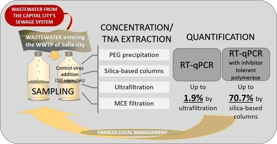
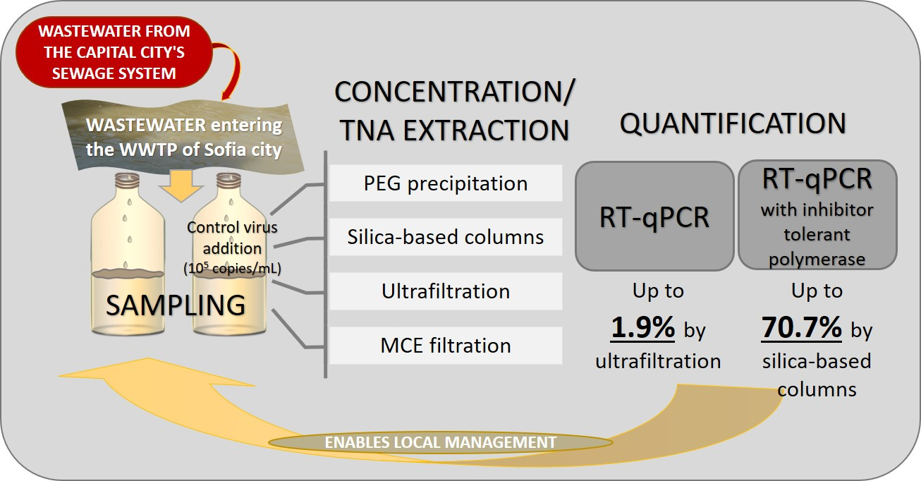
The study described in this communication explored the optimal approach for quantitative detection of SARS-CoV-2 RNA levels in the wastewater of the city of Sofia, Bulgaria. The experimental design of the study is presented in Figure 1.
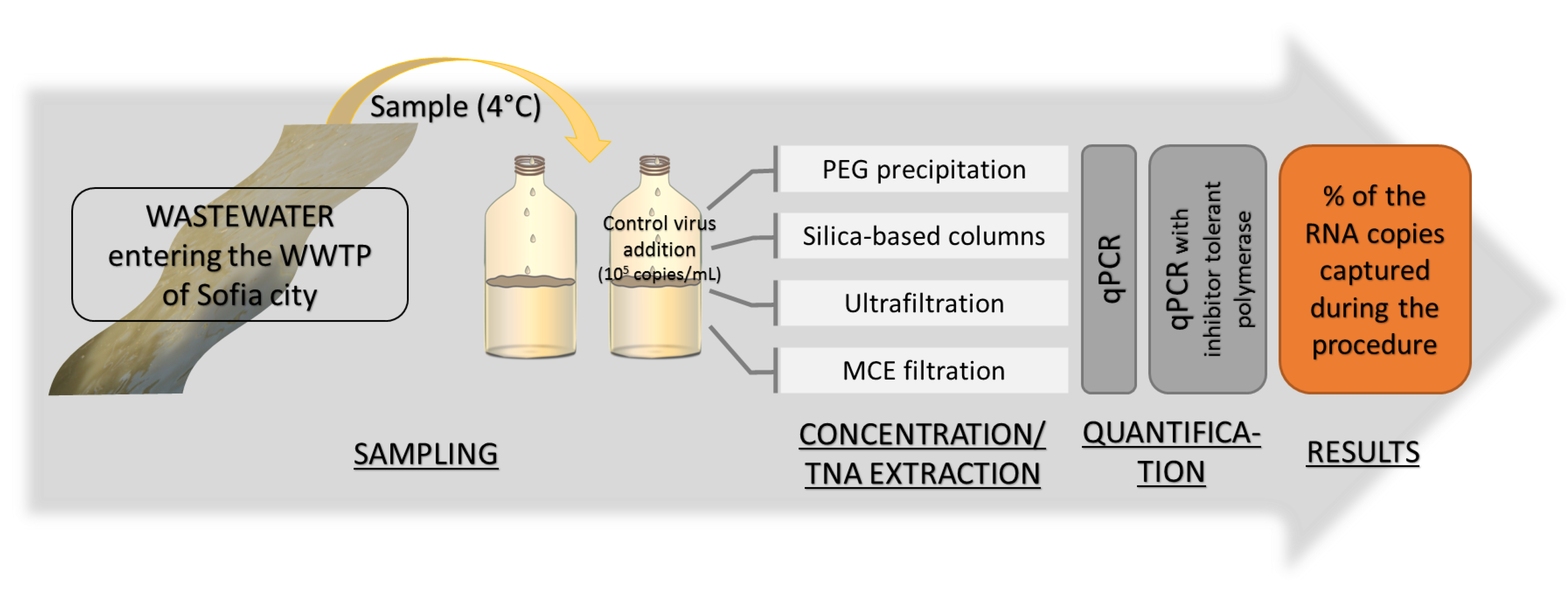
Real wastewater from the city of Sofia, Bulgaria, was used for the analyses. The city is the capital of the country and is inhabited by approximately 1.4 million people. A mixed flow of domestic wastewater, rainwater, and industrial wastewater enters the sewage system of the town, which has a flow rate of 400,000 m3/day. The treatment plant processing these waters uses a technology with primary and secondary treatment. In the primary stage, 10 fine and 10 coarse grids, 3 sand filters, and 4 primary clarifiers separate solid waste and suspended particles (Figure 2). In the biological treatment, 6 full mixed aeration tanks and 10 secondary clarifiers are used for the biodegradation of the pollutants. The water treatment plant is also equipped with modules for chlorination and complete restoration of the purified water in order to disinfect it before discharging it into the Iskar River.
The 14-day average value for COD in Sofia wastewater at the entrance of the WWTP was 395 mgO2/L and for BOD5—185 mgO2/L. It is known that the biodegradability index (BOD5/COD) for municipal wastewater varies from about 0.4 to about 0.8. At values below 0.3, pollutants are considered practically non-biodegradable [34]. For the wastewaters of the city of Sofia, an index of 0.47–0.48 is typical. The relatively low biodegradability index is due to the fact that Sofia WWTP processes a mixed flow of domestic and industrial wastewater. On average, the recalcitrant pollutants of industrial origin contribute to the formation of biodegradability index of 0.47–0.48. Single-day declines of up to 0.29 were recorded during the COVID-19 wave, which began in early 2022. This is an indication of the entry into the treatment plant of pollutants that are difficult to biodegrade. The described situation is typical of waters with xenobiotics—substances that have the potential to act as PCR inhibitors. These are probably related to industrial wastewater flowing into the city sewers, but also to increased consumption and application by the population of disinfectants and xenobiotics during periods of high COVID-19 incidence.
Four methods were used to concentrate the nucleic acids: PEG precipitation, MCE filtration, ultrafiltration, and concentration by silica columns (Figure 1). The recovery efficiency of each approach was determined by adding a known concentration of control virus and calculating the percent recovery. Quantification of SARS-CoV-2 levels was initially performed using an RT-qPCR kit, which is widely used in Bulgaria for analyzing clinical samples. The data are presented in Table 1. The results indicate low recovery rates of control viruses from the samples, with none of the methods exceeding 2% efficiency. The most likely reason for such values was the presence of inhibitors in the wastewater, which can compromise the efficiency of the RT-qPCR reactions.
To limit the influence of such substances, two experimental approaches were applied: dilution of the extracted TNAs and their treatment with a specific kit for PCR inhibitor removal. Dilution of the eluates did not improve recovery efficiency in our experiments. Treating them with a specialized inhibitor removal kit increased the recovery efficiency to 10% when concentrating the samples by ultrafiltration. However, these results had low reproducibility when repeating the experiments with wastewater collected at different dates from the same sampling point. The registered effect is probably due to a quantitative and/or qualitative variation of the inhibiting substances in the composition of the wastewater of the city of Sofia.
As an alternative strategy to remove the influence of inhibitors found in the wastewater, all experiments were also repeated using an RT-qPCR kit containing a polymerase that is resistant to inhibitors. The data is shown in Table 1. In this case, much higher recovery efficiencies were observed, ranging from 9.48% (MCE filtration) to 70.66% (silica-based columns). The increase in efficiency compared to detection with a normal, unadapted polymerase was from 8-fold (ultrafiltration) to 158-fold (MCE filtration). With PEG precipitation, it was also significant—118 times, and with silica-based columns, it was 70 times. These results indicated that the most efficient and reproducible strategy to overcome the effect of diverse and varying amounts of RT-qPCR inhibitors in wastewater is the application of specialized kits with optimized polymerases. In such a case, all four methods, subject to our analysis, allowed the detection of SARS-CoV-2, the most effective being the application of silica-based columns.
In Table 2, we compared our results to those obtained in other studies exploring the recovery efficiency of the four concentration methods used.
The comparison of the different concentration methods showed viral recovery ranging from 0.06% with MCE filtration to 70.66% with the use of silica columns. Such a significant difference is also described in numerous studies by other authors [17,20,23,36,37]. The varying composition of the wastewaters is considered one of the main causes [9,40]. The presence of potential inhibitors in samples is further suggested as an important factor for altering the results of PCR detection of SARS-CoV-2 in studies [41,42]. The present study attaches key importance to this factor. The presence of similar substances in the joint flow of domestic wastewater, industrial wastewater, and water generated is inevitable during the period of increased consumption of medicinal preparations and disinfectants. For this reason, our team registered low values of the biodegradability index, going down to 0.29. Such an index indicates that the concentration of non-biodegradable and toxic substances in the water is so high for biological systems and processes that biological treatment is not an effective option. On the other hand, it is known that these substances can have an inhibitory effect on PCR reactions.
In the complex matrix of urban wastewater (containing xenobiotics, desinfectants, antibiotics, etc.), identifying potential PCR inhibitors and determining their amounts can be quite a tedious task. Nonetheless, such compounds are not only present in wastewaters, but their concentrations increase during the COVID-19 pandemic. Moreover, some combinations of inhibitors have an additive inhibitory effect on the amplification step. All these, taken together, represent the major obstacle to the wider adoption of RT-PCR-based viral quantification as an early warning mechanism.
The main approach offered in these cases is the use of different types of controls in the analyses. This has provided indirect information about whether there is a change in the results due to polymerase inhibitors. The experiments described in the present study differ in one aspect: they give an assessment of the effect on the analysis, caused by the choice of PCR system and the influence of inhibitors.
By using PEG precipitation and a standard coronavirus detection kit, a recovery efficiency of 0.16% was obtained. The most likely reason for the low detection rate of the coronavirus RNA is the presence of inhibitors. It is known that during the concentration of the nucleic acids in polyethylene glycol, the extraction of the PCR inhibitors also takes place [43]. Thus, the data reported for this method can vary in a very wide range—from less than 1% [25,43,44] to over 60% efficiency [45,46]. Table 2 illustrates the differences in data reported by many research teams—Wang et al. [19] and Ye et al. [36] described a low recovery rate of SARS-CoV-2 RNA (below 6%), while Blanco et al. [35], Ahmed et al. [17], and Pellegrinelli et al. [37] reported values above 44%. When using a polymerase that is adapted to the presence of inhibitors, an increase in efficiency of up to 23% was identified. Similar recovery efficiency was also observed by other authors—Flood et al. [47], Zheng et al. [15].
Another standard method for concentrating virus particles is MCE filtration. Its main advantages are related to the large volumes of wastewater that can be treated, the low cost, and the use of materials that are typical for other analyses in water laboratories [44]. However, the results of these assays can be strongly affected by the presence of inhibitors. This leads to a high percentage of false negative samples [42] and a low viral RNA recovery efficiency [23]. This has been identified in our study as the most likely reason for the very low efficiency of this type of analysis (0.06%). When using an RT-qPCR kit with an inhibitor-adapted polymerase, the RNA recovery rate increased to 9.48%. However, this method showed the lowest sensitivity to the presence of SARS-CoV-2 genetic material in the wastewater of the city of Sofia (Table 1).
Ultrafiltration is also among the most widespread methods for concentrating SARS-CoV-2 RNA. When spiking coronavirus in clean water, this method can reach a recovery efficiency of over 90% [39]. At the same time, when using a complex matrix such as wastewater, ultrafiltration also produces results varying in a very wide range—from 0.2–73% (Table 1). Qiu et al. [39] demonstrated that without removing the inhibitors from the samples, the recovery efficiency of the model human coronavirus was 0.20–2.4%. A similar result of 1.88% was recorded in the present study, when using polymerases that are not resistant to inhibitors. After removing part of the inhibitory effect of the substances in the wastewater, Qiu et al. [39] recorded an efficiency of up to 17%. We have also obtained an efficiency close to this—15%—with polymerase resistant to inhibitors (Table 1).
Numerous authors have described ultrafiltration as a fast, simple, and accurate method for the detection of SARS-CoV-2 [21,44], and it is widely used in research on the water-based epidemiology of the COVID-19 pandemic. Its main disadvantages, as described in Table 3, are related to the high price of the filtration devices and their difficult supply, as well as to the risk of clogging the membrane, which significantly lowers productivity. Unlike this method, the concentration and extraction of SARS-CoV-2 RNA based on silica columns achieve high productivity, efficiency, and an acceptable cost (Table 3) [45]. Our data shows a 5-fold higher recovery efficiency using silica columns compared to the ultrafiltration-based method (Table 1). Whitney et al. [46] reported similar results—6 times more viral RNA was isolated using silica columns, and the authors indicated that this method allows one technician to process up to 20 samples in 2 h. Because of these advantages, direct capture of SARS-CoV-2 RNA by silica columns was used by Mondal et al. for regular monitoring of three treatment plants in Wisconsin, USA [26].
4. Discussion
The quantitative determination of SARS-CoV-2 levels in wastewater has become one of the main methods of tracking and predicting the course of the COVID-19 pandemic worldwide due to its relatively low cost and easily accessible samples without the need for ethical approval. Despite its attractiveness, this approach has some serious limitations, such as the lack of a single optimal method for concentrating, extracting, and detecting the viral particles. There are numerous studies, each applying a different experimental approach and reporting different efficiencies of SARS-CoV-2 recovery from wastewater.
To facilitate the selection of a suitable concentration technique, a SWOT analysis was applied to the above methods (Table 3).
The SWOT analysis illustrates that many factors need to be taken into account when choosing a method for the detection of SARS-CoV-2 in wastewater. These factors include the effectiveness of the method, the reproducibility of the results, the analysis time, the cost of the equipment and materials, the complexity of the protocol, the possibilities of providing materials and chemicals, etc.
PEG precipitation is a classic method utilized in virological studies. As such, it has a well-established methodology, and the materials used are low-cost and widely available. The resulting concentrate is in sufficient quantity for additional analyses.
Ultrafiltration is a method for concentrating substances in a specific molecular weight range. The method is widely used in chemical and biological research. The protocol is very simple and time-efficient. The inhibitors in the resulting concentrate can be further washed away. It is also possible to use membranes with different pore sizes to obtain an optimal result for a given wastewater.
MCE filtration is also carried out by a simple procedure. This method uses low-cost materials that are available in any laboratory that processes water samples. The concentration of nucleic acids can be performed on a significantly larger sample volume, i.e., to detect the presence of the target genetic material at a lower concentration. The above advantages and possibilities make this method interesting for specialists in the field. In contrast, filtration through silica columns is primarily used in molecular biology and less often in the practice-oriented field of wastewater. Nevertheless, this method has serious advantages—it is fast and gives a high quality of the concentrated nucleic acids, which is related to the high reproducibility and efficiency of virus recovery that we have registered (Table 1). Furthermore, it saves the cost of a separate nucleic acid extraction kit. Besides, it is the only method that allows complete automation of viral RNA detection and quantification. This makes it particularly suitable for routine monitoring.
The experiments we conducted with the wastewater of the city of Sofia indicated the use of silica-based columns as the most suitable approach for these purposes. Regardless of the higher cost per sample, compared to methods such as PEG precipitation or MCE filtration, this method is characterized by the highest efficiency and very good reproducibility. These advantages draw the choice towards it, and the slightly higher cost of the necessary consumables is not so decisive when monitoring the wastewater of a city with a population of approximately 1.4 million people.
The method for concentrating and extracting the viral RNA based on the silica columns has very few disadvantages—a slightly higher price and the need to use more specific materials than routine water analyses. When working with other concentration methods, we encountered various difficulties and challenges. PEG precipitation made the concentration of samples laborious and time-consuming. With ultrafiltration, the main challenge that our team encountered was the availability of the filtration devices due to delayed production and deliveries. MCE filtration demonstrated unsatisfactory efficiency.
It is also notable that the percentages shown in Table 2 vary significantly and indicate a high degree of heterogeneity for each of the methods studied. A logical explanation for this is the highly varying composition of the wastewater. Therefore, before starting a program to monitor virus levels in a given location, it is recommendable to conduct a proof-of-principle experiment with the specific wastewater in order to establish the most optimal approach for concentrating the virus particles.
A major advantage of the approach successfully used by us, involving inhibitor-resistant PCR polymerase, is that it provides stable and sensitive detection of viral genetic material in the complex wastewater matrix. The described studies can serve as a reference point for laboratories that introduce or optimize their methods for monitoring the spread of SARS-CoV-2. The approach has the limitations characteristic of this type of method. It is possible to lose the signal from the nucleic acids of the target viruses in cases of reduced spread and, accordingly, low concentrations in the sample of SARS-CoV-2 RNA. It should be mentioned that our team did not encounter a similar problem with the minimum number of registered cases between the epidemic waves of 2021. Also, with regular monitoring, there may be fluctuations in the data depending on factors related to the wastewater—degree of pollution, mixing of different pollutants, and fluctuations in pollutant concentrations due to periods of heavy rainfall or drought. Among the method’s limitations are the requirement for highly qualified experimenters and experience working with RNA. The materials used are specific, so careful supply planning is required. It is necessary to designate areas in the wastewater laboratory for working with samples containing high levels of biological risk, as well as others—for high cleanliness. The latter is a challenge when the main activities are wastewater-related.
The four described methods for concentration and extraction of SARS-CoV-2 RNA demonstrated significant differences in the efficiency of the procedures. The data obtained by our team show that it can vary up to 7.5 times when using a polymerase that has been adapted to the presence of inhibitors. The differences when performing RT-PCR detection with a kit that is not optimized for working with wastewater reached 31 times, and the efficiency of recovery of viral RNA didn’t exceed 1.9%. These results were due to the presence of inhibitors in the wastewater. This highlights the need to evaluate the role of such compounds in COVID-19 monitoring in similar samples.
The most important finding from our experiments is that the optimal strategy to avoid the inhibitors in wastewater is the use of specialized RT-qPCR systems with polymerases, specifically optimized for this sample type. The alternative approaches, such as dilution of the isolated TNAs or the application of specialized kits for sample purification, showed significantly worse and, most importantly, non-reproducible results.
The results obtained in the present study open new possibilities for a reliable detection of SARS-CoV-2 in the wastewaters of big cities despite the various inhibitors present there (desinfectants, antibiotics, steroid hormones, dyes, microplastics and other xenobiotics). It demonstrates that inhibitors sometimes have a crucial role in the detection of viral genetic material. Our results provide evidence that when selecting a methodology for testing SARS-CoV-2 levels, this factor should be of leading importance. The data presented in the present study indicate that it has the potential to carry more weight than the choice of sample concentration method.
Author Contributions
Conceptualization, Y.T. and M.B.; methodology, M.B. and S.P.; validation, M.B., S.P. and V.S.; investigation, M.B., S.P. and V.S.; resources, Y.T., S.P. and V.S.; data curation, M.B. and S.P.; writing—original draft preparation, M.B., S.P. and Y.T.; writing—review and editing, Y.T., M.B., S.P. and V.S.; supervision, Y.T. and V.S.; funding acquisition, Y.T. All authors have read and agreed to the published version of the manuscript.
Funding
This investigation has been supported financially by the Operational Program ‘Science and education for smart growth’, co-financed by the European Union through the European structural and investment funds, project BG05M2OP001-1.002-0019: ‘Clean Technologies for Sustainable Environment—Waters, Waste, Energy for a Circular Economy’ and by Grant D01-217/30 November 2018 and agreement D01-278/3 December 2021 under the National Research Programme “Innovative Low-Toxic Bioactive Systems for Precision Medicine (BioActiveMed)” approved by DCM # 658/14 September 2018.
Data Availability Statement
Data is contained within the article.
Acknowledgments
The authors of this study express their gratitude to Olivia Fechtnerand Murat Kirtay (Promega GmbH) for their help in supplying the PCR inhibitor-resistant materials.
Conflicts of Interest
The authors declare that they have no conflict of interest. The funders had no role in the design of the study; in the collection, analyses, or interpretation of data; in the writing of the manuscript; or in the decision to publish the results.
References
- Safford, H.R.; Shapiro, K.; Bischel, H.N. Wastewater Analysis Can Be a Powerful Public Health Tool—If It’s Done Sensibly. Proc. Natl. Acad. Sci. USA 2022, 119, e2119600119. [Google Scholar] [CrossRef] [PubMed]
- Medema, G.; Heijnen, L.; Elsinga, G.; Italiaander, R.; Brouwer, A. Presence of SARS-Coronavirus-2 RNA in Sewage and Correlation with Reported COVID-19 Prevalence in the Early Stage of the Epidemic in the Netherlands. Environ. Sci. Technol. Lett. 2020, 7, 511–516. [Google Scholar] [CrossRef]
- Randazzo, W.; Truchado, P.; Cuevas-Ferrando, E.; Simón, P.; Allende, A.; Sánchez, G. SARS-CoV-2 RNA in Wastewater Anticipated COVID-19 Occurrence in a Low Prevalence Area. Water Res. 2020, 181, 115942. [Google Scholar] [CrossRef] [PubMed]
- Sherchan, S.P.; Shahin, S.; Ward, L.M.; Tandukar, S.; Aw, T.G.; Schmitz, B.; Ahmed, W.; Kitajima, M. First Detection of SARS-CoV-2 RNA in Wastewater in North America: A Study in Louisiana, USA. Sci. Total Environ. 2020, 743, 140621. [Google Scholar] [CrossRef]
- Zhu, Y.; Oishi, W.; Saito, M.; Kitajima, M.; Sano, D. Early Warning of COVID-19 in Tokyo via Wastewater-Based Epidemiology: How Feasible It Really Is? J. Water Environ. Technol. 2021, 19, 170–183. [Google Scholar] [CrossRef]
- Sanjuán, R.; Domingo-Calap, P. Reliability of Wastewater Analysis for Monitoring COVID-19 Incidence Revealed by a Long-Term Follow-Up Study. Front. Virol. 2021, 1, 776998. [Google Scholar] [CrossRef]
- Daughton, C.G. Wastewater Surveillance for Population-Wide Covid-19: The Present and Future. Sci. Total Environ. 2020, 736, 139631. [Google Scholar] [CrossRef]
- Sharara Id, N.; Id, N.E.; Duvallet Id, C.; Ghaeli, N.; Matus, M.; Heussner, J.; Olesenid, S.W.; Alm, E.J.; Chaiid, P.R.; Ericksonid, T.B. Wastewater Network Infrastructure in Public Health: Applications and Learnings from the COVID-19 Pandemic. PLOS Glob. Public Health 2021, 1, e0000061. [Google Scholar] [CrossRef]
- Barceló, D. Wastewater-Based Epidemiology to Monitor COVID-19 Outbreak: Present and Future Diagnostic Methods to Be in Your Radar. Case Stud. Chem. Environ. Eng. 2020, 2, 100042. [Google Scholar] [CrossRef]
- Feng, S.; Roguet, A.; McClary-Gutierrez, J.S.; Newton, R.J.; Kloczko, N.; Meiman, J.G.; McLellan, S.L. Evaluation of Sampling, Analysis, and Normalization Methods for SARS-CoV-2 Concentrations in Wastewater to Assess COVID-19 Burdens in Wisconsin Communities. ACS ES&T Water 2021, 1, 1955–1965. [Google Scholar] [CrossRef]
- Pulicharla, R.; Kaur, G.; Brar, S.K. A Year into the COVID-19 Pandemic: Rethinking of Wastewater Monitoring as a Preemptive Approach. J. Environ. Chem. Eng. 2021, 9, 106063. [Google Scholar] [CrossRef]
- Rusiñol, M.; Martínez-Puchol, S.; Forés, E.; Itarte, M.; Girones, R.; Bofill-Mas, S. Concentration Methods for the Quantification of Coronavirus and Other Potentially Pandemic Enveloped Virus from Wastewater. Curr. Opin. Environ. Sci. Health 2020, 17, 21–28. [Google Scholar] [CrossRef]
- Dimitrakopoulos, L.; Kontou, A.; Strati, A.; Galani, A.; Kostakis, M.; Kapes, V.; Lianidou, E.; Thomaidis, N.; Markou, A. Evaluation of Viral Concentration and Extraction Methods for SARS-CoV-2 Recovery from Wastewater Using Droplet Digital and Quantitative RT-PCR. Case Stud. Chem. Environ. Eng. 2022, 6, 100224. [Google Scholar] [CrossRef]
- Pérez-Cataluña, A.; Cuevas-Ferrando, E.; Randazzo, W.; Falcó, I.; Allende, A.; Sánchez, G. Comparing Analytical Methods to Detect SARS-CoV-2 in Wastewater. Sci. Total Environ. 2021, 758, 143870. [Google Scholar] [CrossRef]
- Zheng, X.; Deng, Y.; Xu, X.; Li, S.; Zhang, Y.; Ding, J.; On, H.Y.; Lai, J.C.C.; In Yau, C.; Chin, A.W.H.; et al. Comparison of Virus Concentration Methods and RNA Extraction Methods for SARS-CoV-2 Wastewater Surveillance. Sci. Total Environ. 2022, 824, 153687. [Google Scholar] [CrossRef]
- Philo, S.E.; Ong, A.Q.W.; Keim, E.K.; Swanstrom, R.; Kossik, A.L.; Zhou, N.A.; Beck, N.K.; Meschke, J.S. Development and Validation of the Skimmed Milk Pellet Extraction Protocol for SARS-CoV-2 Wastewater Surveillance. Food Environ. Virol. 2022, 1, 1–9. [Google Scholar] [CrossRef]
- Ahmed, W.; Bertsch, P.M.; Bivins, A.; Bibby, K.; Farkas, K.; Gathercole, A.; Haramoto, E.; Gyawali, P.; Korajkic, A.; McMinn, B.R.; et al. Comparison of Virus Concentration Methods for the RT-QPCR-Based Recovery of Murine Hepatitis Virus, a Surrogate for SARS-CoV-2 from Untreated Wastewater. Sci. Total Environ. 2020, 739, 139960. [Google Scholar] [CrossRef]
- Sangkham, S. A Review on Detection of SARS-CoV-2 RNA in Wastewater in Light of the Current Knowledge of Treatment Process for Removal of Viral Fragments. J. Environ. Manag. 2021, 299, 113563. [Google Scholar] [CrossRef] [PubMed]
- Wang, X.W.; Li, J.S.; Guo, T.K.; Zhen, B.; Kong, Q.X.; Yi, B.; Li, Z.; Song, N.; Jin, M.; Xiao, W.J.; et al. Concentration and Detection of SARS Coronavirus in Sewage from Xiao Tang Shan Hospital and the 309th Hospital. J. Virol. Methods 2005, 128, 156–161. [Google Scholar] [CrossRef]
- Peinado, B.; Martínez-García, L.; Martínez, F.; Nozal, L.; Sánchez, M.B. Improved Methods for the Detection and Quantification of SARS-CoV-2 RNA in Wastewater. Sci. Rep. 2022, 12, 1–11. [Google Scholar] [CrossRef] [PubMed]
- Fonseca, M.S.; Machado, B.A.S.; Rolo, C.d.A.; Hodel, K.V.S.; Almeida, E.d.S.; de Andrade, J.B. Evaluation of SARS-CoV-2 Concentrations in Wastewater and River Water Samples. Case Stud. Chem. Environ. Eng. 2022, 6, 100214. [Google Scholar] [CrossRef]
- Juel, M.A.I.; Stark, N.; Nicolosi, B.; Lontai, J.; Lambirth, K.; Schlueter, J.; Gibas, C.; Munir, M. Performance Evaluation of Virus Concentration Methods for Implementing SARS-CoV-2 Wastewater Based Epidemiology Emphasizing Quick Data Turnaround. Sci. Total Environ. 2021, 801, 149656. [Google Scholar] [CrossRef] [PubMed]
- Gonzalez, R.; Curtis, K.; Bivins, A.; Bibby, K.; Weir, M.H.; Yetka, K.; Thompson, H.; Keeling, D.; Mitchell, J.; Gonzalez, D. COVID-19 Surveillance in Southeastern Virginia Using Wastewater-Based Epidemiology. Water Res. 2020, 186, 116296. [Google Scholar] [CrossRef] [PubMed]
- Michael-Kordatou, I.; Karaolia, P.; Fatta-Kassinos, D. Sewage Analysis as a Tool for the COVID-19 Pandemic Response and Management: The Urgent Need for Optimised Protocols for SARS-CoV-2 Detection and Quantification. J. Environ. Chem. Eng. 2020, 8, 104306. [Google Scholar] [CrossRef]
- Pecson, B.M.; Darby, E.; Haas, C.N.; Amha, Y.M.; Bartolo, M.; Danielson, R.; Dearborn, Y.; di Giovanni, G.; Ferguson, C.; Fevig, S.; et al. Sars-Cov-2 Interlaboratory Consortium. Reproducibility and Sensitivity of 36 Methods to Quantify the SARS-CoV-2 Genetic Signal in Raw Wastewater: Findings from an Interlaboratory Methods Evaluation in the U.S. Environ. Sci. 2021, 7, 504–520. [Google Scholar] [CrossRef]
- Mondal, S.; Feirer, N.; Brockman, M.; Preston, M.A.; Teter, S.J.; Ma, D.; Goueli, S.A.; Moorji, S.; Saul, B.; Cali, J.J. A Direct Capture Method for Purification and Detection of Viral Nucleic Acid Enables Epidemiological Surveillance of SARS-CoV-2. Sci. Total Environ. 2021, 795, 148834. [Google Scholar] [CrossRef]
- Kabdaşlı, I.; Tünay, O. Concentration Techniques Tailored for the Detection of SARS-CoV-2 Genetic Material in Domestic Wastewater and Treatment Plant Sludge: A Review. J. Environ. Chem. Eng. 2021, 9, 106296. [Google Scholar] [CrossRef]
- Giraud-Billoud, M.; Cuervo, P.; Altamirano, J.C.; Pizarro, M.; Aranibar, J.N.; Catapano, A.; Cuello, H.; Masachessi, G.; Vega, I.A. Monitoring of SARS-CoV-2 RNA in Wastewater as an Epidemiological Surveillance Tool in Mendoza, Argentina. Sci. Total Environ. 2021, 796, 148887. [Google Scholar] [CrossRef]
- Ahmed, W.; Simpson, S.L.; Bertsch, P.M.; Bibby, K.; Bivins, A.; Blackall, L.L.; Bofill-Mas, S.; Bosch, A.; Brandão, J.; Choi, P.M.; et al. Minimizing Errors in RT-PCR Detection and Quantification of SARS-CoV-2 RNA for Wastewater Surveillance. Sci. Total Environ. 2022, 805, 149877. [Google Scholar] [CrossRef]
- Kumblathan, T.; Piroddi, N.; Hrudey, S.E.; Li, X.F. Wastewater Based Surveillance of SARS-CoV-2: Challenges and Perspective from a Canadian Inter-Laboratory Study. J. Environ. Sci. 2022, 116, 229. [Google Scholar] [CrossRef]
- Wu, F.; Zhang, J.; Xiao, A.; Gu, X.; Lin Lee, W.; Armas, F.; Kauffman, K.; Hanage, W.; Matus, M.; Ghaeli, N.; et al. SARS-CoV-2 Titers in Wastewater Are Higher than Expected from Clinically Confirmed Cases. mSystems 2020, 5, e00614-20. [Google Scholar] [CrossRef]
- Whitney, O. Direct Wastewater RNA Capture and Purification via the “Sewage, Salt, Silica and SARS-CoV-2 (4S)” Method; protocols.io. Available online: https://doi.org/10.17504/protocols.io.biwekfbe (accessed on 1 April 2022). [CrossRef]
- Google Maps. Available online: https://maps.google.com/ (accessed on 20 December 2022).
- Srinivas, T. Environmental Biotechnology. New Age International Pvt Ltd. Publishers: New Delhi, India, 2008. [Google Scholar]
- Blanco, A.; Abid, I.; Al-Otaibi, N.; Pérez-Rodríguez, F.J.; Fuentes, C.; Guix, S.; Pintó, R.M.; Bosch, A. Glass Wool Concentration Optimization for the Detection of Enveloped and Non-Enveloped Waterborne Viruses. Food Environ. Virol. 2019, 11, 184–192. [Google Scholar] [CrossRef]
- Ye, Y.; Ellenberg, R.M.; Graham, K.E.; Wigginton, K.R. Survivability, Partitioning, and Recovery of Enveloped Viruses in Untreated Municipal Wastewater. Environ. Sci. Technol. 2016, 50, 10. [Google Scholar] [CrossRef]
- Pellegrinelli, L.; Castiglioni, S.; Cocuzza, C.E.; Bertasi, B.; Primache, V.; Schiarea, S.; Salmoiraghi, G.; Franzetti, A.; Musemeci, R.; Tilola, M.; et al. Evaluation of Pre-Analytical and Analytical Methods for Detecting SARS-CoV-2 in Municipal Wastewater Samples in Northern Italy. Water 2022, 14, 833. [Google Scholar] [CrossRef]
- Sapula, S.A.; Whittall, J.J.; Pandopulos, A.J.; Gerber, C.; Venter, H. An Optimized and Robust PEG Precipitation Method for Detection of SARS-CoV-2 in Wastewater. Sci. Total Environ. 2021, 785, 147270. [Google Scholar] [CrossRef]
- Qiu, Y.; Yu, J.; Pabbaraju, K.; Lee, B.E.; Gao, T.; Ashbolt, N.J.; Hrudey, S.E.; Diggle, M.; Tipples, G.; Maal-Bared, R.; et al. Validating and Optimizing the Method for Molecular Detection and Quantification of SARS-CoV-2 in Wastewater. Sci. Total Environ. 2022, 812, 151434. [Google Scholar] [CrossRef]
- Ahmed, W.; Smith, W.J.M.; Metcalfe, S.; Jackson, G.; Choi, P.M.; Morrison, M.; Field, D.; Gyawali, P.; Bivins, A.; Bibby, K.; et al. Comparison of RT-qPCR and RT-dPCR Platforms for the Trace Detection of SARS-CoV-2 RNA in Wastewater. ACS Environ. Sci. Technol. Water 2022. [Google Scholar] [CrossRef]
- Mazumder, P.; Dash, S.; Honda, R.; Sonne, C.; Kumar, M. Sewage Surveillance for SARS-CoV-2: Molecular Detection, Quantification, and Normalization Factors. Curr. Opin. Environ. Sci. Health 2022, 28, 100363. [Google Scholar] [CrossRef]
- Alhama, J.; Maestre, J.P.; Martín, M.Á.; Michán, C. Monitoring COVID-19 through SARS-CoV-2 Quantification in Wastewater: Progress, Challenges and Prospects. Microb. Biotechnol. 2022, 15, 1719–1728. [Google Scholar] [CrossRef]
- Philo, S.E.; Keim, E.K.; Swanstrom, R.; Ong, A.Q.W.; Burnor, E.A.; Kossik, A.L.; Harrison, J.C.; Demeke, B.A.; Zhou, N.A.; Beck, N.K.; et al. A Comparison of SARS-CoV-2 Wastewater Concentration Methods for Environmental Surveillance. Sci. Total Environ. 2021, 760, 144215. [Google Scholar] [CrossRef]
- LaTurner, Z.W.; Zong, D.M.; Kalvapalle, P.; Gamas, K.R.; Terwilliger, A.; Crosby, T.; Ali, P.; Avadhanula, V.; Santos, H.H.; Weesner, K.; et al. Evaluating Recovery, Cost, and Throughput of Different Concentration Methods for SARS-CoV-2 Wastewater-Based Epidemiology. Water Res. 2021, 197, 117043. [Google Scholar] [CrossRef] [PubMed]
- Dumke, R.; Barron, M.d.l.C.; Oertel, R.; Helm, B.; Kallies, R.; Berendonk, T.U.; Dalpke, A. Evaluation of Two Methods to Concentrate SARS-CoV-2 from Untreatedwastewater. Pathogens 2021, 10, 195. [Google Scholar] [CrossRef] [PubMed]
- Whitney, O.N.; Kennedy, L.C.; Fan, V.B.; Hinkle, A.; Kantor, R.; Greenwald, H.; Crits-Christoph, A.; Al-Shayeb, B.; Chaplin, M.; Maurer, A.C. Sewage, Salt, Silica, and SARS-CoV-2 (4S): An Economical Kit-Free Method for Direct Capture of SARS-CoV-2 RNA from Wastewater. Environ. Sci. Technol. 2021, 55, 4880–4888. [Google Scholar] [CrossRef] [PubMed]
- Flood, M.T.; D’Souza, N.; Rose, J.B.; Aw, T.G. Methods Evaluation for Rapid Concentration and Quantification of SARS-CoV-2 in Raw Wastewater Using Droplet Digital and Quantitative RT-PCR. Food Environ. Virol. 2021, 13, 303–315. [Google Scholar] [CrossRef] [PubMed]
Figure 1.
Experimental design.

Figure 2.
Satellite image of the WWTP of Sofia city [33].
Figure 2.
Satellite image of the WWTP of Sofia city [33].


Table 1.
Efficiency of recovering viral RNA gene copies with different concentration methods by using conventional RT-qPCR and RT-qPCR with polymerase optimized for wastewater detection.
Table 1.
Efficiency of recovering viral RNA gene copies with different concentration methods by using conventional RT-qPCR and RT-qPCR with polymerase optimized for wastewater detection.
| PEG * Concentration | Silica-Based Columns | Ultrafiltration | MCE Filtration | ||
|---|---|---|---|---|---|
| qPCR with inhibitor tolerant polymerase | Initial volume of the sample, mL | 100 mL | 40 mL | 50 mL | 50 mL |
| Need for a separate TNA extraction kit | yes | no | yes | yes | |
| Efficiency of recovery, % | 22.46% | 70.66% | 14.69% | 9.48% | |
| qPCR with conventional polymerase | Initial volume of the sample, mL | 100 mL | 50 mL | 50 mL | 50 mL |
| Need for a separate TNA extraction kit | yes | no | yes | yes | |
| Efficiency of recovery, % | 0.19% | 1.02% | 1.88% | 0.06% |
Note(s): * PEG—polyethylene glycol; MCE—mixed cellulose esters; TNA—total nucleic acids.
Table 2.
Comparison of the data obtained with other studies.
| Concentration Method | Virus | Type of Matrix | Initial Volume of the Sample, mL | Efficiency of Virus Recovery, % | Reference |
|---|---|---|---|---|---|
| PEG precipitation | Severe acute respiratory syndrome Coronavirus 2 (SARS-CoV-2) | Wastewater from municipal WWTP | 105 mL | 22.46% | This study * |
| Severe acute respiratory syndrome Coronavirus (SARSCoV) | Hospital wastewater | 100 mL | 1.02% | Wang, 2005 [19] | |
| Transmissible gastroenteritis virus (Coronaviridae) | Cell culture | Artificial wastewater with 1.78 × 10⁶ TCID50/L | 51% | Blanco, 2019 [35] | |
| Murine hepatitis virus | Wastewater from municipal WWTP | 250 mL | ~6% | Ye, 2016 [36] | |
| Murine hepatitis virus | Wastewater from municipal WWTP | 50 mL | 44.0% | Ahmed, 2020 [17] | |
| Severe acute respiratory syndrome Coronavirus 2 (SARS-CoV-2) | Wastewater from municipal WWTP | 80–250 mL | 1%–76% | Pellegrinelli, 2022 [37] | |
| Severe acute respiratory syndrome Coronavirus 2 (SARS-CoV-2) | Wastewater from municipal WWTP | 100 mL | 27.5%–56.7% | Sapula, 2021 [38] | |
| Ultrafiltration | Severe acute respiratory syndrome Coronavirus 2 (SARS-CoV-2) | Wastewater from municipal WWTP | 50 ml | 14.69% | This study * |
| Murine hepatitis virus | Wastewater from municipal WWTP | 50 mL | 56.0% | Ahmed, 2020 [17] | |
| Mengovirus, added as a surrogate for SARS-CoV-2 | Wastewater from municipal WWTP | 100 mL | 5.74–17.59% | Peinado, 2022 [20] | |
| Human coronavirus 229E | Wastewater from municipal WWTP | 100 mL | 0.02–73.0% | Qiu, 2022 [39] | |
| Severe acute respiratory syndrome Coronavirus 2 (SARS-CoV-2) | Wastewater from municipal WWTP | 40 mL | 46.2–63.8% | Fonseca, 2022 [21] | |
| Electronegative filters | Severe acute respiratory syndrome Coronavirus 2 (SARS-CoV-2) | Wastewater from municipal WWTP | 50 mL | 9.48% | This study * |
| Murine hepatitis virus | Wastewater from municipal WWTP | 50 mL | Up to 65.7% | Ahmed, 2020 [17] | |
| Bovine coronavirus | Wastewater from municipal WWTP | 100 mL | 4.8% | Gonzalez, 2020 [23] | |
| Severe acute respiratory syndrome Coronavirus 2 (SARS-CoV-2) | Wastewater from municipal WWTP | 40 mL | 21.1–34.5% | Fonseca, 2022 [21] | |
| Silica-based columns | Severe acute respiratory syndrome Coronavirus 2 (SARS-CoV-2) | Wastewater from municipal WWTP | 40 ml | 70.66% | This study * |
| Severe acute respiratory syndrome Coronavirus 2 (SARS-CoV-2) | Wastewater from municipal WWTP | 50 mL | 38–39% | Dimitrakopoulos, 2022 [13] |
Note(s): * RT-qPCR results using an inhibitor resistant polymerase.
Table 3.
SWOT analysis of the concentration methods used in the study.
| Concentration Method | Strengths | Weaknesses | Opportunities | Threats |
|---|---|---|---|---|
| PEG precipitation | - many virological studies have applied this method, which allows for good comparability of results - low price - requires only basic equipment - the necessary components are easily accessible | - more time-consuming - laborious - requires the use of a separate kit for nucleic acid extraction, which prolongs, complicates, and makes testing more expensive concentration not only of nucleic acids but also of inhibitors in the samples | - possibility to change the starting volume of the samples - possibility of flexible use of the concentrate (adding steps for additional purification of the inhibitors) | - risk of errors due to a longer protocol - lower reproducibility of results |
| Ultrafiltration | - simple method - quick to execute - only basic equipment is needed - does not require the use of specialized chemicals - reproducible | - high price of consumables - concentration of some inhibitors together with nucleic acids - limited volume of processed samples requires the use of a separate kit for the extraction of nucleic acids, which prolongs, complicates, and makes the testing more expensive | - possibility of washing the concentrate in order to decrease the concentration of lower molecular weight inhibitors; - possibility of using membranes with a different molecular range, optimized for specific wastewaters | - high risk of shortage of specific ultrafiltration devices due to difficulties in purchaseand supply |
| Electronegative filters | - simple to implement - inexpensive - materials are routine for laboratories working with wastewater | - low efficiency of viral RNA recovery - requires the use of a separate kit for nucleic acids extraction, which prolongs, complicates, and makes the testing more expensive - substantial influence of inhibitors. | - concentration can be done from significantly larger volumes of the sample | - risk of low reproducibility due to the many steps that are not standardized and depend on the investigator - risk of filter clogging; |
| Silica-based columns | - fast method - concentration and extraction of viral RNA are combined in one protocol—no separate kit for TNA extraction is required - high efficiency - high reproducibility of the results | - a method that requires specific equipment - higher price of consumables | - possibility of automation - ready-made kits are available on the market, which makes the work even easier | - risk of supply shortages for the specific materials (columns, etc.) |
Disclaimer/Publisher’s Note: The statements, opinions and data contained in all publications are solely those of the individual author(s) and contributor(s) and not of MDPI and/or the editor(s). MDPI and/or the editor(s) disclaim responsibility for any injury to people or property resulting from any ideas, methods, instructions or products referred to in the content. |
© 2023 by the authors. Licensee MDPI, Basel, Switzerland. This article is an open access article distributed under the terms and conditions of the Creative Commons Attribution (CC BY) license (https://creativecommons.org/licenses/by/4.0/).
Share and Cite
MDPI and ACS Style
Belouhova, M.; Peykov, S.; Stefanova, V.; Topalova, Y. Comparison of Two Methods for SARS-CoV-2 Detection in Wastewater: A Case Study from Sofia, Bulgaria. Water 2023, 15, 658. https://doi.org/10.3390/w15040658
AMA Style
Belouhova M, Peykov S, Stefanova V, Topalova Y. Comparison of Two Methods for SARS-CoV-2 Detection in Wastewater: A Case Study from Sofia, Bulgaria. Water. 2023; 15(4):658. https://doi.org/10.3390/w15040658
Chicago/Turabian StyleBelouhova, Mihaela, Slavil Peykov, Vesela Stefanova, and Yana Topalova. 2023. "Comparison of Two Methods for SARS-CoV-2 Detection in Wastewater: A Case Study from Sofia, Bulgaria" Water 15, no. 4: 658. https://doi.org/10.3390/w15040658
Note that from the first issue of 2016, this journal uses article numbers instead of page numbers. See further details here.