Article

Treatment of Fly-Ash-Contaminated Wastewater Loaded with Heavy Metals by Using Fly-Ash-Synthesized Iron Oxide Nanoparticles

by
Virendra Kumar Yadav
1,2,3,*,
Abdelfattah Amari
4,5,*,
Amel Gacem
6,
Noureddine Elboughdiri
7,8,
Lienda Bashier Eltayeb
9 and
M. H. Fulekar
10,11
1
Department of Biosciences, School of Liberal Arts & Sciences, Mody University of Science and Technology, Lakshmangarh, Sikar 332311, Rajasthan, India
2
Biological and Environmental Science and Engineering Division, King Abdullah University of Science and Technology, Makkah 23955, Saudi Arabia
3
School of Nanosciences, Central University of Gujarat, Gandhinagar 382030, Gujarat, India
4
Department of Chemical Engineering, College of Engineering, King Khalid University, Abha 61411, Saudi Arabia
5
Research Laboratory of Processes, Energetics, Environment and Electrical Systems, National School of Engineers of Gabes, Gabes University, Omar Ibn Khattab Street, Gabes 6029, Tunisia
6
Department of Physics, Faculty of Sciences, University 20 Août 1955, Skikda 21000, Algeria
7
Chemical Engineering Department, College of Engineering, University of Ha’il, Ha’il 81441, Saudi Arabia
8
Chemical Engineering Department, Modelling Analysis and Control of Systems, National School of Engineering Gabes, University of Gabes, Gabes 6029, Tunisia
9
Department of Medical Laboratory Sciences, College of Applied Medical Sciences, Prince Sattam Bin Abdulaziz University- Al-Kharj, Riyadh 11942, Saudi Arabia
10
Center of Research for Development, Parul University, Vadodara 391760, Gujarat, India
11
School of Environment & Sustainable Development, Central University of Gujarat, Gandhinagar 382030, Gujarat, India
*
Authors to whom correspondence should be addressed.
Water 2023, 15(5), 908; https://doi.org/10.3390/w15050908
Submission received: 8 February 2023 / Revised: 14 February 2023 / Accepted: 22 February 2023 / Published: 27 February 2023
(This article belongs to the Special Issue Recent Advances in Monitoring and Treatment of Drinking Water Quality)

Abstract

:
Every year, a huge amount of water is polluted by various sources, out of which coal fly ash (CFA) is one of the major pollutants. CFA has a large number of toxic metals, which reaches water bodies by coming in contact with water or rain. Due to heavy-metal contamination, water becomes unfit for drinking for human beings, which in long term may cause several disorders. Thus, iron oxide nanoparticles (IONPs) recovered from waste, such as CFA, could be the most promising material for treating wastewater, due to their low-cost, recyclable nature and magnetic property. The synthesis of IONPs from CFA involves three sequential steps. The first step involves extraction of ferrous materials from CFA, followed by acidic treatment of ferrous materials to obtain acidic leachate, and lastly the precipitation of iron oxides by an alkali. The particle size of the synthesized IONPs varied from 30–70 nm and purity was about 90–93%, as confirmed by transmission electron microscope (TEM) and electron diffraction spectroscopy (EDS). Further, the synthesized IONPs were used for the remediation of various heavy metals, especially Pb and Cr ions from 20% CFA aqueous solutions. The heavy-metal removal efficiency of IONPs varied from 40–70%. The developed method suggests heavy-metal removal from wastewater by using an economical and greener route.

1. Introduction

Every year, various potable water sources are polluted due to various anthropological activities. Industries discharge dyes, heavy metals, and other pollutants into drinkable water bodies, and make them unfit for drinking [1]. Heavy metal is one such inorganic pollutant from industrial activities and several byproducts, that causes toxicity in living beings.
Though there are numerous sources of heavy metals discharged into the environment and water bodies, coal fly ash is one of the major concerns in the whole world. CFA is considered one of the major global pollutants, which is produced in millions of tonnes every year [2,3]. It is also considered a hazardous material due to the presence of a high concentration of heavy metals [4]. CFA is formed in coal-fired thermal power plants (TPPs) during the combustion of pulverized coal, at the time of generation of electricity. These coal-fired TPPs utilize coal as the major fuel, which in turn forms CFA as a major byproduct. Coal being a geological material has all the elements present in the soil, so these elements are also found in the CFA. The CFA has numerous toxic heavy metals, such as As2+, Pb2+, Cd2+, Cr6+, Cr4+, Ni2+, Co2+, Cu2+, Cu3+, etc., in addition to other elements [5]. The majority of countries follow a conventional practice of dumping heavy-metal-loaded CFAs in fly ash ponds in the near vicinity of the TPPs. Once the CFA present in these fly ash ponds comes in contact with water during a rainy season, then these toxic heavy metals could percolate into the groundwater or in agricultural land. Further, the heavy metals from these contaminated sites may reach nearby water bodies, such as lakes, rivers, and ponds [6]. The consumption of such heavy-metal-loaded contaminated water, either by humans or aquatic animals, may lead to several harmful effects. In the long run, there may be heavy-metal accumulation in aquatic animals, which could enter the food chain and lead to a threat towards its consumers. The consumption of such contaminated water may lead to several disorders associated with heavy metals [7].
Therefore, there is a need for the treatment of such heavy-metal-contaminated water, but the majority of the current treatment techniques (precipitation, coagulation, electroplating, etc.) are ineffective. Thus, the remediation of these heavy metals from the contaminated water requires an efficient technique, such as nanotechnology [8]. The nanoparticles developed by applying nanotechnology have shown potential as an adsorbent due to their high surface area to volume ratio (SVR) and high efficiency. Nevertheless, this technology is very expensive, which will make the whole wastewater treatment process non-economical [9]. Among all the nanoparticles, alumina, silica [10], zeolites and magnetic nanoparticles have gained huge attention in recent years as an adsorbent for heavy metal removal. Out of these, NPs, magnetic nanoparticles, such as iron oxide nanoparticles, are most preferred due to their magnetic behavior being so easily recovered or manipulated from the outside. In addition to this, IONPs are also non-toxic, reusable and easily recoverable after the experiment, leading to a lesser loss of NPs. The reusability of iron-oxide-based nanomaterial leads to a decrease in the economic burden. All these unique and noble properties of IONPs make them a suitable candidate for the uptake of heavy metals from wastewater [11]. Currently, most of the NPs, including IONPs, are synthesized by expensive techniques from precursors, which not only makes the NPs costly but also the whole wastewater treatment. Therefore, to solve the expensiveness of NPs, as well as heavy-metal treatment techniques, there is an immediate requirement for the synthesis of IONPs from iron-rich waste materials generated in various industries [12]. CFA [13], incense sticks ash [14,15,16], bauxite [17], red mud, and iron scraps from steel-based industries, are some of the industrial wastes which are rich in ferrous fractions and easily available around the globe [18,19].
From various pieces of literature, it is found that CFA has a 5–15% ferrous fraction [20], depending on the types of fly ash and their geographical origin [21,22]. A detailed investigation of numerous investigators has shown that these ferrous particles in CFA are mainly associated with small amounts of Mg2+, Ca2+, Na2+, Si, Ti, and Al3+ traces as an impurity, which makes them unsuitable for several industrial and research-based applications, where purity is the first priority. Investigators have also reported that these iron oxides in the CFA are present on the ferrospheres in crystalline form, as a mixture of magnetite, hematite, or goethite.
Accordingly, there is a need for an approach for the synthesis of highly pure IONPs from the ferrous fractions isolated from CFA, and utilizing them as an economical nano-adsorbent for the remediation of heavy metals from contaminated water.
In the present research work, coal fly ash was collected from the Gandhinagar thermal power plant. Further, ferrous fractions were isolated by a wet slurry method. Here, firstly, ferrous fractions were collected from the CFA slurry by using a strong neodymium magnet. This was later treated with strong mineral acid under sonication, to obtain ferrous leachate. Further, this ferrous leachate was used as a precursor material for the synthesis of highly pure IONPs by the co-precipitation method. The synthesized IONPs were then analyzed by sophisticated instruments for their detailed information, along with their purity. The objective was to use CFA as an alternative and economical source of ferrous materials. Another objective was to synthesize pure iron oxides from the extracted ferrous fraction, by using a chemical approach, such as the sonochemical and co-precipitation methods. Another objective was to assess the potential of the synthesized IONPs for the removal of heavy metals from wastewater. The synthesis, as well as applications of IONPs, as nano adsorbents, not only suggest a suitable substitute, but at the same, also minimizes pollution in the form of solid waste arising due to CFA. The synthesis of IONPs from CFA and their application as nano-adsorbents for the remediation of heavy metals or wastewater treatment, makes the process green and economical.

2. Materials and Methods

2.1. Materials

Coal fly ash (Gandhinagar Thermal power plant, Gandhinagar, Gujarat, India), Neodymium magnet (circular shape (5 cm × 2 cm), procured from A to Z magnet, Chandni Chowk, Delhi, India), conc. HCl (37%) (RENKEM, Gujarat, India), sodium hydroxide pellets (Himedia, Gujarat, India), ferrous sulfate heptahydrate (FeSO4.7H2O, Merck, Gujarat, India), 100 mL round bottom flask, ethanol (SRL, New Delhi, India), and Erlenmeyer flasks were used as materials in this study.

2.2. Methods

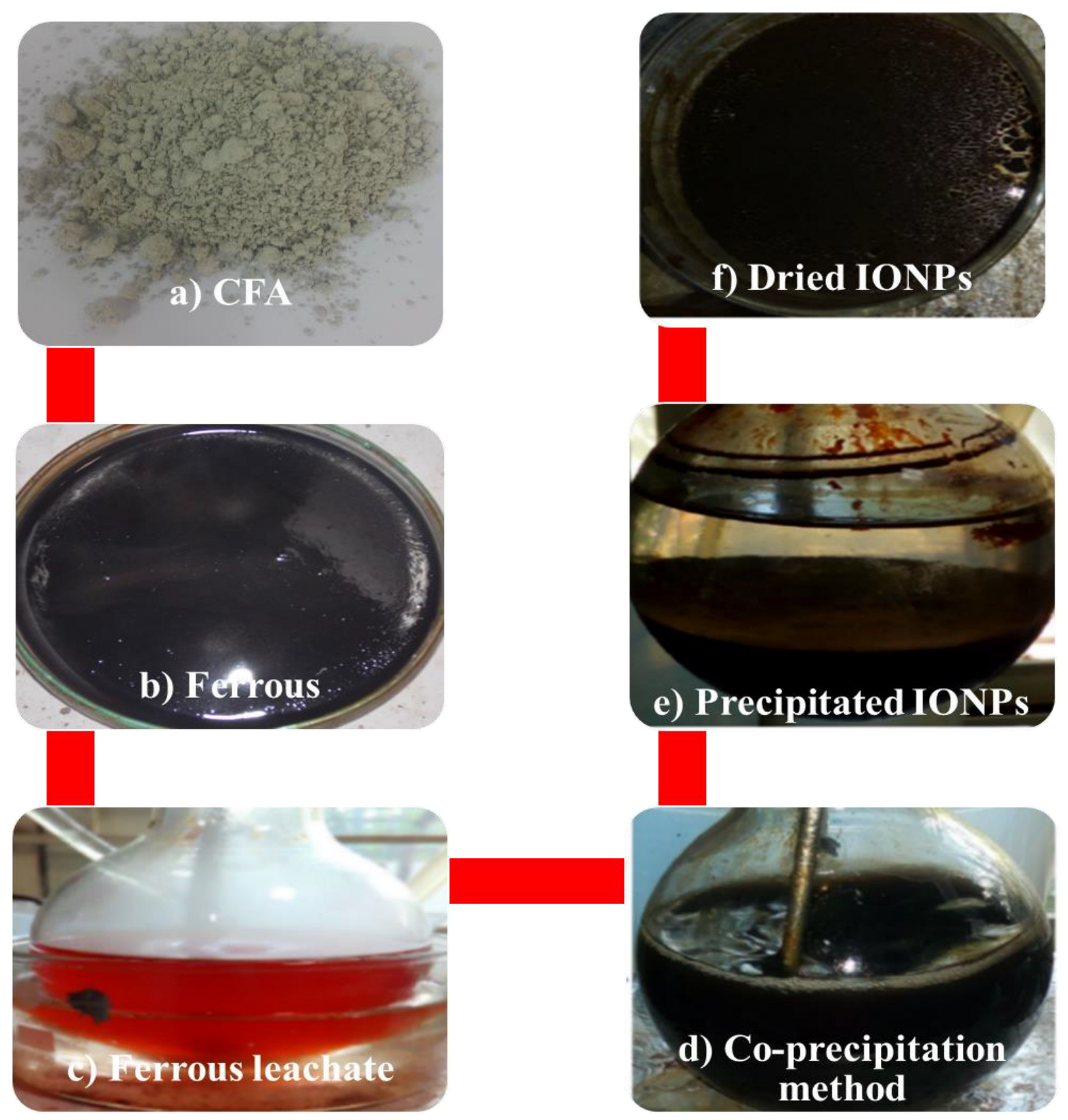
2.2.1. Synthesis of Iron Oxide Nanoparticles from Fly Ash

Firstly, CFA was added to distilled water in a plastic beaker to obtain a slurry. By using a strong external neodymium magnet, ferrous particles were extracted from the slurry, as shown in CFA (Figure 1a). The extracted ferrous particles (Figure 1b) were dried in an oven at 60–70 °C. Further, a weighed amount of dried ferrous particles was added to 37% HCl in a round bottom flaks, keeping a solid-to-liquid ratio of 1:5. Further, the mixture was subjected to sonication by placement in an ultrasonicator (40 KHz), at 60–70 °C, in a round bottom flask (Figure 1c). Once the experiment was over, the reaction mixture was allowed to cool at room temperature. In order to obtain the acidic solution of ferrous material, the mixture was centrifuged at 5000 rpm for 5 min. Further, the acidic leachate was filtered by using Whatman Filter paper 42. About 20 mL of ferrous leachate was taken in a 100 mL RB, and there was continuous addition of 8M NaOH with continuous stirring at 200–300 rpm, along with heating at 60–70 °C (Figure 1d). The NaOH addition was stopped when a brown-to-black color precipitate started to appear, and at pH 11–13, and the reaction was carried out for 60–90 min (Figure 1e). When the mixture was cooled, the precipitate was washed several times with distilled water and ethanol, to remove any impurities. Finally, the obtained iron oxide particles were oven-dried at 40–50 °C and analyzed by various techniques, as shown in Figure 1f.

2.2.2. Preparation of 20% Fly Ash Aqueous Solution

The 20% aqueous solution of CFA was prepared by adding 200 g of CFA to one liter of double-distilled water (ddw) in a plastic container. The plastic container was sealed with paraffin and kept for shaking in a horizontal shaker at 150 rpm, at 25–26 °C. The shaking was carried out for 24 h. After that, the mixture was left undisturbed for 12 h, so that the solid CFA particles could settle down. When the solution appeared clean, leachate was collected [20,21]. After that, a few drops of nitric acid were added and kept in a refrigerator at 4 °C, to prevent any additional chemical reactions.

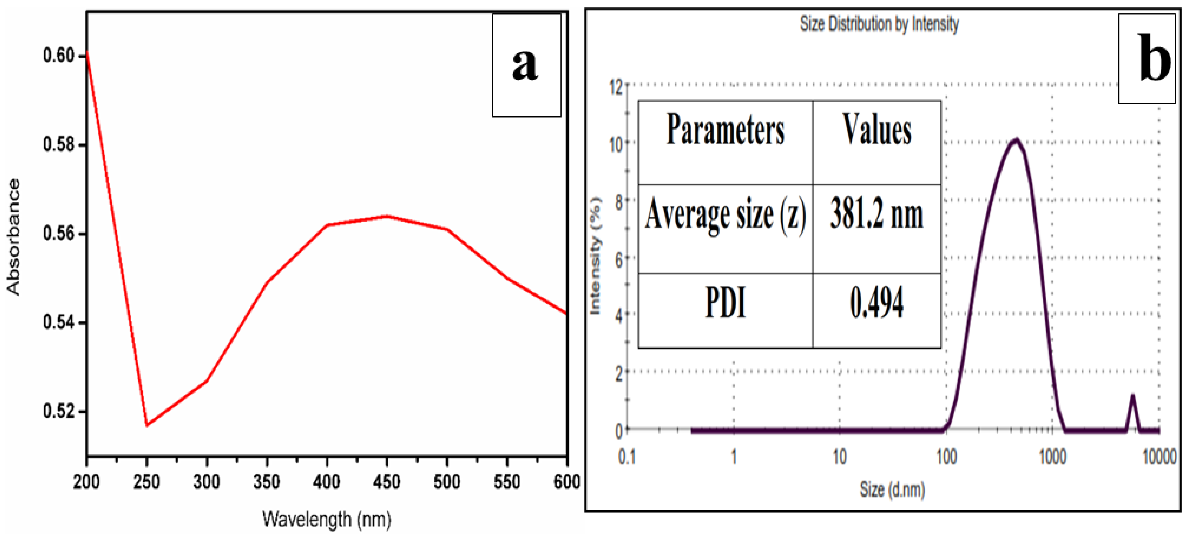
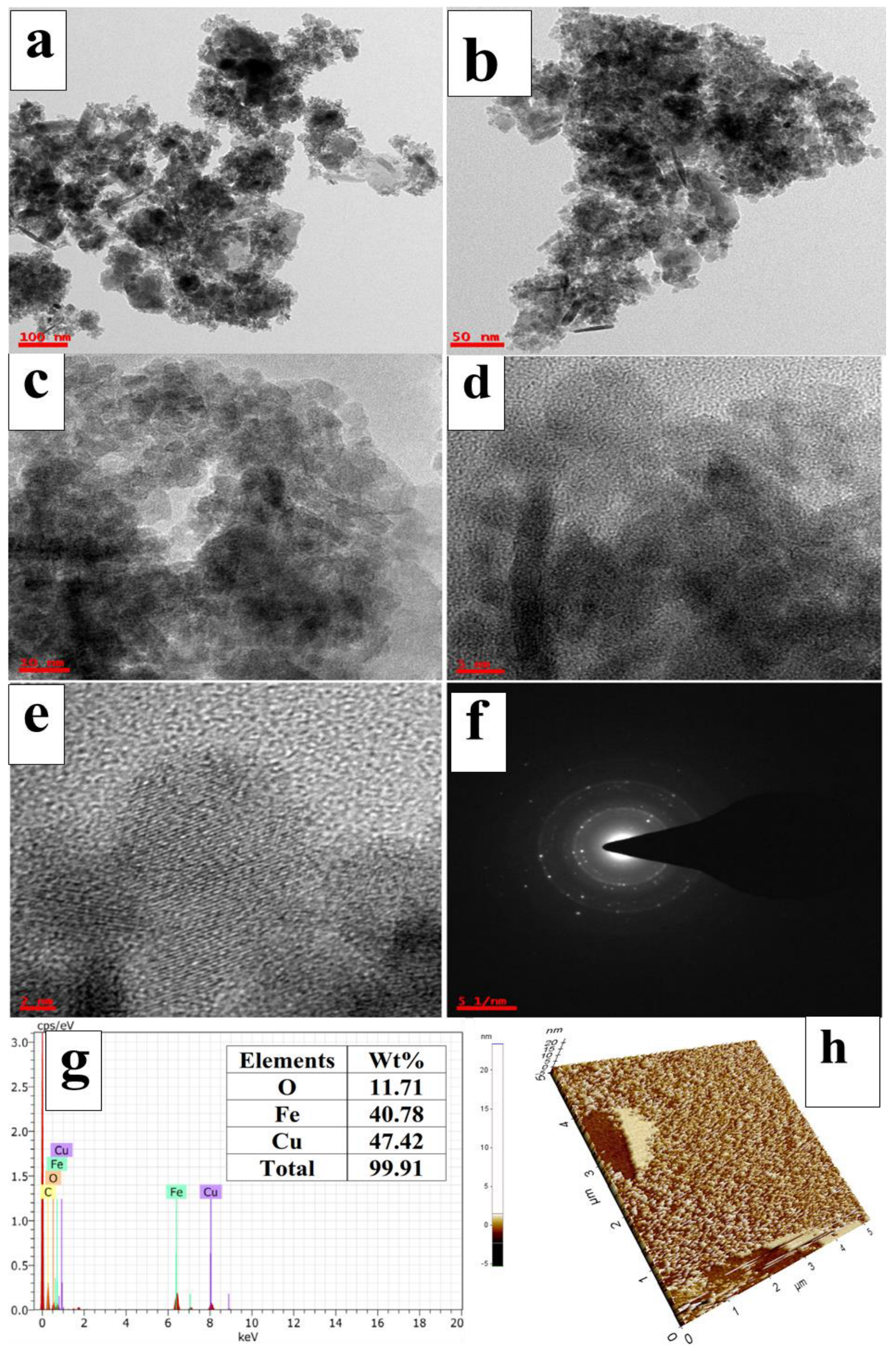
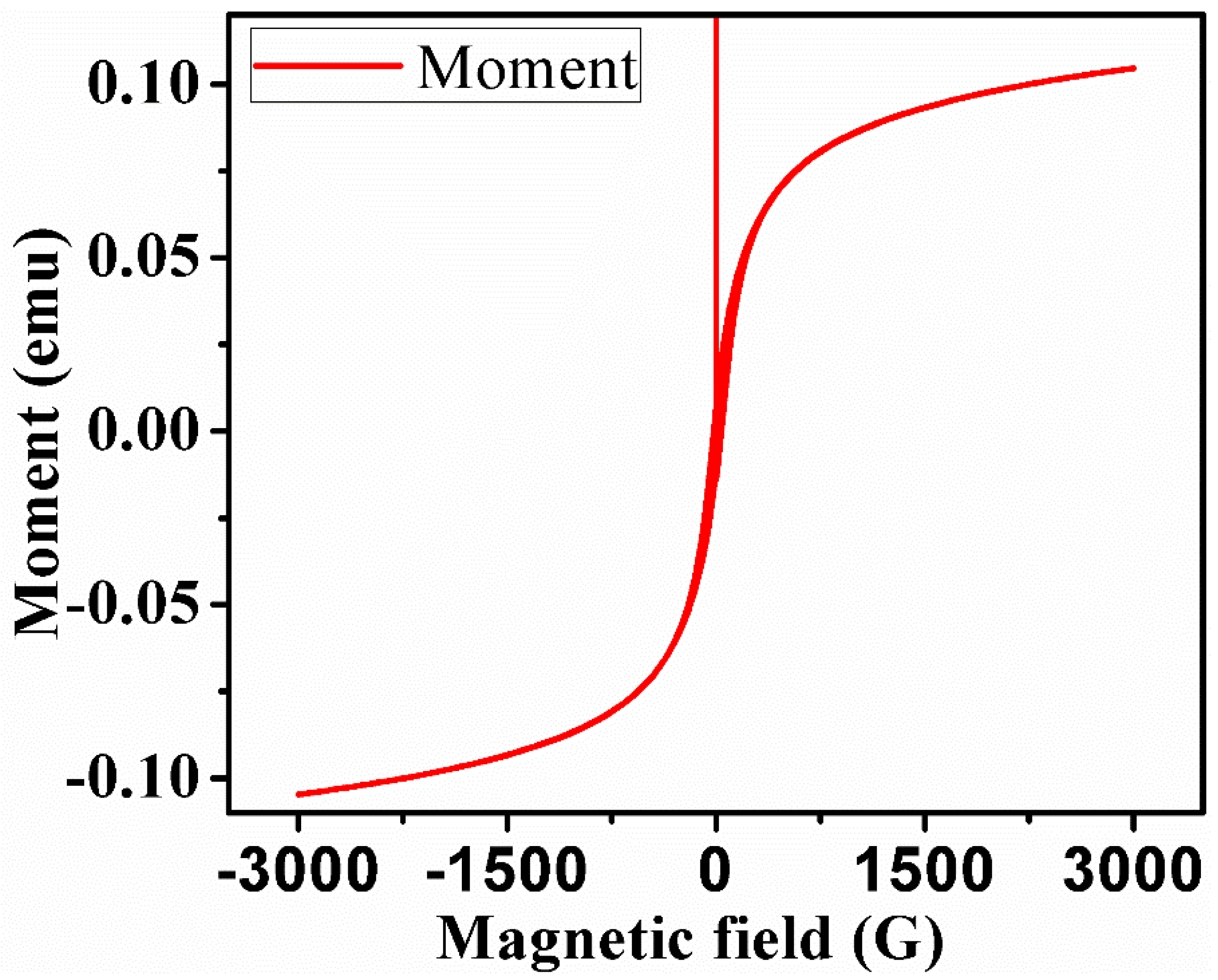
2.2.3. Characterization of Iron Oxide Nanoparticles

The UV-Vis measurement was done by dispersing the IONPs in deionized water, followed by sonication for 10 min in the range of 200–800 nm by using a UV-1800 Double Beam spectrophotometer, (Shimadzu, Japan), operated at a resolution of 1 nm. The FTIR spectra were obtained by preparing (IONPs (2mg) + KBr (198 mg)) the KBr-based solid pellet, using the Spectra SP65, (Perkin Elmer, Waltham, USA) instrument. The FTIR measurement was done in the transmittance mode, in the mid-IR region of 400–4000 cm−1, at a resolution of 2 cm−1. The Raman spectroscopy measurement, in the region of 200–4000 cm−1, was done by placing the sample on the glass coverslips and passing a beam of a laser at 560 nm and 650 nm for 5–10 s, with the help of a Witec Alpha300S+ SNOM Raman Module (Witec, Ulm, Germany) instrument. The particle size distribution of the synthesized IONPs was done by using a particle size analyzer (PSA) Nano S90 (Malvern Zetasizer, Malvern WR14 1XZ, UK), at a temperature of 25 °C, by dispersing samples in the distilled water, followed by sonication for 10 min. The XRD patterns of IONPs were recorded using a D-8 Advance Bruker (Germany) instrument equipped with an X’celerometer, in the 2-theta range of 20–70, with a step size of 0.02 and a time of 5 s per step at, 40 kV voltage and a current of 30 mA. The surface morphology, purity, and elemental composition of the synthesized IONPs were analyzed by the FESEM, Nova, NanoSEM, and FEI-450 FESEM (Netherlands). The synthesized IONPs were loaded onto the carbon tape, which in turn was placed on the aluminum stub holder. The gold coating of the sample was done with 10 min of sputtering, to make the sample conductive. The electron diffraction spectroscopy (EDS) analysis of the IONPs was analyzed by an Oxford EDS analyzer attached to the FESEM, at variable magnifications and 20 kV. Transmission electron microscope (TEM) analysis was done for the internal size and shape determination of the synthesized IONPs. The analysis was done by using the FEI Model Tecnai G2 20 Twin (USA), operated at a voltage of 200 kV. For TEM and HR-TEM measurements, the sample was prepared by a drop-casting technique, where the IONPs were suspended in an aqueous medium and loaded onto carbon-coated copper grids. For obtaining the 3D structure of the synthesized IONPs, atomic force microscopy (AFM) was done. The IONPs dispersed in ddw for UV-Vis were sonicated, and a drop of the sample was loaded on a clean glass cover slip with the help of a micropipette. The cover slip was allowed to dry in the oven at 50–55 °C, till complete dryness. Further, the excess sample was removed by washing the slide, and dried again. Finally, the 3D and 2D measurements of the IONPs were taken by using the Perk-System model: XE-70 (Perkin Elmer, NY, USA) instrument. The magnetic measurements of the synthesized IONPs were analyzed by the physical property measurement system (PPMS) instrument, with model no. PPMS, at 300 k, i.e., room temperature, in the magnetic field of −7000 k to +7000 k. About 30 mg of powdered IONPs were taken, wrapped in paraffin tape and placed in the quartz tube.

2.2.4. Batch Experiment for Remediation of Heavy Metals

The remediation of heavy metals by the adsorption process in a multi-component system (Cd2+, Mn2+, Zn2+, Pb2+, Al, Cu2+, Co2+, Cr6+, Cr4+ and Ni2+) was performed by the shake-flask batch adsorption process. In a typical batch experiment, 50 mg and 100 mg of IONPs were dispersed in 150 mL of 20% w/v CFA solution in a 250 mL Erlenmeyer flask. The sample was shaken thoroughly in an incubator shaker at 150 pm, at 30 °C and at neutral pH (7). An aliquot of ~10 mL sample was collected from the flask after every regular interval of time, i.e., 0, 10, 30, 60, 90, 120 minutes, and analyzed by inductively coupled plasma-optical emission spectroscopy (ICP-OES) (Model: 7300 DV, Make: Perkin Elmer, Netherlands), for the detection of heavy-metal concentration. Among all nine heavy metals selected for the removal from the CFA solution, the per cent removal of heavy metals was calculated by the following formula:
Removal   efficiency   ( % ) = C 0 C t C 0 × 100
where,
  • C0 = initial concentration of heavy metal
  • Ct = heavy-metal concentration at a particular time

3. Results and Discussion

In the present study, CFA-extracted ferrous particles have been used for the synthesis of pure IONPs. The HCl-based acidic treatment causes the dissolution of Fe from ferrous oxides into the HCl solution, and forms mixtures of ferrous chloride and ferric chloride. As Fe, Al, Na and other alkali metals have better solubility in an acidic medium, such as HCl, they also get dissolved into it, which was confirmed by the ICP-OES. The formation of IONPs takes place through the reactions given below. The CFA-extracted ferrospheres have different phases of iron oxides, such as magnetite (λ-Fe3O4), maghemite (β-Fe2O3), and hematite (α-Fe2O3). Therefore, during the exposure of HCl to such ferrospheres, there is the dissolution of Fe from such iron oxides into the acidic medium, and the formation of ferric chloride and ferrous chloride according to reactions [2,3]. Moreover, Si, being insoluble in acid, remained in the ferrospheres. The ferrospheres were ruptured, along with the formation of the grooves and pits on their surface due to the Fe leaching into the medium, as confirmed by FESEM. Further, according to reaction [4], the NaOH precipitates these chlorides formed in the reactions (2&3) [23,24,25]. These synthesized IONPs were analyzed after drying for confirmation and purity by sophisticated instruments.
Reactions
Fe 3 O 4 + 8 HCl FeCl 2 + 2 FeCl 3 + 4 H 2 O
Fe 2 O 3 + 6 HCl 2 FeCl 3 + 3   H 2 O  
FeCl 2 + 2 FeCl 3 + 8 NaOH   Fe 3 O 4   ( Pure ) + 8 NaCl + 4   H 2 O  

Characterization

The preliminary characterization of synthesized nanoparticles by UV-Vis spectroscopy has proven to be a very useful technique for the analysis of nanoparticles [26].
The synthesized IONPs were dispersed in ddw water, followed by sonication for 10 min by an ultrasonicator (Sonar, 40 KHz). The well-dispersed sample was divided into two parts: one part was analyzed by UV-Vis, while the second part was analyzed by the PSA at 25 °C. The UV-Vis measurement peak at 210 nm was the characteristic feature of the presence of Fe2+ ions and was a preliminary indication of the formation of IONPs, but did not distinguish among the phases of the iron oxides. Figure 2b exhibits the particle size distribution of IONPs, where the average particle size is 381.2 nm, with a polydispersity index (PDI) of 0.494. The PDI and size were in good agreement with size [27,28].
Figure 3a exhibits the FTIR spectrum of the as-synthesized and after-use IONPs. The as-synthesized IR spectra of IONPs exhibit characteristic bands of iron oxides [29,30]. The band at 422 cm−1 and 576 cm−1 is attributed to the vibrational modes of Fe-/Fe-O-Fe (magnetite and maghemite) of IONPs. The band at 3400 cm−1 and 1600 cm−1 indicates the presence of OH groups of either water molecules or ferric hydroxides (Fe-O-OH) [31]. Previously, Gupta and their team and Yadav et al. also obtained bands in the same region for the synthesized IONPs from ISA, and the results were in close agreement [14,32].
A typical XRD diffractogram is shown for the as-synthesized IONPs and 100 mg residue in Figure 3b. The as-synthesized IONPs show small intensity peaks at 33° and 35°, which are due to the hematite and magnetite, respectively. The XRD confirms the mixture of hematite and magnetite phases in the synthesized IONPs. The mixed phases could be because of either partial transformation of ferrous chlorides into a single phase during the synthesis. A team led by Yadav [14] obtained similar results for the synthesized IONPs from ISA. They also had a peak in the range of 33–36°, attributed to the magnetite or maghemite [14].
A typical Raman spectrum of IONPs and residue of 100 mg is shown in Figure 4. It was applied to determine the nature of the IONPs. The bands at 293 cm−1, 481 cm−1 and 633 cm−1 are attributed to the Fe–O–Fe bonds in the as-synthesized IONPs. Similar observations were also reported by Yadav and their team in 2020 [33], and in the year 2017 by a team led by Justin [29], which determined that IONPs are excited by the 560 nm wavelength laser excitation. These bands confirm the synthesis of crystalline IONPs from the CFA-extracted ferrospheres.
The TEM micrographs in Figure 5a,b reveals that the particle size varied from 10 to 30 nm, and the particles were showing aggregation. The particles were spherical in shape, along with a few rod-shaped particles. On the other hand, Figure 5c,d shows HRTEM images of IONPs, which are mainly aggregated and spherical-shaped. Figure 5e shows the lattice fringes of IONPs, where the d-spacing of the IONPs is 0.24 nm. Figure 5f shows the scattering area diffraction pattern (SAED) pattern of the synthesized IONPs. The SAED pattern shows the polycrystalline nature of the IONPs. In 2018, Yadav and Fulekar [18], and a team led by Yadav in 2021 [14], showed similarly shaped IONPs synthesized from ferrous fractions, collected from iron precursors and incense sticks ash, respectively. The synthesized IONPs were amorphous in nature in both cases.
The EDS spectra (Figure 5g) also show peaks for Fe and O only, which indicates the purity of the synthesized ferrous carbonate and Cu peaks, which are due to the copper grids. The AFM image (Figure 5h) reveals the 3D images of the synthesized IONPs, which was also justified by the FESEM and TEM images [14].
While PPMS spectra (Figure 6) reveal the magnetic strength of the IONPs. The magnetic moment was calculated against the magnetic field [34,35]. During the year 2020, a team led by Yadav also showed similar magnetic properties of the polyhedral-shaped IONPs from ISA [14].
The FESEM micrographs in Figure 7a,b show that the synthesized IONPs are generally spherical in shape, but they are aggregated to form lumps. In Figure 7b, an aggregated lump of IONPs is shown to have an aggregation of numerous rod-shaped particles inside a large structure. In addition to this, there are several spherical-shaped, white-color depositions of small particles on their surface. Figure 7d shows aggregations of numerous spherical-shaped particles at a 200 nm scale. The size of spherical particles varied from 40 to 70 nm, while rod-shaped particles were more than 70 nm in length and 10–20 nm in width. The EDS spectra in Figure 8a have peaks for Fe, O and C, where Fe was 63% and O was 31%, which indicates the high purity of the synthesized IONPs [33]. While carbon is present at up to 4%, it is the only impurity which may be associated with the ferrous particles, or due to the carbon tape used in the stub. The total Fe and O content was 94%, which indicates all the trace elements have been removed during the acidic treatment of the ferrous material. In 2021, a team led by Yadav also obtained similar results with IONPs synthesized from incense sticks ash (ISA).

4. Effect on Nano-Adsorbent after Remediation of Heavy Metal

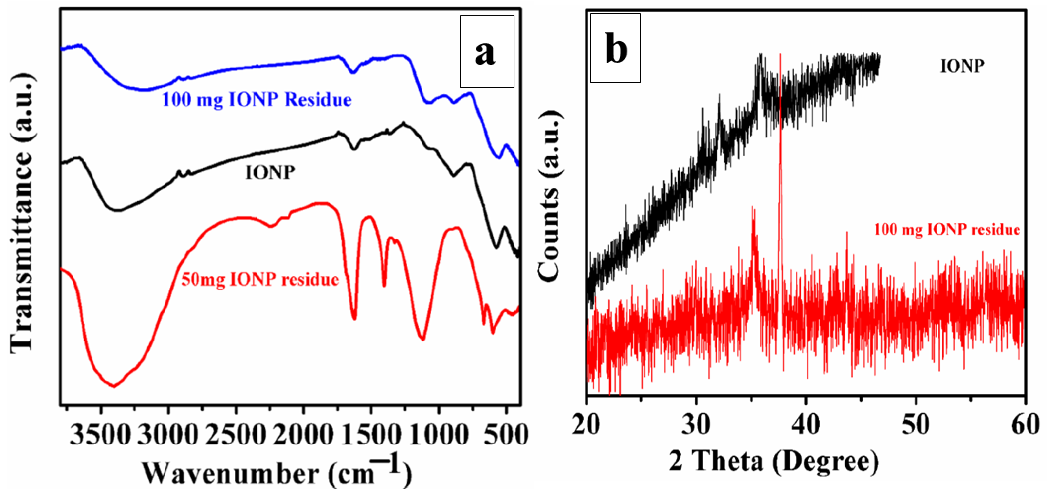
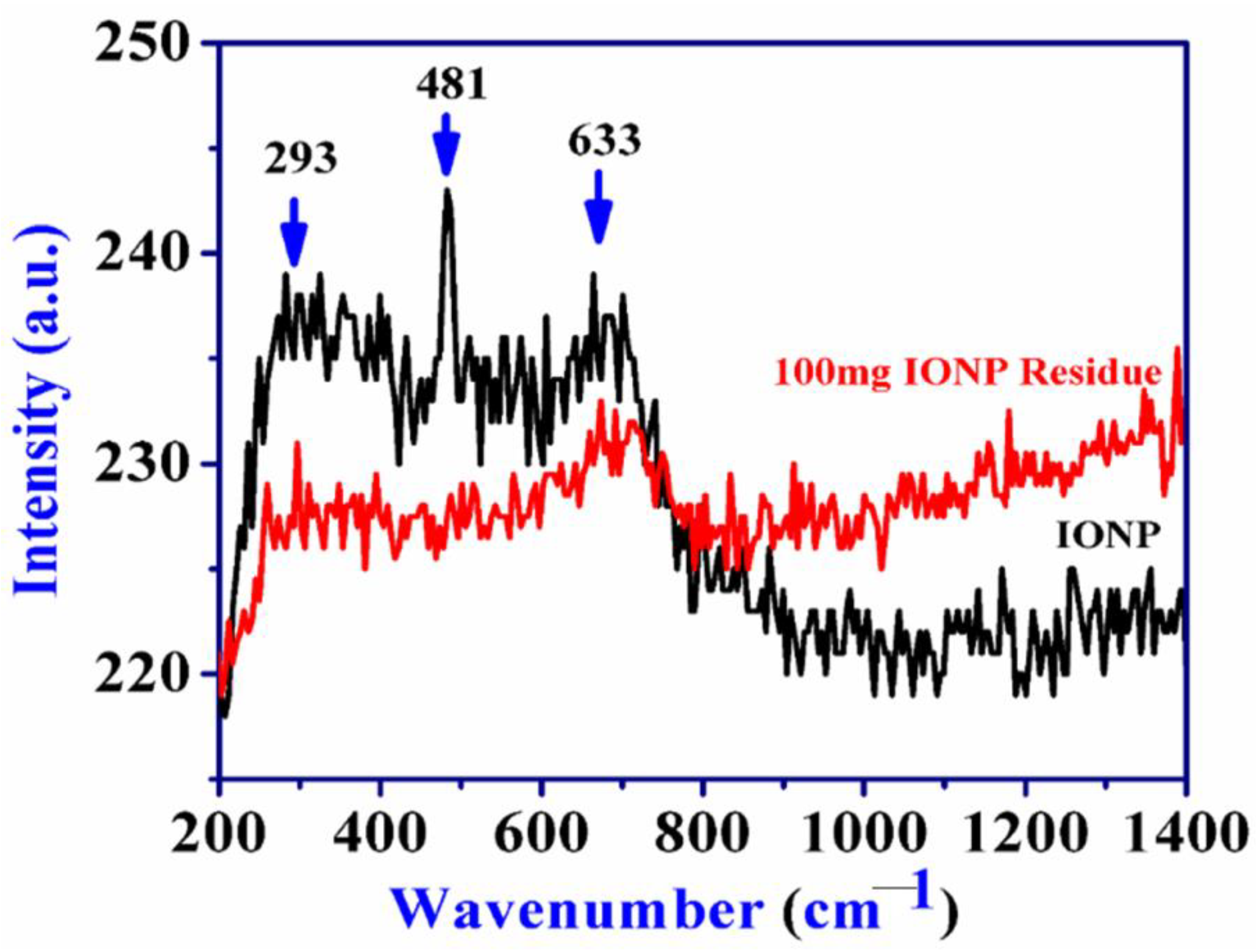
4.1. Mineralogical Changes: XRD, FTIR and Raman

FTIR analysis of residue IONPs was done to determine the functional groups involved in the remediation of heavy metals. The FTIR spectra of IONPs, 50 and 100 mg residue in Figure 3a, reveal that there are no significant changes in the bands of IONPs and 100 mg residue, as all the bands remain more or less the same. The bands in the region of 400–700 cm−1, in all the IONPs, are attributed to Fe–O or Fe–O–Fe (magnetite and maghemite) bonds. After heavy-metal removal in 50 mg IONPs, some differences in the locations of the absorbance peaks were observed. There was a formation of sharp and prominent new bands in the 50 mg residue at 1127, 1398 and 1625 cm−1. These bands could be attributed to the carbonates of Ca and Mg in the sample, which is also evident in the EDS data. The band at 1127 cm−1 could also be attributed to silicate adsorption on the IONPs, after use of the aqueous solution [36].
This could be also due to the formation of hydro complexes (FeOH, Fe–OH–Fe) of metal hydroxides, which were previously not present in the sample. While the band at 1622 cm−1 may be due to the bending vibrations of water molecules H–O–H and Fe–OH complexes, which became sharper and more intense in the 50 mg residue after the adsorption of heavy metals. The band in 50 mg residue at 3400 cm−1 was attributed to water molecules or ferrous hydroxides, having become broader than the IONPs and 100 mg residue. Initially, the peaks for carbonates and silicates were not present in the IONPs, which were adsorbed from the CFA aqueous solution on the surface of IONPs after the reaction, also supported by the EDS data [37,38]. It is assumed that the formation of new absorption bands, the change in absorption intensity and the shift in the wavenumber of functional groups might be due to the complexation between metal ions and the binding sites of the nano-adsorbents. The binding mechanism involved electron pair-sharing between electron donor atoms (O) and metal ions [39]. The XRD pattern in Figure 3b reveals that there are no major changes in the mineralogy of the IONPs before and after the remediation of heavy metals. The peaks at 33° and 35° remains as such, which are the characteristic peaks of iron oxide. Additionally, there is one prominent and sharp peak at 37°, which could be due to the CaO adsorption on the surface of 100 mg IONPs, which is also further supported by EDS and FTIR data [40,41].
The Raman spectra in Figure 4 reveal that there is a shift in the peaks of IONPs before and after used IONPs. There are changes in the peaks, i.e., the peak is shifted to a lower wavenumber in the residue. The bands of as-synthesized IONPs from 293 cm−1 and 674 cm−1 shifted to 287 cm−1 and 633 cm−1, respectively. There was also a decrease in the intensity of the bands, while the sharp and major band at 481 cm−1 completely disappeared in the residue. This could be because of the oxidation of the sample, or because of the deposition of elements from the aqueous solutions, which may respond to a weak signal. There are also changes in the crystallinity of the 100 mg residual sample, where its crystallinity was affected after being used as an adsorbent. This could be due to the adsorption of heavy metals and carbonates, and the silicates on them [42,43].

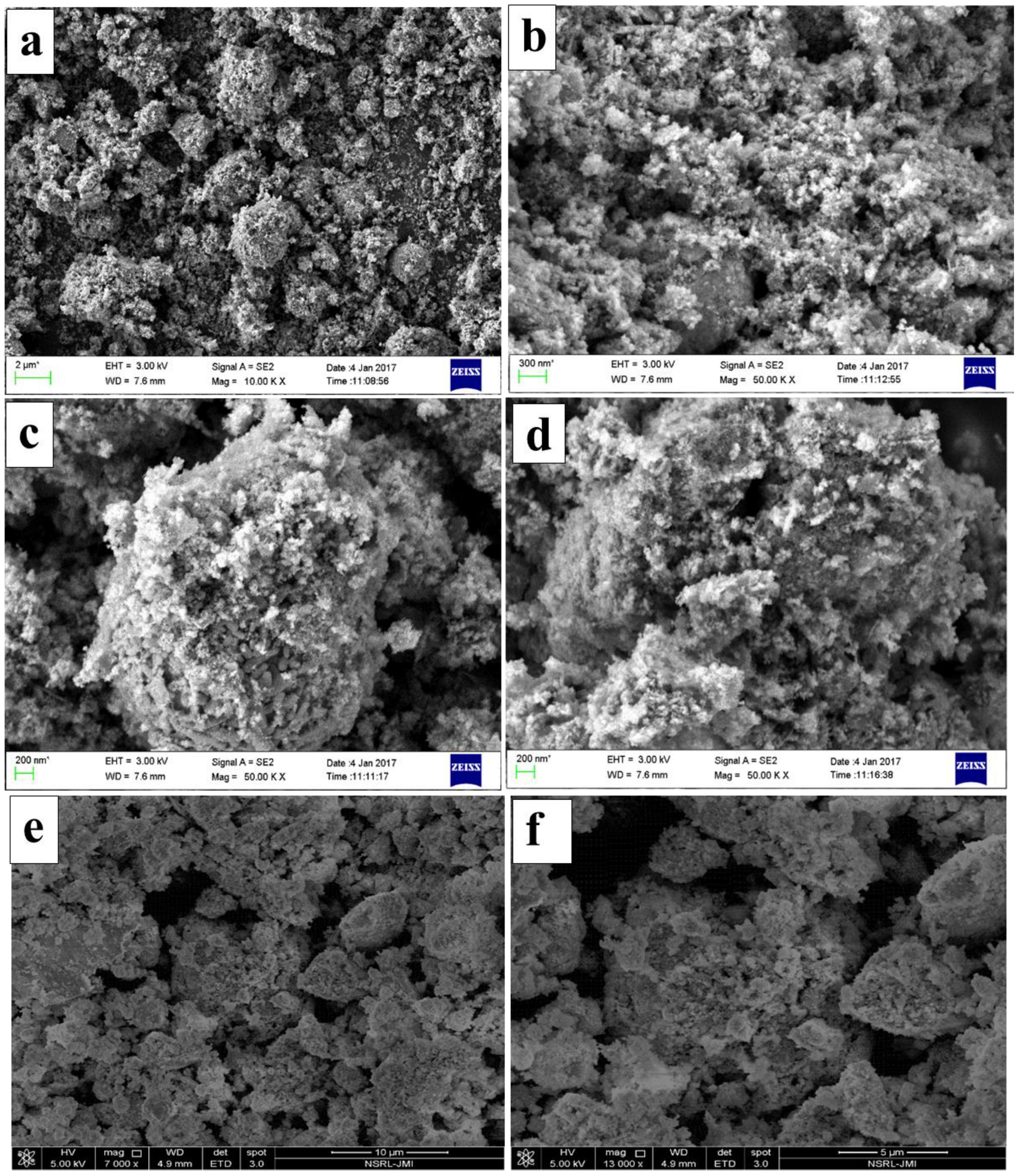
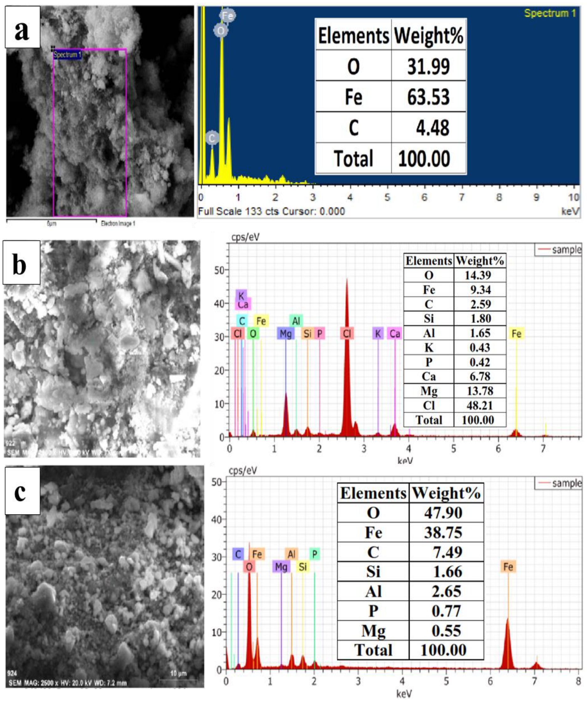
4.2. Morphological and Elemental Changes: FESEM-EDS and ICP-OES

From the EDS spectra, it is revealed that there is adsorption of Si, Al3+, P, and Mg2+, in both the samples of IONPs, i.e., in 50 mg and 100 mg residues. However, 50 mg IONPs have adsorbed more elements than the 100 mg IONPs besides heavy metals on their surface, as it has Cl-, Ca2+ and K+, in addition to the above-mentioned elements [44]. In the 50 mg IONP residue EDS spectra, Cl-, Mg2+, and Ca2+ alone comprise about 68.77%, which indicates the formation and deposition of chlorides on the surface of the 50 mg residue. This is because the fresh aqueous solutions of CFA have higher Ca and Mg, and both have a high solubility in normal room-temperature water. The higher solubility and concentration of Ca2+ and Mg2+ in the 20% CFA solution is also supported by the ICP-OES data. This indicates that when the IONP dose was 50 mg, then the adsorption of heavy metals and other elements was more effective [45,46,47].
The FESEM micrographs of IONPs before and after remediation of heavy metals are shown in Figure 8a–d. Figure 8a,b shows the FESEM micrographs of as-synthesized IONPs, while Figure 8e,f are images of 100 mg IONPs after use. The FESEM micrographs reveal that there are no distinguishable morphological changes, i.e., either in their shape or size before and after the use of IONPs. However, the aggregation of the particles remained as such. There was hardly any positive impact of a higher dose of IONPs on the rate of adsorption. The data was also supported by FTIR and ICP-OES. These findings, as related to EDS analysis, showed the involvement of the ion exchange mechanism in the removal of metal ions by the IONPs [48]. About 3 mg of the 100 mg IONPs, after use, was digested in aqua regia at 220 °C, and after digestion, it was filtered and make-up volume was prepared by adding ddw. The sample was analyzed for the detection of trace elements present on its surface after adsorption, as shown in Table 1.
The following elements were identified on its surface, which were initially not present in the IONP sample: Ca2+, Al3+, Si, P, K+, Li3+, Mg2+, Na+, Cu2+, Cd2+, Co2+, Cr6+, Cr4+ Pb2+, Mn3+, and Zn2+, have adsorbed on the IONP surface, which confirms the adsorption of heavy metals. All these were not present initially in the original sample, i.e., IONPs, which indicates that the source of these elements was the aqueous solutions of CFA. All these elements were adsorbed on the surface of IONPs during the reaction. Among non-heavy metals, the maximum adsorption was of Al3+, Mg2+, Ca2+ and Na+, as these elements were present in a large concentration in the CFA [49].

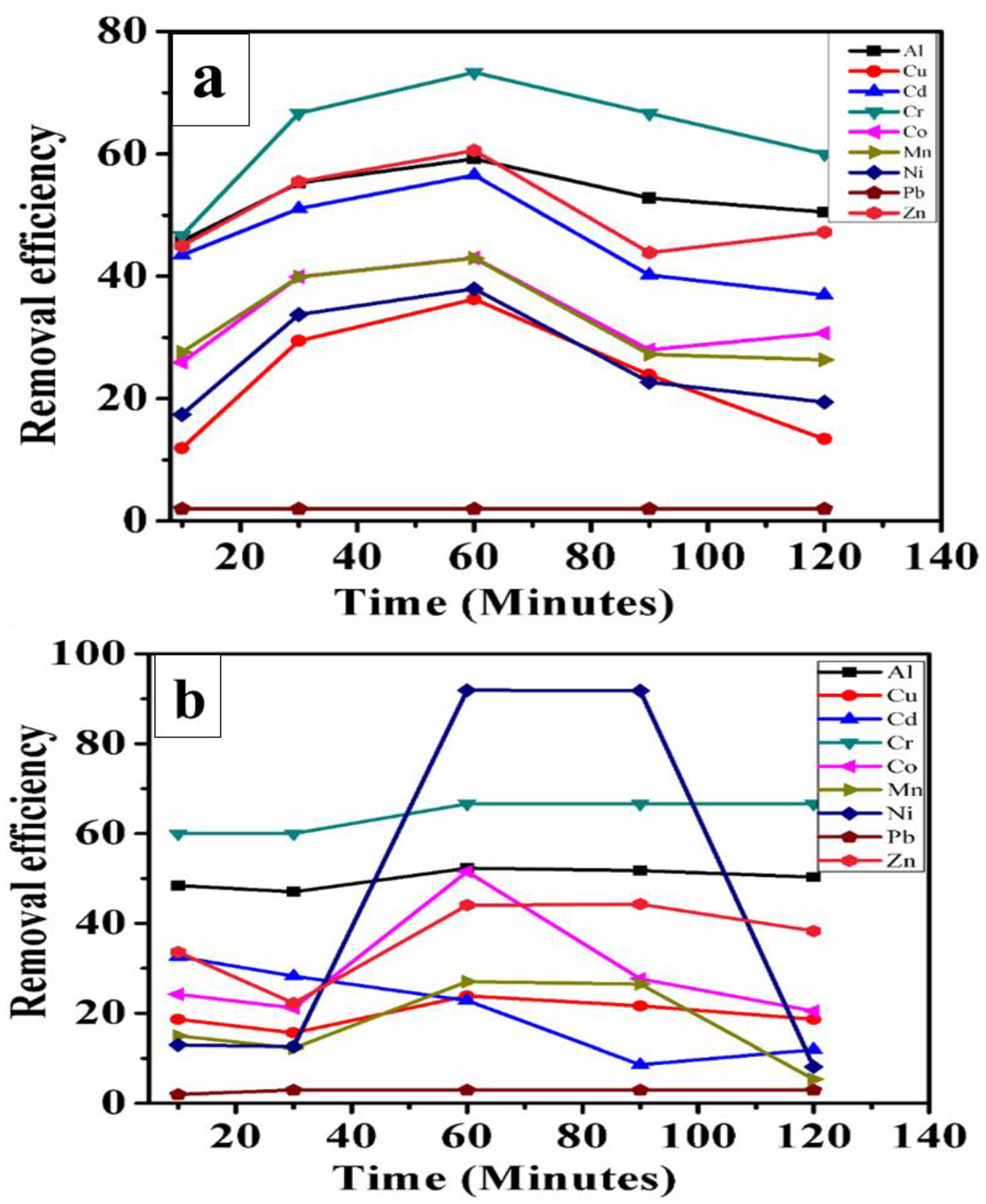
4.3. The Remediation of Heavy Metals by 50 mg IONPs

The removal efficiency of all the nine heavy metals [Al3+, Cu2+, Cd2+, Co2+, Cr6+, Cr4+, Pb2+, Mn3+, Ni2+ and Zn2+] continuously increased, in Figure 9a from 10 min up to 60 min after which there was a decrease in the removal efficiency for all the heavy metals at 90 min. A further decrease was noticed at 120 min in all the heavy metals except for Co and Zn. This indicates that the removal efficiency was maximum at 60 min for all the heavy metals, where they reached equilibrium and after which desorption occurred on the surface of IONPs. This desorption process continued until 120 min except for Co2+ and Zn2+. While the Pb2+ was not detected after 10 min, in the solution and remained as such for up to 120 min. This was because Pb2+ was present in a much lesser concentration than the other heavy metals in the solution. Therefore, within 10 min only its value reached below the sensitivity level of ICP-OES. The desorption is due to the non-availability of free adsorption sites on the IONP surface [50,51].

4.4. The Remediation of Heavy Metals by 100 mg IONPs

The removal efficiency of Ni2+, Cr6+ and Cr4+ remained constant for up to 30 min, and a sudden increase was noticed at 60 min, after which it remained as such until 90 min, as shown in Figure 9b. A drastic decrease in the removal efficiency was seen in Ni2+ at 120 min, while the Cr6+ and Cr4+ reached saturation after 90 min to 120 min. In the case of Cu2+, Co2+, Zn2+, Al3+ and Mn3+, there was an initial decrease in the efficiency from 10 min to 30 min, after which an increase was noticed in all of them at 60 min. After this, a marginal decrease was noticed at 90 min, which continued to decrease until 120 min. The Cd2+ value continuously decreased from 10 min and continued up to 90 min, and a minor increase was noticed at 120 min. Likewise, at 50 mg, the Pb2+ value increased at 10 min, after which the Pb2+ was not detected up to 120 min, due to the above reason. Initially, the Pb2+ concentration was less in the solution, and after 10 min, its value reached below the sensitivity level of ICP-OES.
At the higher dose of IONPs, the remediation did not increase drastically, possibly due to agglomeration and precipitation of nano-photocatalyst, which may have increased the size of the IONPs, leading to a reduction in the specific surface area and ultimately lowering the number of surface-active sites [52,53].

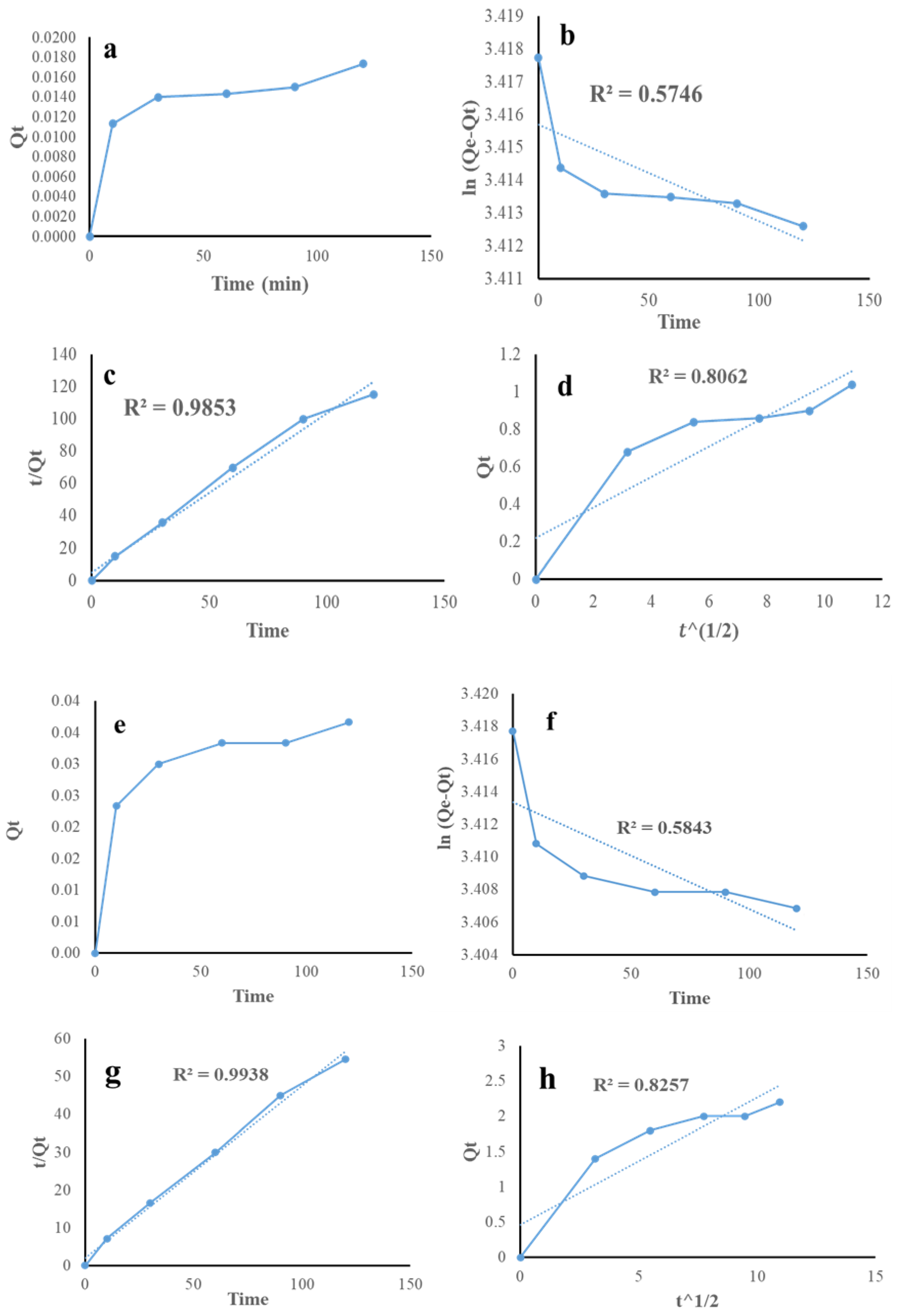
4.5. Kinetics Study of Pb and Cr

To find out the remediation of Pb2+, Cr6+ and Cr4+ ions from the CFA 20% simulated wastewater by iron IONPs, three different kinetic models (Pseudo-first order (1), Pseudo-second order (2) and intra-particle diffusion (3)) were applied to perform the kinetic studies for the removal of Pb2+ and Cr ions by IONPs. Figure 10a–d shows kinetic studies for Pb removal and Figure 10e–h shows kinetic studies for Cr ion removal by IONPs. Table 2 and Table 3 show the kinetic parameters of Pb removal and Cr ions removal, respectively. From the above data, it can be summarized that both Pb removal, as well as Cr ions removal, follows a Pseudo-second order reaction.
The slandered equation is given below to calculate the kinetics parameters for the suitable kinetic orders:
ln ( Q e Q t ) = ln Q e k 1 t 2.303
where Q e and Q t are the amounts of metal (Pb, Cr) adsorbed (mg/g) at equilibrium and at time; t (min), respectively, and   k 1 (min−1) is the rate constant in the pseudo-first order kinetic model.
t Q t = 1 k 2 × Q e 2 + t Q e
where k 2 is the rate constant (g/mg. min) in the pseudo-second order kinetic model for adsorption.
Q t = k i d t 1 2 + C
where k i d is the intra-particle diffusion rate constant (mg/g. min3/2), and the values of C (mg/g) depict the boundary thickness.
Table 2 shows the kinetic parameters of the nano-adsorbent for Pb2+. The highest R 2 value for the pseudo-second order is closer to 1 compared to others (Figure 10c), having Q e = 1.01 (mg/g) and   k 2 = 0.019 × 10⁻¹ (g/mg. min), which attribute to the pseudo-second order adsorption kinetics followed for Pb2+ adsorption. However, the same trend (psuedo-second order) adsorption kinetics was obtained for Cr2+ metal adsorption with the highest R 2 value ≈ 0.993 (Figure 10g), having Q e = 2.19 (mg/g) and   k 2 = 0.0101 × 10⁻¹ (g/mg. min) (Table 3).

4.6. Adsorption Mechanism

Here the adsorption of heavy metals on the surface of IONPs was via electrostatic attraction and coordination. At neutral or slightly acidic pH on the surface of IONPs has a positive charge, which produces PZC repulsion to cations, and the adsorption of cations on the IONPs is due to the coordination between CH on the surface of IONPs and the bivalence metal ions. Such a phenomenon of adsorption and deposition was seen because the CFA aqueous solutions are a multicomponent system, where several heavy metals and non-heavy metals compete for the limited adsorption sites. Once these binding sites are occupied, there is competition among ions and nonmetallic ions, which results in the desorption of the heavy metals. The concentration of Pb2+ reached below the detection level of ICP-OES within 30 min only, in comparison to the other cations. This could be because Pb2+ has a higher electronegativity, as electronegativity plays an important role in controlling the adsorption of metal ions. The higher the electronegativity, the stronger the covalent formation of the metals with oxygen atoms located on the surface of IONPs [18].

5. Conclusions

The present study showed that it is possible to synthesize pure IONPs from CFA, and they could be used as alternative to nano-adsorbents for the removal of heavy metals from wastewater or CFA aqueous solutions. The CFA-based synthesized IONPs can effectively and efficiently remediate the CFA heavy metals from a multi-component CFA solution, and only the remediation time of different heavy metals varies. Whether heavy metals reach equilibrium early or later depends on the intrinsic properties of the respective ions and the conditions for adsorption. After reaching equilibrium, there is desorption of heavy metals from the binding sites of IONPs. This is due to the reason that initially, most of the adsorption sites are occupied by the ions and there are no further free sites available for adsorption. The efficiency of heavy metal removal is better with a lower dose of IONPs than with a higher dose, i.e., 50 mg. When the dose of IONPs is 50 mg, then most of the heavy metals are being remediated more efficiently. This is the case because there is an agglomeration of the IONPs at higher doses. Moreover, at higher doses, there are no vacant adsorption sites. Besides heavy metals, IONPs also remove other elements, such as K+, P, Na+, Be, Ca2+, Mg2+, etc., that were present on the residue during EDS analysis. They can also act as an adsorbent for the removal of several non-heavy metals, as revealed by the residual analysis by ICP-OES. Therefore, IONPs can effectively remove heavy metals, as well as non-heavy metals, from the solution.

Author Contributions

Conceptualization, V.K.Y., A.A. and A.G.; data curation, A.G., N.E., M.H.F. and V.K.Y.; methodology, V.K.Y., A.A. and A.G.; validation, V.K.Y., L.B.E., A.A., A.G. and N.E.; formal analysis, V.K.Y., A.A., L.B.E. and M.H.F.; resources, A.A., M.H.F. and A.G.; writing—original draft preparation, V.K.Y. and N.E.; writing—review and editing, V.K.Y., A.A. and M.H.F.; supervision, V.K.Y., A.A., L.B.E. and M.H.F.; project administration V.K.Y., A.A. and N.E.; funding acquisition, A.G., N.E., L.B.E. and A.A.; investigation, V.K.Y., A.A. and N.E.; software, V.K.Y., A.A., M.H.F. and L.B.E., visualization, V.K.Y., A.A. and M.H.F. All authors have read and agreed to the published version of the manuscript.

Funding

This research was funded by the Deanship of Scientific Research at King Khalid University under grant number RGP.2/182/43.

Institutional Review Board Statement

Not applicable.

Informed Consent Statement

Not applicable.

Data Availability Statement

Not applicable.

Acknowledgments

The authors extend their appreciation to the Deanship of Scientific Research at King Khalid University for funding this work through the Research Groups Program under grant number RGP.2/182/43. The authors are thankful to the UGC, CIF of CUG, Gandhinagar, Jamia Millia Islamia, New Delhi, CIMPAP-CSIR Lucknow, SRM University Chennai and SAIF-IIT Madras for extending their characterization and instrument facilities.

Conflicts of Interest

The authors declare no conflict of interest.

References

  1. Patel, H.; Yadav, V.K.; Yadav, K.K.; Choudhary, N.; Kalasariya, H.; Alam, M.M.; Gacem, A.; Amanullah, M.; Ibrahium, H.A.; Park, J.-W.; et al. A Recent and Systemic Approach Towards Microbial Biodegradation of Dyes from Textile Industries. Water 2022, 14, 3163. [Google Scholar] [CrossRef]
  2. Gadore, V. Ahmaruzzaman Fly ash–based nanocomposites: A potential material for effective photocatalytic degradation/elimination of emerging organic pollutants from aqueous stream. Environ. Sci. Pollut. Res. 2021, 28, 46910–46933. [Google Scholar] [CrossRef] [PubMed]
  3. Ankrah, A.F.; Tokay, B.; Snape, C.E. Heavy Metal Removal from Aqueous Solutions Using Fly-Ash Derived Zeolite NaP1. Int. J. Environ. Res. 2022, 16, 17. [Google Scholar] [CrossRef]
  4. Yadav, V.K.; Fulekar, M.H. The current scenario of thermal power plants and fly ash: Production and utilization with a focus in India. Int. J. Adv. Eng. Res. Dev. 2018, 5, 768–777. [Google Scholar]
  5. Park, H.; Wang, L.; Yun, J.-H. Coal beneficiation technology to reduce hazardous heavy metals in fly ash. J. Hazard. Mater. 2021, 416, 125853. [Google Scholar] [CrossRef]
  6. Yadav, V.K.; Gnanamoorthy, G.; Cabral-Pinto, M.M.S.; Alam, J.; Ahamed, M.; Gupta, N.; Singh, B.; Choudhary, N.; Inwati, G.K.; Yadav, K.K. Variations and similarities in structural, chemical, and elemental properties on the ashes derived from the coal due to their combustion in open and controlled manner. Environ. Sci. Pollut. Res. 2021, 28, 32609–32625. [Google Scholar] [CrossRef]
  7. Qasem, N.A.A.; Mohammed, R.H.; Lawal, D.U. Removal of heavy metal ions from wastewater: A comprehensive and critical review. Npj Clean Water 2021, 4, 36. [Google Scholar] [CrossRef]
  8. Puri, N.; Gupta, A.; Mishra, A. Recent advances on nano-adsorbents and nanomembranes for the remediation of water. J. Clean. Prod. 2021, 322, 129051. [Google Scholar] [CrossRef]
  9. Guerra, F.D.; Attia, M.F.; Whitehead, D.C.; Alexis, F. Nanotechnology for Environmental Remediation: Materials and Applications. Molecules 2018, 23, 1760. [Google Scholar] [CrossRef] [Green Version]
  10. Nicola, R.; Costişor, O.; Ciopec, M.; Negrea, A.; Lazău, R.; Ianăşi, C.; Picioruş, E.-M.; Len, A.; Almásy, L.; Szerb, E.I.; et al. Silica-Coated Magnetic Nanocomposites for Pb2+ Removal from Aqueous Solution. Appl. Sci. 2020, 10, 2726. [Google Scholar] [CrossRef] [Green Version]
  11. Wegmann, M.; Scharr, M. Chapter 8—Synthesis of Magnetic Iron Oxide Nanoparticles. In Precision Medicine; Deigner, H.-P., Kohl, M., Eds.; Academic Press: Cambridge, MA, USA, 2018; pp. 145–181. [Google Scholar] [CrossRef]
  12. Sokol, E.; Kalugin, V.; Nigmatulina, E.; Volkova, N.; Frenkel, A.; Maksimova, N. Ferrospheres from fly ashes of Chelyabinsk coals: Chemical composition, morphology and formation conditions. Fuel 2002, 81, 867–876. [Google Scholar] [CrossRef]
  13. Yadav, V.K.; Amari, A.; Wanale, S.G.; Osman, H.; Fulekar, M.H. Synthesis of Floral-Shaped Nanosilica from Coal Fly Ash and Its Application for the Remediation of Heavy Metals from Fly Ash Aqueous Solutions. Sustainability 2023, 15, 2612. [Google Scholar] [CrossRef]
  14. Yadav, V.K.; Yadav, K.K.; Gnanamoorthy, G.; Choudhary, N.; Khan, S.H.; Gupta, N.; Kamyab, H.; Bach, Q.-V. A novel synthesis and characterization of polyhedral shaped amorphous iron oxide nanoparticles from incense sticks ash waste. Environ. Technol. Innov. 2020, 20, 101089. [Google Scholar] [CrossRef]
  15. Gupta, N.; Yadav, V.K.; Yadav, K.K.; Alwetaishi, M.; Gnanamoorthy, G.; Singh, B.; Jeon, B.-H.; Cabral-Pinto, M.M.; Choudhary, N.; Ali, D.; et al. Recovery of iron nanominerals from sacred incense sticks ash waste collected from temples by wet and dry magnetic separation method. Environ. Technol. Innov. 2021, 25, 102150. [Google Scholar] [CrossRef]
  16. Yadav, V.K.; Gnanamoorthy, G.; Ali, D.; Bera, S.P.; Roy, A.; Kumar, G.; Choudhary, N.; Kalasariya, H.; Basnet, A. Cytotoxicity, Removal of Congo Red Dye in Aqueous Solution Using Synthesized Amorphous Iron Oxide Nanoparticles from Incense Sticks Ash Waste. J. Nanomater. 2022, 2022, 5949595. [Google Scholar] [CrossRef]
  17. Wang, C.; Zhou, J.; Wang, Y.; Yang, M.; Li, Y.; Meng, C. Synthesis of zeolite X from low-grade bauxite. J. Chem. Technol. Biotechnol. 2012, 88, 1350–1357. [Google Scholar] [CrossRef]
  18. Yadav, V.K.; Fulekar, M. Biogenic synthesis of maghemite nanoparticles (γ-Fe2O3) using Tridax leaf extract and its application for removal of fly ash heavy metals (Pb, Cd). Mater. Today: Proc. 2018, 5, 20704–20710. [Google Scholar] [CrossRef]
  19. Blaha, U.; Sapkota, B.; Appel, E.; Stanjek, H.; Rösler, W. Micro-scale grain-size analysis and magnetic properties of coal-fired power plant fly ash and its relevance for environmental magnetic pollution studies. Atmospheric Environ. 2008, 42, 8359–8370. [Google Scholar] [CrossRef]
  20. Joos, A.; Rümenapp, C.; Wagner, F.E.; Gleich, B. Characterisation of iron oxide nanoparticles by Mössbauer spectroscopy at ambient temperature. J. Magn. Magn. Mater. 2016, 399, 123–129. [Google Scholar] [CrossRef]
  21. Adewuyi, Y.G. Recent Advances in Fly-Ash-Based Geopolymers: Potential on the Utilization for Sustainable Environmental Remediation. ACS Omega 2021, 6, 15532–15542. [Google Scholar] [CrossRef]
  22. Khan, M.A.; Memon, S.A.; Farooq, F.; Javed, M.F.; Aslam, F.; Alyousef, R. Compressive Strength of Fly-Ash-Based Geopolymer Concrete by Gene Expression Programming and Random Forest. Adv. Civ. Eng. 2021, 2021, 6618407. [Google Scholar] [CrossRef]
  23. Zhao, Y.; Qiu, Z.; Huang, J. Preparation and Analysis of Fe3O4 Magnetic Nanoparticles Used as Targeted-drug Carriers. Chin. J. Chem. Eng. 2008, 16, 451–455. [Google Scholar] [CrossRef]
  24. Hariharan, S.; Ramar, V.; Joshi, S.P.; Balaya, P. Developing a light weight lithium ion battery—An effective material and electrode design for high performance conversion anodes. RSC Adv. 2013, 3, 6386–6394. [Google Scholar] [CrossRef]
  25. Ma, J.; Guo, X.; Yan, Y.; Xue, H.; Pang, H. FeOx-Based Materials for Electrochemical Energy Storage. Adv. Sci. 2018, 5, 1700986. [Google Scholar] [CrossRef]
  26. You, S.; Ho, S.W.; Li, T.; Maneerung, T.; Wang, C.-H. Techno-economic analysis of geopolymer production from the coal fly ash with high iron oxide and calcium oxide contents. J. Hazard. Mater. 2018, 361, 237–244. [Google Scholar] [CrossRef] [Green Version]
  27. Wang, H.; Wang, M.; Zhang, J.; Wang, N.; Wang, J.; Yang, J. Preparation of fly ash-based cobalt-iron silicate as supercapacitor electrode material. Chem. Eng. J. 2022, 434, 134661. [Google Scholar] [CrossRef]
  28. Li, X.; Ma, B.; Wang, C.; Chen, Y.; Yang, W.; Zhang, W. A sustainable process to recycle aluminum from coal fly ash for simultaneous removal of iron: Solid waste management and evaluation. Miner. Eng. 2022, 184, 107638. [Google Scholar] [CrossRef]
  29. Justin, C.; Philip, S.A.; Samrot, A.V. Synthesis and characterization of superparamagnetic iron-oxide nanoparticles (SPIONs) and utilization of SPIONs in X-ray imaging. Appl. Nanosci. 2017, 7, 463–475. [Google Scholar] [CrossRef]
  30. Kumar, P.; Mathpal, M.C.; Ghosh, S.; Inwati, G.K.; Maze, J.R.; Duvenhage, M.-M. Plasmonic Au nanoparticles embedded in glass: Study of TOF-SIMS, XPS and its enhanced antimicrobial activities. J. Alloys Compd. 2022, 909, 164789. [Google Scholar] [CrossRef]
  31. Mahmoudi, M.; Sahraian, M.A.; Shokrgozar, M.A.; Laurent, S. Superparamagnetic Iron Oxide Nanoparticles: Promises for Diagnosis and Treatment of Multiple Sclerosis. ACS Chem. Neurosci. 2011, 2, 118–140. [Google Scholar] [CrossRef] [Green Version]
  32. Modi, S.; Inwati, G.K.; Gacem, A.; Saquib Abullais, S.; Prajapati, R.; Yadav, V.K.; Syed, R.; Alqahtani, M.S.; Yadav, K.K.; Islam, S.; et al. Nanostructured Antibiotics and Their Emerging Medicinal Applications: An Overview of Nanoantibiotics. Antibiotics 2022, 11, 708. [Google Scholar] [CrossRef]
  33. Yadav, V.; Ali, D.; Khan, S.; Gnanamoorthy, G.; Choudhary, N.; Yadav, K.; Thai, V.; Hussain, S.; Manhrdas, S. Synthesis and Characterization of Amorphous Iron Oxide Nanoparticles by the Sonochemical Method and Their Application for the Remediation of Heavy Metals from Wastewater. Nanomaterials 2020, 10, 1551. [Google Scholar] [CrossRef] [PubMed]
  34. Rafieizonooz, M.; Khankhaje, E.; Rezania, S. Assessment of environmental and chemical properties of coal ashes including fly ash and bottom ash, and coal ash concrete. J. Build. Eng. 2022, 49, 104040. [Google Scholar] [CrossRef]
  35. Dash, S.; Chaudhuri, H.; Gupta, R.; Nair, U.G. Adsorption study of modified coal fly ash with sulfonic acid as a potential adsorbent for the removal of toxic reactive dyes from aqueous solution: Kinetics and thermodynamics. J. Environ. Chem. Eng. 2018, 6, 5897–5905. [Google Scholar] [CrossRef]
  36. Correia, L.M.; Saboya, R.M.A.; Campelo, N.D.S.; Cecilia, J.A.; Rodríguez-Castellón, E.; Cavalcante, C.L.; Vieira, R.S. Characterization of calcium oxide catalysts from natural sources and their application in the transesterification of sunflower oil. Bioresour. Technol. 2014, 151, 207–213. [Google Scholar] [CrossRef] [PubMed]
  37. Konopacka-Łyskawa, D.; Czaplicka, N.; Kościelska, B.; Łapiński, M.; Gębicki, J. Influence of Selected Saccharides on the Precipitation of Calcium-Vaterite Mixtures by the CO2 Bubbling Method. Crystals 2019, 9, 117. [Google Scholar] [CrossRef] [Green Version]
  38. Yadav, V.K.; Yadav, K.K.; Gacem, A.; Gnanamoorthy, G.; Ali, I.H.; Khan, S.H.; Jeon, B.-H.; Kamyab, H.; Inwati, G.K.; Choudhary, N.; et al. A novel approach for the synthesis of vaterite and calcite from incense sticks ash waste and their potential for remediation of dyes from aqueous solution. Sustain. Chem. Pharm. 2022, 29, 100756. [Google Scholar] [CrossRef]
  39. Gnanamoorthy, G.; Yadav, V.K.; Ali, D.; Ramar, K.; Ali, H.; Narayanan, V. New designing (NH4) 2SiP4O13 nanowires and effective photocatalytic degradation of Malachite Green. Chem. Phys. Lett. 2022, 803, 139817. [Google Scholar] [CrossRef]
  40. Ganesh, A.B.; Shekar, B.C. Synthesis, characterization and plant growth assessment of hybrid calcium oxide nanoparticles. Int. J. pharma Bio Sci. 2017, 8, 193–198. [Google Scholar] [CrossRef]
  41. Bharathiraja, B.; Sutha, M.; Sowndarya, K.; Chandran, M.; Yuvaraj, D.; Kumar, R.P. Calcium Oxide Nanoparticles as An Effective Filtration Aid for Purification of Vehicle Gas Exhaust. In Advances in Internal Combustion Engine Research. Energy, Environment, and Sustainability; Srivastava, D., Agarwal, A., Datta, A., Maurya, R., Eds.; Springer: Singapore, 2018; pp. 181–192. [Google Scholar] [CrossRef]
  42. Gnanamoorthy, G.; Yadav, V.K.; Ali, D.; Parthiban, E.; Kumar, G.; Narayanan, V. New orchestrated of X-CuTiAP (en, trien, ETA and DMA) nanospheres with enhanced photocatalytic and antimicrobial activities. J. Ind. Eng. Chem. 2022, 110, 503–519. [Google Scholar] [CrossRef]
  43. Gnanamoorthy, G.; Ramar, K.; Ali, D.; Yadav, V.K.; Sureshbabu, K.; Narayanan, V. A series of ZnCo2O4/rGO/Pt nanocubes with excellent photocatalytic activity towards visible light. Chem. Phys. Lett. 2020, 759, 137988. [Google Scholar] [CrossRef]
  44. Yadav, V.K. Nano based approaches techniques and method development for separation of ferrous alumina and silica from waste fly ash. Nanosciences. Ph.D. Thesis, Central University of Gujarat, Gandhinagar, India, 14 October 2019. [Google Scholar]
  45. Pan, J.; Hassas, B.V.; Rezaee, M.; Zhou, C.; Pisupati, S.V. Recovery of rare earth elements from coal fly ash through sequential chemical roasting, water leaching, and acid leaching processes. J. Clean. Prod. 2020, 284, 124725. [Google Scholar] [CrossRef]
  46. Rezaei, H.; Shafaei, S.Z.; Abdollahi, H.; Shahidi, A.; Ghassa, S. A sustainable method for germanium, vanadium and lithium extraction from coal fly ash: Sodium salts roasting and organic acids leaching. Fuel 2021, 312, 122844. [Google Scholar] [CrossRef]
  47. Ma, Y.; Stopic, S.; Xakalashe, B.; Ndlovu, S.; Forsberg, K.; Friedrich, B. A cleaner approach for recovering Al and Ti from coal fly ash via microwave-assisted baking, leaching, and precipitation. Hydrometallurgy 2021, 206, 105754. [Google Scholar] [CrossRef]
  48. Sahay, D.K.; Bansal, S. Use of Fly Ash—A Resourceful Byproduct in Road Embankment: A Review. In Advances in Construction Materials and Sustainable Environment; Gupta, A.K., Shukla, S.K., Azamathulla, H., Eds.; Springer: Singapore, 2022; pp. 539–550. [Google Scholar]
  49. Ishwari, B.K.; Kumar, P.S.; Shivaprasad, K.H. Adsorption Studies of Heavy Metals on Coal Fly Ash Samples from Aqueous Solutions. Appl. Ecol. Environ. Sci. 2022, 10, 25–33. [Google Scholar] [CrossRef]
  50. Chen, Y.; Chen, F.; Zhou, F.; Lu, M.; Hou, H.; Li, J.; Liu, D.; Wang, T. Early solidification/stabilization mechanism of heavy metals (Pb, Cr and Zn) in Shell coal gasification fly ash based geopolymer. Sci. Total. Environ. 2021, 802, 149905. [Google Scholar] [CrossRef]
  51. Zhang, Z.; Wang, Y.; Zhang, Y.; Shen, B.; Ma, J.; Liu, L. Stabilization of heavy metals in municipal solid waste incineration fly ash via hydrothermal treatment with coal fly ash. Waste Manag. 2022, 144, 285–293. [Google Scholar] [CrossRef]
  52. Ebrahimi, R.; Hossienzadeh, K.; Maleki, A.; Ghanbari, R.; Rezaee, R.; Safari, M.; Shahmoradi, B.; Daraei, H.; Jafari, A.; Yetilmezsoy, K.; et al. Effects of doping zinc oxide nanoparticles with transition metals (Ag, Cu, Mn) on photocatalytic degradation of Direct Blue 15 dye under UV and visible light irradiation. J. Environ. Heal. Sci. Eng. 2019, 17, 479–492. [Google Scholar] [CrossRef]
  53. Degefa, A.; Bekele, B.; Jule, L.T.; Fikadu, B.; Ramaswamy, S.; Dwarampudi, L.P.; Nagaprasad, N.; Ramaswamy, K. Green Synthesis, Characterization of Zinc Oxide Nanoparticles, and Examination of Properties for Dye-Sensitive Solar Cells Using Various Vegetable Extracts. J. Nanomater. 2021, 2021, 3941923. [Google Scholar] [CrossRef]
Figure 1. Steps involved in the synthesis of IONPs from ferrospheres of CFA.
Water 15 00908 g001
Figure 2. UV-Vis spectrum (a) and particle size distribution (b) of IONPs.
Water 15 00908 g002
Figure 3. FTIR spectra (a) and XRD spectra (b) of as-synthesized and after-use IONPs.
Water 15 00908 g003
Figure 4. Raman spectra of as-synthesized and 100 mg after use IONPs.
Water 15 00908 g004
Figure 5. TEM images (a,b), HRTEM (ce) SAED (f) EDS spectra (g) and AFM image (h) of IONPs.
Water 15 00908 g005
Figure 6. Magnetic strength of the synthesized IONPs.
Water 15 00908 g006
Figure 7. FESEM micrographs of as-synthesized (ad), and after-use (e,f) IONPs.
Water 15 00908 g007
Figure 8. EDS spectra of as-synthesized (a), 50 mg residue (b), and 100 mg after use (c) IONPs.
Water 15 00908 g008
Figure 9. Effect of contact time on the adsorption of Al, Cr, Co, Mn, Ni, Pb and Zn by (a) 50 mg (b) 100 mg IONPs.
Water 15 00908 g009
Figure 10. Contact time study (a), Pseudo-first order (b), Pseudo-second order (c) Intra-particle diffusion study of Pb2+ removal by IONPs (d). Contact time study (e) Pseudo -first order (f) Pseudo-second order (g) Intra-particle diffusion study of Cr6+ and Cr4+ ions by IONPs (h).
Water 15 00908 g010
Table 1. Elements present in the IONP residue by ICP-OES.
ElementsConcentration (mg/L)
Be0.10
Ca195.8
Cr1.771
Cd0.002
Co0.305
Cu3.602
Fe378.0
Li0.259
Mg323.4
Mn24.98
Mo0.056
Ni1.284
Pb0.156
Sb0.164
Se0.014
Sr5.327
Ti24.86
V0.378
Zn2.825
Table 2. Kinetics parameters for Pb removal.
KineticParameterValue
Pseudo-first order Q e (mg/g)30.4382
k 1 (min−1)2.5 × 10⁻⁷
R 2 0.5746
Pseudo-second order Q e (mg/g)1.01
k 2 (g/mg. min)0.019 × 10⁻¹
R 2 0.9853
Intra-particle diffusion k i d (mg/g. min3/2)0.0813
C (mg/g)0.2209
R 2 0.8062
Table 3. Kinetics parameters for Cr removal.
KineticParameterValue
Pseudo-first order Q e (mg/g)20.368
k 1 (min−1)5.8333 × 10⁻⁷
R 2 0.5843
Pseudo-second order Q e (mg/g)2.19
k 2 (g/mg. min)0.0101 × 10⁻¹
R 2 0.993
Intra-particle diffusion k i d (mg/g. min3/2)0.1805
C (mg/g)0.4587
R 2 0.8257
Disclaimer/Publisher’s Note: The statements, opinions and data contained in all publications are solely those of the individual author(s) and contributor(s) and not of MDPI and/or the editor(s). MDPI and/or the editor(s) disclaim responsibility for any injury to people or property resulting from any ideas, methods, instructions or products referred to in the content.

