Article

Microplastic Toxicity and Trophic Transfer in Freshwater Organisms: Ecotoxicological and Genotoxic Assessment in Spirodela polyrhiza (L.) Schleid. and Echinogammarus veneris (Heller, 1865) Treated with Polyethylene Microparticles

1
ENEA, Italian National Agency for New Technologies, Energy and Sustainable Economic Development C.R. Casaccia, Via Anguillarese, 301, 00123 Roma, Italy
2
Research Institute on Terrestrial Ecosystems (IRET), National Research Council of Italy (CNR), Via Salaria km 29.300, Monterotondo Scalo, 00015 Roma, Italy
*
Author to whom correspondence should be addressed.
Water 2023, 15(5), 921; https://doi.org/10.3390/w15050921
Submission received: 19 January 2023 / Revised: 21 February 2023 / Accepted: 23 February 2023 / Published: 27 February 2023
(This article belongs to the Special Issue Aquatic Ecotoxicology and Environmental Contamination)

Abstract

:
The widespread occurrence of microplastics (MPs) has resulted in their interaction with biological processes. Thus, there is a great concern about the potential toxicity of MPs on animal and plant cells and on the possibility that MPs reach humans through the food web. In order to shed light on both issues, laboratory assays were performed for evaluating the effects of polyethylene (PE) microparticles on the aquatic plant Spirodela polyrhiza (L.) Schleid. and the gammarid Echinogammarus veneris (Heller, 1865). Moreover, a stock of MP-treated Spirodela plants was used to feed gammarid individuals, and the presence of MP particles in their digestive tracts was analyzed. Results evidenced the lack of toxic effects of MPs on plants, evaluated at growth and physiological level by biometric parameters, pigment content, and photosynthetic performance estimated by chlorophyll fluorescence imaging through the ETPT (EcoTox Photosystem Tool). Only a slight reduction in pigment-related indices in MP-treated plants was observed. A remarkable genotoxic effect was instead highlighted by Comet assay in the hemocytes of gammarid individuals exposed to MPs, with three times more DNA damage (expressed as Tail Moment) in MP-treated individuals compared to control ones. Finally, the gut content of the gammarids fed with MP-treated plants revealed the presence of 7.6 MP particles/individual, highlighting the occurrence of trophic transfer of MPs among freshwater ecosystem organisms. Novel indications about the potential impact of the PE microparticles in the aquatic compartment are provided. Notably, the transfer of MP particles between primary producer and primary consumer organisms of the freshwater trophic chain and the genotoxic effects associated with the ingestion of such particles by gammarids are issues of concern for the aquatic ecosystem and the food web leading to the human diet.

1. Introduction

The release and accumulation of plastic debris in both terrestrial and aquatic compartments represent environmental threats of growing concern for the ecosystem and human health. In fact, beyond its occurrence and persistence in the environment [1], even extreme [2,3], the physical or biological degradation of plastic debris into smaller particles is of great interest to the potential bioavailability and toxicity to marine [4,5] and freshwater organisms [6]. Microplastics (MPs), either native or resulting from the above reported processes, can be categorized by their size (1 μm to 5 mm) [7] or, beyond their size, by other characteristics such as shape, color, origin, and chemical composition [8]. At the research level, a primary focus on the presence and the hazards associated with MPs has been placed on the marine environment, and much of the literature is devoted to the matter [9,10]. On the contrary, the extensive MP contamination and adverse effects on soil [11,12] have been largely overlooked, and only recently has there been a surge of freshwater studies evidencing MP abundance and dispersion in freshwater compartments [13,14]. The accumulation of MPs in natural habitats has been evidenced as a threat to the resident animal organisms, which can ingest them by mistaking them as food [2,15], or the MPs can accumulate in the animal organisms’ bodies by filtration [16,17]. There is a lack of understanding of their possible biological effects and consequences on their associated food webs [18] and, despite the recent literature on Lemna minor [19], Spirodela polyrhiza [20], and Gammarus duebeni [21,22], the fate and toxicity of MPs in freshwater primary producers are still to be elucidated. An expansion of knowledge about the impact of these materials on the aquatic ecosystem is therefore needed. In the present work, two model organisms (belonging to duckweeds and gammarids) of the freshwater compartment are targeted for measuring the effects of PE microparticles at the ecotoxicological and genotoxic levels and highlighting a possible trophic transfer between them.
Duckweeds (Lemnaceae family) are free-floating aquatic tiny plant species characterized by high multiplication rates, susceptibility to pollutants, and importance in the aquatic food web as primary producers. They are commonly used as model aquatic plants in toxicity testing procedures of various inorganic and organic chemicals and their mixtures [23,24] and as bioindicators of water quality [25,26]. A growing interest in duckweeds as a tool for evaluating MP toxicity and trophic transfer in the freshwater ecosystem has recently emerged [6]. In this regard, Kalčíková et al. [27] observed that in Lemna minor the leaf growth rate and content of photosynthetic pigments were not affected by polyethylene (PE) microbeads whereas the exposure to MPs significantly reduced the root growth by mechanical blocking, a result also highlighted by Kokalj et al. [28] for aged and pristine MPs. Moreover, Dovidat et al. [20] did not highlight any significant effects of MPs and nanoplastics (NPs) on growth and chlorophyll production in Spirodela polyrhiza. No frond growth impairment, following treatment with different MP types, on L. minor was also reported [29]. Finally, Xiao et al. [30] suggested that in L. minor the long-term presence of NPs might induce variations in the transcription level representing a potential threat to floating microphytes and aquatic ecosystems.
Beyond a direct effect of MPs on biological organisms, MPs are also suspected of acting as a carrier for other contaminants, especially heavy metals that can be sorbed and released within the cells (i.e., the “Trojan horse effect”) [31]. A recent review [32] highlighted that simultaneous exposure to MPs and heavy metals affects chlorophyll content, photosynthetic activity, and reactive oxygen species levels in plants.
In the aquatic environment, the combined effects of PE and Cu on macrophytes was recently reported by Zhou et al. [33], discussing the role of PE in transferring Cu and its toxic effects to plants. The direct or indirect influence on the physiology and growth of S. polyrhiza and L. minor plants by the concomitant exposure of MPs and the antibiotic ciprofloxacin was reported by Mao et al. [34]. Ecotoxicological effects on the aquatic macrophyte Salvinia cucullata were also reported by Yu et al. [35] as a synergistic action between MPs and the herbicide glyphosate.
Although aquatic macrophytes are hypothesized to be a major temporary sink for MPs, and MP ingestion is mainly reported in grazer and filter feeder organisms, contamination in primary producers is rarely investigated [36]. In this context, Mateos-Cardenas et al. [21] reported that PE microparticles strongly adsorbed to L. minor fronds, without toxic effects, and a small number of them were transferred to the gammarid Gammarus duebeni with no apparent impact on mortality or mobility.
Gammarid amphipods (Crustacea, Amphipoda) are primary consumers, detritivorous and polyphagous, representing a key link in trophic webs [37]. They are often dominant as biomass, significantly contributing to the energy flow by decomposing organic material, as in leaf litter breakdown [38], and also being an important source of food for many species of invertebrates, fishes, and birds. Due to their trophic roles, they can interact with MPs and consume them by mistaking them for food particles, thus constituting entry points for MPs, making them available to the higher levels of the food web, potentially up to humans [39,40]. MP ingestion in amphipods has already been documented in several taxa, in the deep sea [41], polar areas [2], and semiterrestrial species [17], and studies have tested MP toxicity on them. Specifically, a significant reduction in growth of the amphipod Hyalella azteca after polypropylene (PP) exposure [42] and in the assimilation efficiency of Gammarus fossarum after polyamide (PA) treatment was reported [43]. However, polyethylene terephthalate (PET) and polylactic acid (PLA) foils caused no effects on mortality, feeding, and behavior in the same species [44]. Amphipods are commonly used as model organisms in ecotoxicology, being sensitive to different types of aquatic pollution [45], and are bioindicators for assessing water quality [46]. Their utilization in the present experimental approach is of great relevance for the ecological implications in the freshwater compartment of the novel findings reported in this work.
The evaluation of the toxicity of MPs on aquatic primary producers and primary consumers/decomposers, and their potential role as entry points for MPs in the trophic chain of the freshwater ecosystem, is certainly an issue of extreme importance [19,47]. Nonetheless, to our knowledge, only one work dealing with this matter is present in literature [21] focusing on L. minor and Gammarus duebeni. Therefore, in the present work, an expansion of knowledge on the role of MPs as potential toxic agents entering the freshwater food web was targeted by evaluating the possible MP adsorption on duckweed fronds and trophic transfer to a gammarid amphipod, paralleling the MP ecotoxicity and genotoxicity. For this scope, by using a particular experimental set-up [48], the direct effect of MPs on duckweed (Spirodela polyrhiza L.) fronds at the morpho physiological level was assessed. Specifically, biometric parameters, pigment content and photosynthetic performance estimated by chlorophyll fluorescence imaging, were analyzed. A stock of plants was subsequently used to feed the gammarid (Echinogammarus veneris) and quantify the possible MP uptake. As a novel methodology, this experimental approach allows for measuring the effects of MPs on the same plant individuals successively put in contact with the amphipods. After direct exposure of E. veneris to MPs, we assessed the DNA damage induced through Comet assay on hemocytes. The genotoxic effects of MPs have been rarely explored, and there are no data on amphipods, although they are increasingly used as model organisms in ecotoxicological studies [49,50].
In this work, an expansion of knowledge on the role of MPs as potential toxic agents entering the freshwater food web was targeted, with a slight reduction in pigment-related indices in plants and a notable genotoxic effect in gammarids put in evidence. As a representative component of the MPs in the aquatic environment, PE was chosen as one of the most diffused in the Italian freshwater compartment [51].
The responses of these two model species will help to understand their vulnerability to MP exposure and the mechanisms underpinning organismal responses to MP pollution in freshwater ecosystems. This information is of the utmost importance to more precisely understand the impact of the MP presence in the aquatic environment from an ecological and toxicological point of view. Furthermore, the food web relationship between the two model species used in this work and other organisms within the trophic chain leading to humans may pose great concern as a result of the adverse effects possibly occurring for human health.

2. Material and Methods

2.1. Microplastic, Reagent and Chemical Characteristics

All the experiments were performed using polyethylene as the main polymer produced in the world, the main constituent of MPs found in freshwater ecosystems [14] and more frequently ingested by amphipod species [18,41]. The product (code 434272; Sigma-Aldrich, St. Louis, MO, USA) is characterized by particles of 40–48 μm size, ultra high molecular weight, and density of 0.94 g/mL (Figure S1 (Supplementary Materials)). All the reagents and chemicals (Sigma-Aldrich, St. Louis, MO, USA) used in the experiments are characterized by the analytical grade (97–99% purity).

2.2. Spirodela polyrhiza–Growth and Experimental Treatment

The stock culture of S. polyrhiza fronds were long maintained and prepared for the experiments as previously described [24]. Covered Pyrex glass Petri plates (110 mm diameter, 16 mm height), previously autoclaved, were filled with sterile 50 mL one-fifth strength Hoagland’s nutrient solution (pH 6.5) containing 400 mg/L PE powder (approx. 50,000 MP particles/mL) and 0.0005% Tween 20 [21]. Petri plates without MPs were used as control. As in a preparatory study, no effects of 0.0005% Tween 20 solution on plants were observed; a control thesis without Tween 20 was not arranged. Five homogenous Spirodela fronds were transferred to each plate that was placed on a rotary plate (50 rpm) in a growth chamber (25 °C, 16 h light/8 h dark, 60 µmol m−2 s−1) for 168h (7 days). All the operations were performed under a laminar flow cabinet to minimize accidental contamination.

2.3. Echinogammarus veneris–Growth and Experimental Treatment

The Circum-Mediterranean gammarid Echinogammarus veneris (Heller, 1865), typical of oligo and oligo-mesotrophic waters [52], is considered sensitive to environmental stress and recently it was suggested for ecotoxicological studies [53,54]. Specimens of Echinogammarus veneris were sampled with a hand net from the spring Fontana di Muro (Pontinia, Latium), transferred in laboratory and maintained as in Cosentino et al. [55].

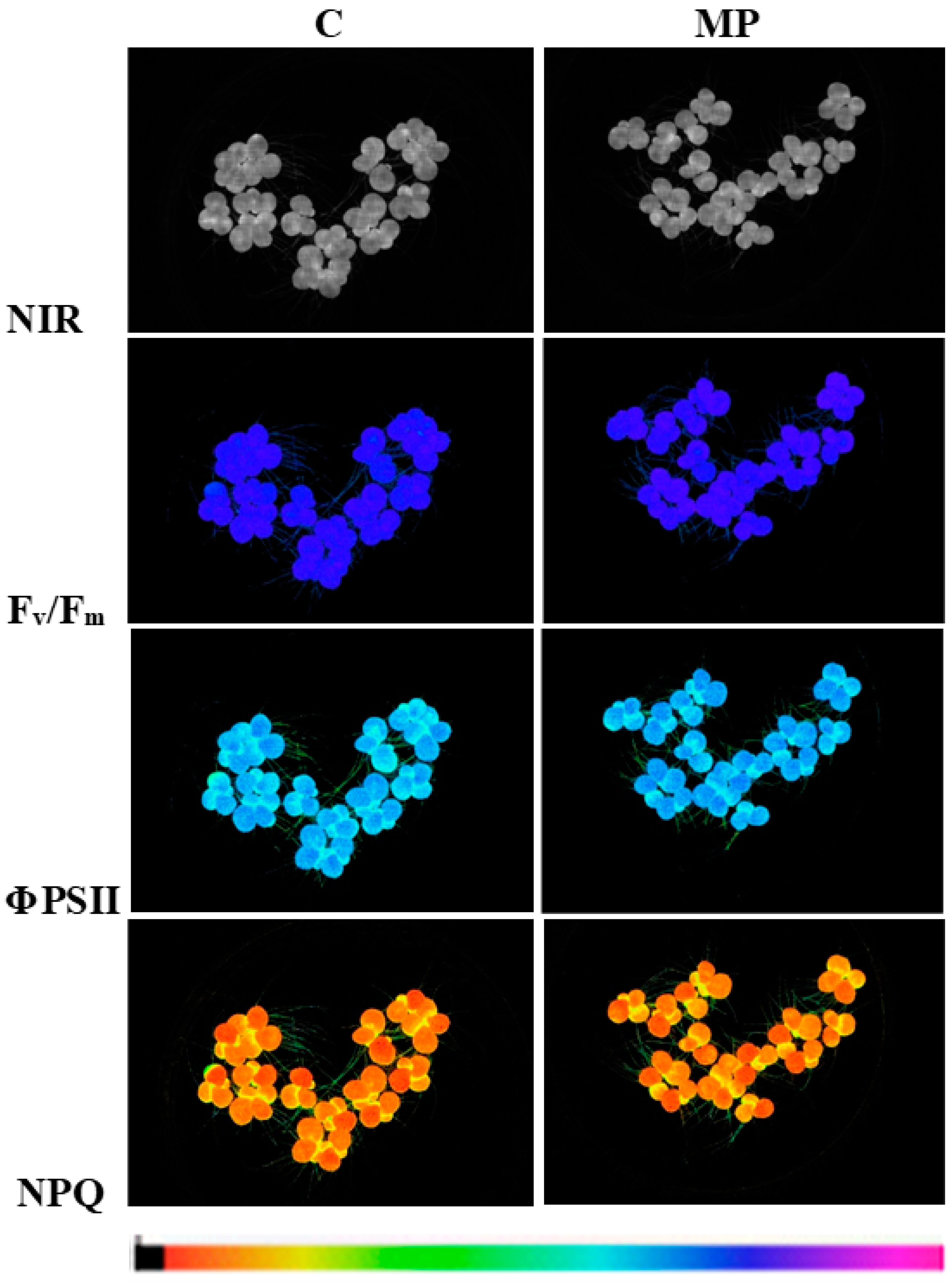
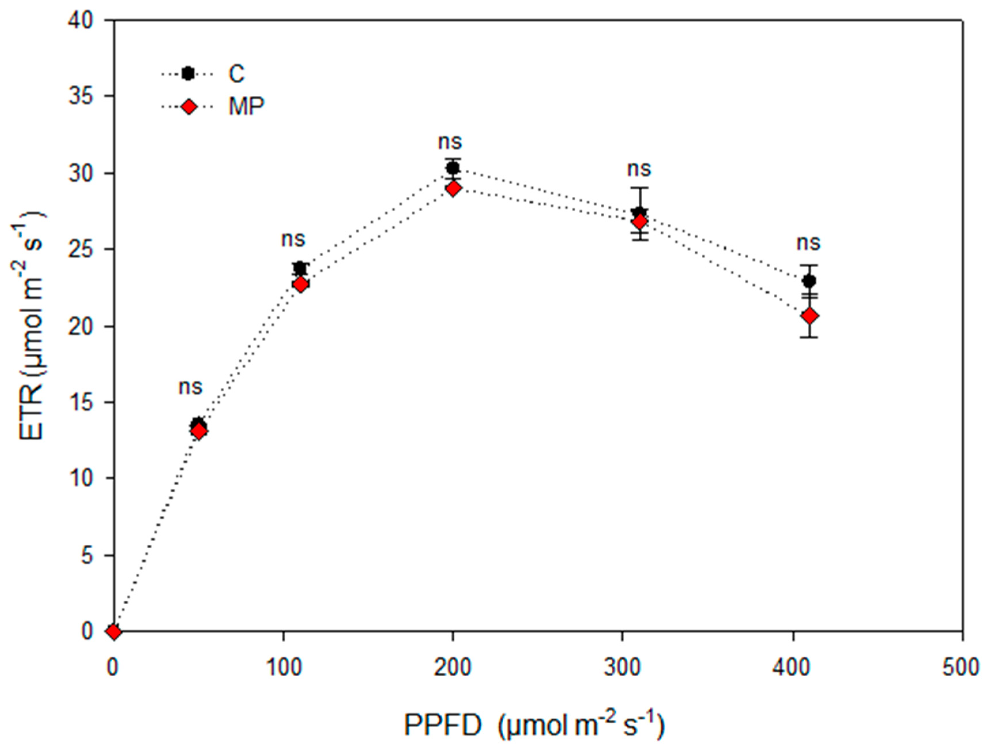
2.4. Plant Biometric and Physiological Traits Evaluation

By using the EcoTox Photosystem Tool (ETPT) [48], the main growth parameters and the physiological state of the photosynthetic apparatus of S. polyrhiza fronds were measured in real time and in a non-destructive way through the acquisition of whole plant imaging for chlorophyll fluorescence (Imaging PAM, Walz, Germany) under different light intensities.
At the beginning (t0) and at the end of the experiment (7 days, t7), a set of biometric parameters was determined through whole plant imaging and data processing by an imaging analysis software (ImageJ, IJ 1.46r, http://imagej.nih.gov/ij/ (accessed on 2 September 2021)) as in Pietrini et al. [56].
Concomitantly, chlorophyll fluorescence parameters and their associated images were measured on S. polyrhiza plants to evaluate the performance of the photosynthetic apparatus [57]. The parameters of chlorophyll fluorescence reported in Figure 1 were measured and calculated as previously reported [58]. Moreover, by applying the modified equation of Evans [59], the chlorophyll content was obtained [60].
Furthermore, to provide detailed information on the saturation characteristics of electron transport, as well as the overall photosynthetic performance of S. polyrhiza plants, the light-response curves (LCs) of the electron transport rate (ETR) were obtained by exposing the plants to five increasing irradiance levels, from 60 to 410 µmol m–2 s–1. By fitting the LCs to the equation of Platt et al. [61], the parameters reported in Figure 2 were calculated.
At the end of the experiment, on the same plant material used for the biometric and chlorophyll analyses, leaf reflectance spectra were obtained in the spectral range 350–1025 nm by using the ASD FieldSpec 3 spectroradiometer (Analytical Spectral Devices Inc., Boulder, CO, USA). Data were sampled at intervals of 1.4 nm with a spectral resolution of 3 nm and a white reference panel (Spectralon) was used to calibrate spectral measurement. An external 50 W halogen lamp was set up with an illumination angle of 45°. Specifically, the gun probe was placed on a stand unit orthogonally to the plate, respecting a fixed distance (0.5 cm) to the plant layer in order to assure a field of view (FOV) of approximately 2 mm (using a 25° bare fiber-optic). Reflectance spectra were recorded as the ratio of sample data to white reference data under the same illumination and viewing conditions. The mean of the five spectra was determined to provide a single spectral value. Reflectance data can be converted into estimates of important plant biophysical and ecophysiological traits by calculating different spectral reflectance indices. Six spectral reflectance indices were derived from the collected data, according to the following equations where R is the reflectance value measured in each band expressed in nm that is indicated by the subscript number:
-
Photochemical Reflectance Index (PRI) = (R531 − R570)/(R531 + R570) [62]
-
Pigment Specific Simple Ratio a (PSSRa) = (R800)/(R680) [63]
-
Pigment Specific Simple Ratio b (PSSRb) = (R800)/(R635) [63]
-
Pigment Specific Simple Ratio c (PSSRc) = (R800)/(R470) [63]
-
Normalized Phaeophytinization Index (NPQI) = (R415 − R435)/(R415 + R435) [64]
-
Anthocyanin Reflectance Index (ARI) = (1/R550)/(1/R700) [65].

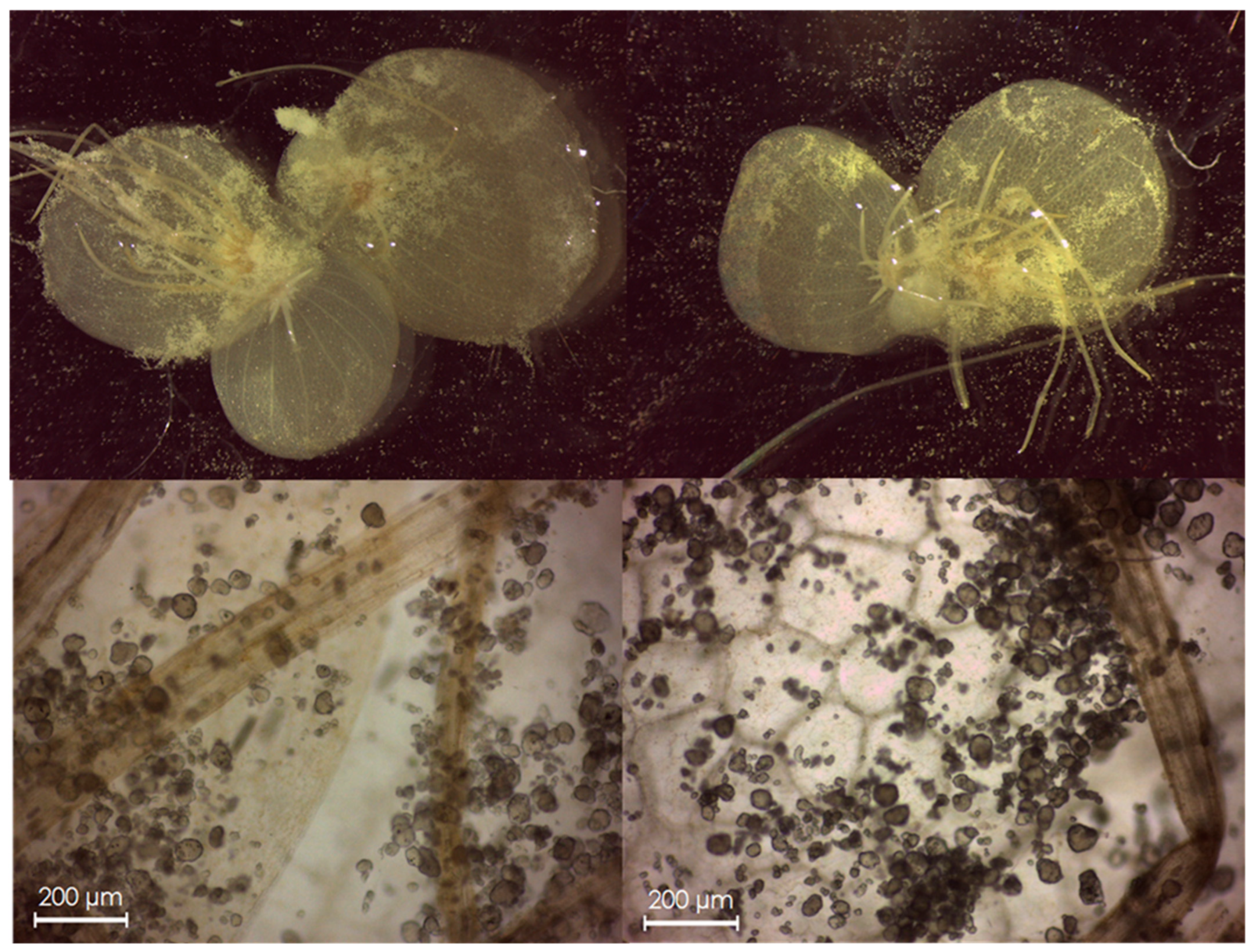
2.5. Evaluation of MP Transfer from S. polyrhiza to E. veneris

Preliminary trials revealed the maximum density of MP particles adsorbed on S. polyrhiza plants after 24 h of exposure (Figure 3). Thus, the trophic transfer experiment was performed by carefully removing the plant colonies from the Petri plates (see 2.2) after 24 h and placing them in a Petri plate with distilled water. Plants were immersed, gently shaken and then put in a glass container to be transferred for the confirmation of the MP adsorption by microscope (Leica DM750; Wetzlar, Germany) at 40× magnification with a Digital HD camera (Leica ICC50HD; Wetzlar, Germany). The same plants were used to feed the amphipod Echinogammarus veneris. Fifty specimens were individually placed in 100 mL beakers filled with 100 mL tap water in the presence of a single S. polyrhiza plant previously treated with MPs for 24 h (Figure S2). As a negative control, 15 specimens were placed in the same conditions with untreated S. polyrhiza plants. Amphipods were kept in the absence of food for 48 h prior to feeding to ensure starvation and overcome the gut resident time, presumably similar to that demonstrated by Ugolini et al. [66] for the MPs ingested by the talitrid amphipod Talitrus saltator (24 h). The procedures for MP staining and observation were previously reported [18]. A procedural blank was also performed to exclude contaminations, and no MPs were observed.

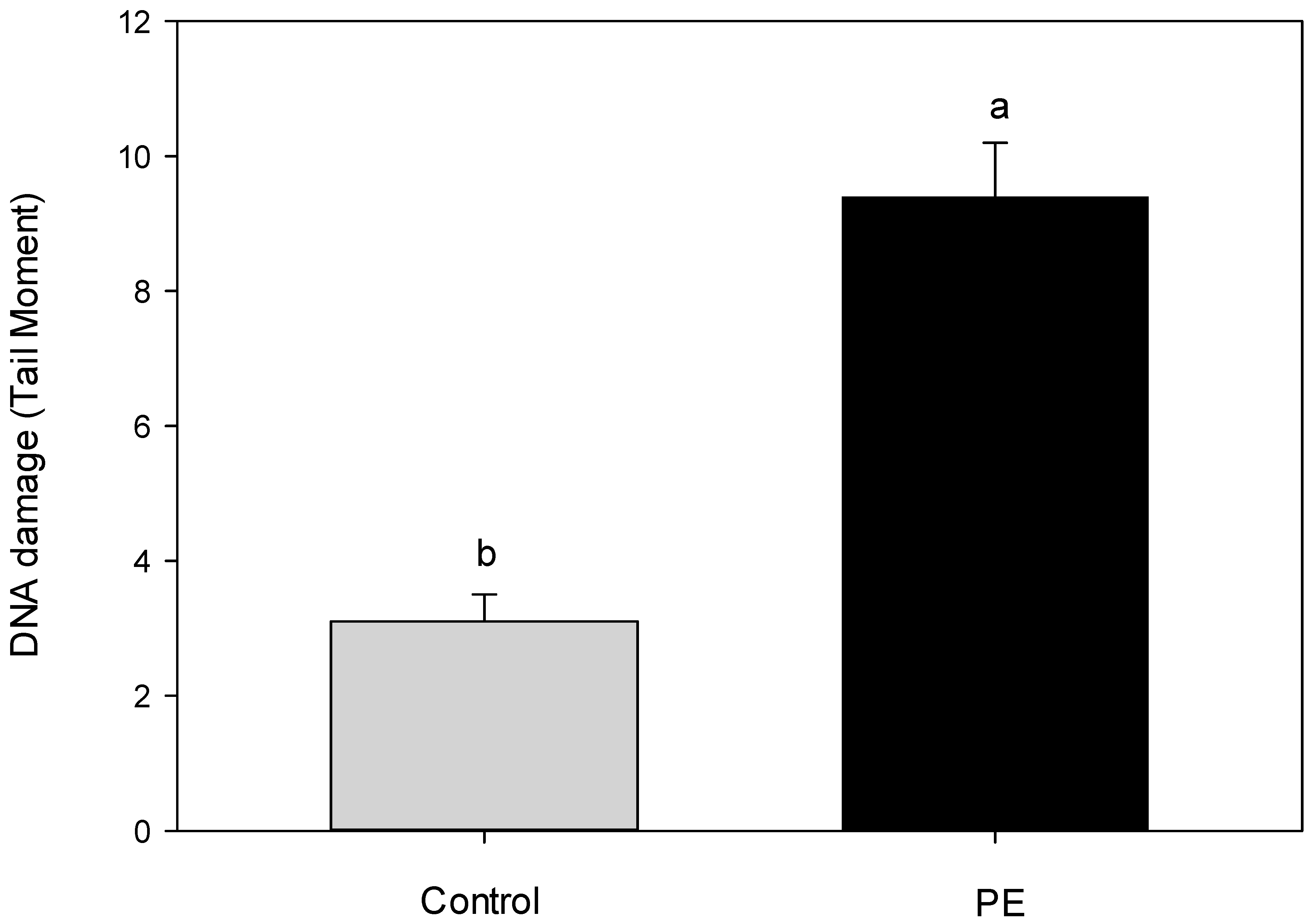
2.6. Genotoxicity Test in E. veneris

Comet assay is utilized as a biomarker of exposure, it is a rapid and sensitive tool that can be performed on different tissues and cell types and it has increasingly been used in genotoxicity testing. This technique quantifies DNA damage through a gel electrophoresis-based method [67]. The alkaline version of the Comet assay that enables the visualization of DNA double and single strand breaks (DNA DSBs and SSBs), but also alkali-labile sites (ALS), was performed in this study [68].
Five specimens were exposed to each condition: MPs in water (6 mg in 150 mL, approx. 5000 MP particles/mL); MPs with water and Triton X-100 (6 mg in 150 mL + 0.5 µL); negative control 1 (water only); negative control 2 (water + Triton X-100). All the experiments were replicated 2 times, using 150 mL glass beakers with 5 specimens each. The Comet test was conducted after 24 h exposure time, analyzing 40 specimens in total, following the procedure described in Cosentino et al. [55]. The images obtained were analyzed by the software © 2017 TriTekCorp™ CometScore (Sumerduck, VA, USA), version 2.0 measuring the Tail Moment (TM), defined as the product of the tail length and the fraction of total DNA in the tail.

2.7. Statistics

Experiments on plants were set up taking into account the OECD 221 guidelines for ecotoxicological test (7-day test, OECD 2006) [69]. Two independent experimental trials were run, each consisting of three independent replicates per thesis, unless otherwise stated. Normally distributed data were processed by one-way ANOVA in order to evaluate the effects of the MP presence on plant endpoints at different time intervals, by using the SPSS (Chicago, IL, USA) software tool. Statistical significance of the mean data was assessed by LSD test (p ≤ 0.05), unless otherwise stated.
The results of the genotoxicity test on amphipods, expressed as TM values, were presented as means ± SE, and analyzed using the statistical analysis program PAST3 (version 1.0.0.0., Oslo, Norway). Since the data were not normally distributed (Shapiro–Wilk test), a nonparametric test (Kruskal–Wallis) was used in order to compare each treatment with the relative control group and considered significant for p ≤ 0.05.

3. Results and Discussion

The effects of MPs on the growth of S. polyrhiza plants were evaluated by calculating biometric indices, commonly used as proxies for toxicity assessment in duckweeds [56,60]. As clearly reported in Table 1, none of the indices showed a significant difference in MP-treated plants in comparison to control ones at the end of the experimental time-course. Specifically, no alteration in the frond area and number of plants was detected because of the MP exposure. Consistently, no visible signs of toxicity were observed in the fronds of MP-treated S. polyrhiza plants (data not shown). The lack of toxic effects on the growth of duckweed plants exposed to MPs is consistent with the few reports dealing with this issue in literature. Dovidat et al. [20] observed that the exposure of S. polyrhiza plants to nano- and micro-sized polystyrene (PS) plastic particles resulted in unaltered growth and Kalčíková et al. [70] reported no effects in the frond growth of L. minor treated with plastic beads. More recently, Rozman et al. [71] showed the absence of toxic effects on the growth of L. minor plants after their treatment with particles of various environmentally relevant plastic types. It must be stressed that under lab experiments MPs cannot exert the role of vector of other contaminants (i.e., the Trojan Horse effect) likely affecting the biological processes on many aquatic organisms.
Chlorophyll content has long been recognized as a valuable physiological endpoint to be analyzed for ecotoxicological assessment in duckweeds [24,69]. Moreover, to better describe the effects of suspected toxicants on the photosynthetic machinery, some eco-physiological traits such as chlorophyll content [58], fluorescence parameters [72,73], and spectral reflectance indices [74] were targeted in the literature. In Figure 1 and Table 2, chlorophyll fluorescence parameters and their associated images were analyzed in S. polyrhiza plants at the end of the MP treatment. Data reported in Table 2 highlighted that, apart from a slight effect on chlorophyll content and leaf-absorptivity, the exposure of plants to MPs did not alter the physiological state of the photosynthetic apparatus. In fact, no significant differences in the chlorophyll fluorescence parameters (Fv/Fm, Φ PSII, NPQ, and ETR) between control and treated plants were observed. It is noteworthy that, as reported by Maxwell and Johnson [57], an Fv/Fm ratio in the range of 0.75 to 0.83 is the approximate optimal value for many plant species, thus confirming that S. polyrhiza plants were not affected by MP treatment. Therefore, the slight reduction in chlorophyll content and leaf absorptivity found in MP-treated plants did not cause any impairment to their photosynthetic apparatus. Moreover, the representative images of the chlorophyll fluorescence parameters (Fv/Fm, Φ PSII, and NPQ) in fronds of Spirodela (Figure 1) showed an almost homogeneous pattern of distribution of chlorophyll fluorescence both in the control and treated plants, highlighting the absence of detrimental effects at the photosynthetic level. Consistent with these findings, no effect of MPs on plant photosynthetic performance was observed after analyzing data of light-response curves (LCs) of electron transport rate (ETR) reported in Figure 2 and Table 3. In fact, the pattern of LCs (Figure 2) was not affected by exposure to MPs as confirmed by unchanged values of photosynthetic efficiency (α), maximum electron transport rate (ETRmax), and the minimum saturating irradiance (Ek) (Table 3). Finally, to further characterize the effects of MPs at the physiological level, the optical properties of S. polyrhiza fronds were investigated by analyzing some spectral reflectance indices obtained at the end of MP treatment by a spectroradiometer (ASD FieldSpec3). Leaf reflectance properties depend on the leaf surface and internal structure characteristics, as well as on the concentration and distribution of biochemical components; therefore, indices measured through reflected light spectra analysis can be used to assess the plant physiological status [75]. In Table 4, in accordance with the data from Table 2, only indices related to the content of pigments, namely PSSRa, PSSRb, and PSSRc associated with the chlorophyll a and b, and carotenoid content, respectively [63], highlighted a moderate effect exerted by the presence of MPs in the nutrient solution. In this regard, a possible effect of the MPs adsorbed on roots on nutrient element uptake, likely impairing the metabolism of pigments, cannot be ruled out. Preliminary experiments allowed the exclusion of a role of MPs in partly shielding the leaf surface, possibly reducing the spectral data acquisition. Consistently, the values of the other spectral reflectance indices such as PRI, used as an index of photosynthetic performance [76]; NPQI, associated with the level of chlorophyll degradation [63] and ARI, related to the anthocyanin content [65], did not show significant differences between the control and MP-treated plants.
Taken together, data on plant physiological performance revealed a substantially unaltered photosynthetic apparatus in MP-treated S. polyrhiza plants in comparison to their control. This feature confirmed previous findings reported in duckweed plants treated with different MP types by Dovidat et al. [20] on S. polyrhiza and Kalčíková et al. [70] and Mateos-Cardenas et al. [6,21] on L. minor, indicating that the adsorption of plastic particles on root and frond surfaces does not result in the impairment of photosynthetic performance in aquatic plants.
To evaluate the transfer of the MPs adsorbed on fronds to the amphipod E. veneris, Nile red staining was utilized to color, highlight, and count in fluorescence the MP particles in the extracted stomach contents (Figure S3). This faster and cost-effective method was used for the recovery of applied MPs on a total of 65 amphipods (n = 15 belonged to the control group, and n = 50 to MP treatments). The gut content of control group specimens, exposed to clean S. polyrhiza plants, was MP-free. On the contrary, all the other processed samples showed the MPs used to contaminate S. polyrhiza, clearly indicating the feeding of E. veneris specimens on these plants. Namely, after 24 h of feeding, a mean value of 7.6 ± 2.6 MPs/individual was observed. This value is higher than that obtained by Mateos-Cardenas et al. [21] in a trophic transfer experiment from Lemna minor to the gammarid Gammarus duebeni, though E. veneris is a smaller species than G. duebeni. However, it is necessary to underline that S. polyrhiza is probably able to adsorb and concentrate a greater number of MPs than L. minor, given the presence of a multiple root system on each frond, with as many as 12 adventitious roots [77], as its specific name indicates (polyrhiza = many roots). Dovidat et al. [20] observed plastic particles adsorbed to the roots of S. polyrhiza after exposure to PS particles and argued about the possible transfer from this species to herbivorous aquatic invertebrates. This process was clearly demonstrated in our experiments.
The role of amphipods as an entry point for MPs in the aquatic trophic chain is highlighted by the finding that they can ingest MPs, and these, after passing through the digestive system, can be expelled into the fecal pellets, thus becoming available again in the debris chain. It must also be emphasized that the gammarids could fragment the particles, as Mateos-Cardenas et al. [22] highlighted in the freshwater amphipod Gammarus duebeni, contributing to the degradation and size reduction in the particles ingested. In the present work, the MPs found in the digestive tracts were of a smaller average size (26.2 ± 1.6 μm) than that of the MPs used to contaminate S. polyrhiza (43.6 ± 1.2 μm). However, because of the irregular shape of the MPs used, it is not possible to infer if this is due to a digestive fragmentation, similar to that observed in G. duebeni, or selection before the ingestion. The predated E. veneris, however, will transfer their MP body burden to higher trophic levels. Since gammarids are often dominant in terms of biomass in macrobenthic communities, the impact of the MP transfer could be very relevant. Gammarids represent one example of the selected prey of some salmonid species such as brown trout [78]. Lollobrigidi et al. [79] reported that Salmo cettii specialized on Echinogammarus sp. (E. veneris, pers. comm.), with Echinogammarus sp. representing 67.22% of the total amount of food items in their stomachs. The availability of the MPs ingested for uptake to the higher levels of the food web makes them also a threat for human populations. The presence of MPs in the edible part of fish (muscle tissue) has already been proven [80,81] although the internalization process of the particles and MPs accumulation have not been completely understood. However, Barboza et al. [80] demonstrated that the main route of entry of small MPs into the muscle of fish is the digestive system. This evidence demonstrates the risk for human consumption of the translocation of MPs in the food chain and the importance of model systems to study these processes.
A genotoxic assessment was performed on gammarid specimens directly exposed or not to MPs in water. On these samples, the DNA-induced damage, resulting in DNA fragmentation, was determined as TM after a 24 h exposure by the Comet assay. Exposure of the gammarids resulted in significant damage to the DNA compared to the control group, with TM values of 9.4 (Figure 4). The DNA-damaging effects of MPs have rarely been investigated in invertebrates [82], and, to our knowledge, the genotoxic effect observed in this study represents the first evidence in amphipods. In fact, in the literature, there has been only one study reported, specifically on crustaceans (the decapod Atydae Neocaridina davidi), in which PS MPs significantly increased the DNA tail moment after 24 h exposure [83]. Polystyrene MPs also increased the DNA damage in Scrobicularia plana hemocytes [84] while the genotoxic effects of PE MPs were only highlighted in hemocytes of Mytilus galloprovincialis, causing a significant enhancement of DNA strand breaks [85]. Studies on vertebrates showed an increase in micronuclei (MN) and nuclear buds (NBUD) in Salmo trutta, at the larval stage, caused by PS, PET, and PE MPs exposure, with PE causing the most damage (Jakubowska et al., 2020) [86]. Moreover, environmental MPs in Japanese Medaka (Oryzias latipes) caused a significant induction of DNA damage [87]. Although the literature on this subject is still limited, all this evidence induces the hypothesis that at least the plastic polymers analyzed or the additives contained therein are directly bioavailable to the organisms, resulting in their ability to exert toxic effects on DNA. Even if the mechanism of DNA damage induction remains unknown, the most widespread hypothesis attributes the cause of such damages to the production of reactive oxygen species (ROS) as a response to foreign particles inside cells or an inflammatory response caused by interaction with cell membranes [82]. This hypothesis is supported by the experimental evidence of Ribeiro et al. [84], who highlighted an oxidative stress response to PS in gills and digestive gland cells of S. plana and by Revel et al. [88] who measured an increase in the antioxidant enzyme and acid phosphatase activities after exposure of Mytilus edulis to PE and PP. Increased ROS production and inflammatory response to MP exposure are linked to biochemical processes and can be responsible for the DNA damage observed in the present study and in those reported in the literature. However, at the present stage, it is not possible to clearly ascribe the induction of such damaging effects to the polymer as well or to additives and plasticizers used in plastic production [55,60].

4. Conclusions

The large presence of MPs in the environmental compartments is demanding increasing efforts for evaluating their impact on biota. Therefore, apart from the direct or indirect biological effects of MPs, a particular concern is posed in relation to the possible transfer and magnification effect along the food web involving humans. In this regard, the mechanisms by which MPs enter and move into the food chain are rarely investigated. In this work, the consequences of the exposure of PE MPs on a primary producer (Spirodela polyrhiza (L) Schleid.) and a primary consumer (Echinogammarus veneris) have been studied at a different level of biological organization. In particular, the lack of a direct toxic effect of MPs on plants was highlighted by a novel experimental approach (using the Eco-Tox Photo system Tool (ETPT)), both at the growth and photosynthetic performance levels. A clear genotoxic effect of PE MPs on gammarids was instead demonstrated for the first time by means of the Comet assay. The transfer of PE MPs from S. polyrhiza to E. veneris was also evidenced, paralleling the observation previously reported for other freshwater organisms. The overall results also put in evidence the complexity of the interaction between MPs and living organisms at the lab scale. Finally, the potential transfer of PE particles from a primary producer to a primary consumer strengthens the demand for further knowledge about the risks to animal and human health associated with the trophic transfer of MPs in the food network of the freshwater ecosystem.

Supplementary Materials

The following supporting information can be downloaded at: https://www.mdpi.com/article/10.3390/w15050921/s1: Figure S1: Microscope images of PE MPs, used to perform the exposures, observed in brightfield (left) and, stained with Nile red, in green fluorescence (ex. 450–490; em 515–565 nm) (right); Figure S2: Specimens of E. veneris individually placed in 100 mL beakers filled with 100 mL tap water in presence of a single S. polyrhiza colony previously treated with PE MPs for 24 h (left); Particular of E. veneris individual clung to plant roots during feeding (right); Figure S3: Microscope images of PE MPs, from digestive tracts of E. veneris, on a filter stained with Nile red, observed in green fluorescence; Figure S4: Representative images of nuclei from Echinogammarus veneris hemocytes, treated (B) or not (A) with 40 mg/L PE MPs in water for 24 h, exhibiting different DNA damage level after Alkaline Comet assay procedure and EtBr staining.

Author Contributions

Conceptualization, V.I., M.Z. and F.P.; Data curation, V.I. and F.P.; Formal analysis, V.I. and F.P.; Investigation, V.I., L.P., S.C., F.L., G.S. and F.P.; Methodology, V.I. and F.P.; Project administration, M.Z.; Resources, L.P., S.C. and F.L.; Supervision, M.Z.; Validation, V.I., M.Z. and F.P.; Visualization, L.P.; Writing—original draft, L.P. and M.Z.; Writing—review and editing, V.I., M.Z. and F.P. All authors have read and agreed to the published version of the manuscript.

Funding

This research received no external funding.

Institutional Review Board Statement

Not applicable.

Informed Consent Statement

Not applicable.

Data Availability Statement

Not applicable.

Conflicts of Interest

The authors declare that they have no competing interest.

References

  1. Maheswaran, B.; Karmegam, N.; Al-Ansari, M.; Subbaiya, R.; Al-Humaid, L.; Raj, J.S.; Govarthanan, M. Assessment, characterization, and quantification of microplastics from river sediments. Chemosphere 2022, 298, 134268. [Google Scholar] [CrossRef]
  2. Iannilli, V.; Pasquali, V.; Setini, A.; Corami, F. First evidence of microplastics ingestion in benthic amphipods from Svalbard. Environ. Res. 2019, 179, 108811. [Google Scholar] [CrossRef]
  3. Mishra, A.K.; Singh, J.; Mishra, P.P. Microplastics in polar regions: An early warning to the world’s pristine ecosystem. Sci. Total Environ. 2021, 784, 147149. [Google Scholar] [CrossRef]
  4. Fagiano, V.; Compa, M.; Alomar, C.; Rios-Fuster, B.; Morató, M.; Capó, X.; Deudero, S. Breaking the paradigm: Marine sediments hold two-fold microplastics than sea surface waters and are dominated by fibers. Sci. Total. Environ. 2023, 858, 159722. [Google Scholar] [CrossRef]
  5. Wright, S.L.; Thompson, R.C.; Galloway, T.S. The physical impacts of microplastics on marine organisms: A review. Environ. Pollut. 2013, 178, 483–492. [Google Scholar] [CrossRef]
  6. Mateos-Cárdenas, A.; van Pelt, F.N.; O’Halloran, J.; Jansen, M.A. Adsorption, uptake and toxicity of micro- and nanoplastics: Effects on terrestrial plants and aquatic macrophytes. Environ. Pollut. 2021, 284, 117183. [Google Scholar] [CrossRef]
  7. Frias, J.; Nash, R. Microplastics: Finding a consensus on the definition. Mar. Pollut. Bull. 2018, 138, 145–147. [Google Scholar] [CrossRef]
  8. Hartmann, N.B.; Hüffer, T.; Thompson, R.C.; Hassellöv, M.; Verschoor, A.; Daugaard, A.E.; Rist, S.; Karlsson, T.M.; Brennholt, N.; Cole, M.; et al. Are We Speaking the Same Language? Recommendations for a Definition and Categorization Framework for Plastic Debris. Environ. Sci. Technol. 2019, 53, 1039–1047. [Google Scholar] [CrossRef] [Green Version]
  9. McIvor, A.J.; Pires, R.; Lopes, C.; Raimundo, J.; Campos, P.F.; Pais, M.P.; Canning-Clode, J.; Dinis, A. Assessing microplastic exposure of the Critically Endangered Mediterranean monk seal (Monachus monachus) on a remote oceanic island. Sci. Total Environ. 2023, 856, 159077. [Google Scholar] [CrossRef]
  10. Peng, L.; Fu, D.; Qi, H.; Lan, C.Q.; Yu, H.; Ge, C. Micro-and nano-plastics in marine environment: Source, distribution and threats—A review. Sci. Total Environ. 2020, 698, 134254. [Google Scholar] [CrossRef]
  11. Wang, J.; Coffin, S.; Sun, C.; Schlenk, D.; Gan, J. Negligible effects of microplastics on animal fitness and HOC bioaccumulation in earthworm Eisenia fetida in soil. Environ. Pollut. 2019, 249, 776–784. [Google Scholar] [CrossRef]
  12. Xu, B.; Liu, F.; Cryder, Z.; Huang, D.; Lu, Z.; He, Y.; Wang, H.; Lu, Z.; Brookes, P.C.; Xu, J. Microplastics in the soil environment: Occurrence, risks, interactions and fate–a review. Crit. Rev. Environ. Sci. Technol. 2020, 50, 2175–2222. [Google Scholar] [CrossRef]
  13. Eerkes-Medrano, D.; Thompson, R. Occurrence, fate, and effect of microplastics in freshwater systems. In Microplastic Contamination in Aquatic Environments; Elsevier: Amsterdam, The Netherlands, 2018; pp. 95–132. [Google Scholar]
  14. Koutnik, V.S.; Leonard, J.; Alkidim, S.; DePrima, F.J.; Ravi, S.; Hoek, E.M.; Mohanty, S.K. Distribution of microplastics in soil and freshwater environments: Global analysis and framework for transport modeling. Environ. Pollut. 2021, 274, 116552. [Google Scholar] [CrossRef]
  15. Iannilli, V.; Di Gennaro, A.; Lecce, F.; Sighicelli, M.; Falconieri, M.; Pietrelli, L.; Poeta, G.; Battisti, C. Microplastics in Talitrus saltator (Crustacea, Amphipoda): New evidence of ingestion from natural contexts. Environ. Sci. Pollut. Res. 2018, 25, 28725–28729. [Google Scholar] [CrossRef]
  16. Corami, F.; Rosso, B.; Iannilli, V.; Ciadamidaro, S.; Bravo, B.; Barbante, C. Occurrence and Characterization of Small Microplastics (<100 μm), Additives, and Plasticizers in Larvae of Simuliidae. Toxics 2022, 10, 383. [Google Scholar] [CrossRef]
  17. Atamanalp, M.; Kokturk, M.; Gündüz, F.; Parlak, V.; Ucar, A.; Alwazeer, D.; Alak, G. The Use of Zebra Mussel (Dreissena polymorpha) as a Sentinel Species for the Microplastic Pollution of Freshwater: The Case of Beyhan Dam Lake, Turkey. Sustainability 2023, 15, 1422. [Google Scholar] [CrossRef]
  18. Iannilli, V.; Corami, F.; Grasso, P.; Lecce, F.; Buttinelli, M.; Setini, A. Plastic abundance and seasonal variation on the shorelines of three volcanic lakes in Central Italy: Can amphipods help detect contamination? Environ. Sci. Pollut. Res. 2020, 27, 14711–14722. [Google Scholar] [CrossRef]
  19. Rozman, U.; Kalčíková, G. The Response of Duckweed Lemna minor to Microplastics and Its Potential Use as a Bioindicator of Microplastic Pollution. Plants 2022, 11, 2953. [Google Scholar] [CrossRef]
  20. Dovidat, L.C.; Brinkmann, B.W.; Vijver, M.G.; Bosker, T. Plastic particles adsorb to the roots of freshwater vascular plant Spirodela polyrhiza but do not impair growth. Limnol. Oceanogr. Lett. 2020, 5, 37–45. [Google Scholar] [CrossRef] [Green Version]
  21. Mateos-Cárdenas, A.; Scott, D.T.; Seitmaganbetova, G.; van Pelt Frank NA, M.; O’Halloran, J.; Jansen Marcel, A.K. Polyethylene microplastics adhere to Lemna minor (L.), yet have no effects on plant growth or feeding by Gammarus duebeni (Lillj.). Sci. Total Environ. 2019, 689, 413–421. [Google Scholar] [CrossRef]
  22. Mateos-Cárdenas, A.; O’Halloran, J.; van Pelt, F.N.A.M.; Jansen, M.A.K. Rapid fragmentation of microplastics by the freshwater amphipod Gammarus duebeni (Lillj.). Sci. Rep. 2020, 10, 1–12. [Google Scholar] [CrossRef]
  23. Baudo, R.; Foudoulakis, M.; Arapis, G.; Perdaen, K.; Lanneau, W.; Paxinou, A.C.; Kouvdou, S.; Persoone, G. History and sensitivity comparison of the Spirodela polyrhiza microbiotest and Lemna toxicity tests. Knowl. Manag. Aquat. Ecosyst. 2015, 416, 23. [Google Scholar] [CrossRef] [Green Version]
  24. Pietrini, F.; Di Baccio, D.; Aceña, J.; Pérez, S.; Barceló, D.; Zacchini, M. Ibuprofen exposure in Lemna gibba L.: Evaluation of growth and phytotoxic indicators, detection of ibuprofen and identification of its metabolites in plant and in the medium. J. Hazard. Mater. 2015, 300, 189–193. [Google Scholar] [CrossRef]
  25. Mkandawire, M.; da Silva, J.A.T.; Dudel, E.G. The Lemna Bioassay: Contemporary Issues as the Most Standardized Plant Bioassay for Aquatic Ecotoxicology. Crit. Rev. Environ. Sci. Technol. 2014, 44, 154–197. [Google Scholar] [CrossRef]
  26. Ceschin, S.; Bellini, A.; Scalici, M. Aquatic plants and ecotoxicological assessment in freshwater ecosystems: A review. Environ. Sci. Pollut. Res. 2020, 28, 4975–4988. [Google Scholar] [CrossRef]
  27. Kalčíková, G. Aquatic vascular plants—A forgotten piece of nature in microplastic research. Environ. Pollut. 2020, 262, 114354. [Google Scholar] [CrossRef]
  28. Kokalj, A.J.; Kuehnel, D.; Puntar, B.; Gotvajn, A.; Kalčikova, G. An exploratory ecotoxicity study of primary microplastics versus aged in natural waters and wastewaters. Environ. Pollut. 2019, 254, 112980. [Google Scholar] [CrossRef]
  29. Rozman, U.; Kokalj, A.J.; Dolar, A.; Drobne, D.; Kalčíková, G. Long-term interactions between microplastics and floating macrophyte Lemna minor: The potential for phytoremediation of microplastics in the aquatic environment. Sci. Total. Environ. 2022, 831, 154866. [Google Scholar] [CrossRef]
  30. Xiao, F.; Feng, L.J.; Sun, X.D.; Wang, Y.; Wang, Z.W.; Zhu, F.P.; Yuan, X.Z. Do Polystyrene Nanoplastics Have Similar Effects on Duckweed (Lemna minor L.) at Environmentally Relevant and Observed-Effect Concentrations? Environ. Sci. Technol. 2022, 56, 4071–4079. [Google Scholar] [CrossRef]
  31. Hildebrandt, L.; Nack, F.; Zimmermann, T.; Pröfrock, D. Microplastics as a Trojan horse for trace metals. J. Hazard. Mater. Lett. 2021, 2, 100035. [Google Scholar] [CrossRef]
  32. Kumar, R.; Ivy, N.; Bhattacharya, S.; Dey, A.; Sharma, P. Coupled effects of microplastics and heavy metals on plants: Uptake, bioaccumulation, and environmental health perspectives. Sci. Total. Environ. 2022, 836, 155619. [Google Scholar] [CrossRef]
  33. Zhou, J.; Liu, X.; Jiang, H.; Li, X.; Li, W.; Cao, Y. Antidote or Trojan horse for submerged macrophytes: Role of microplastics in copper toxicity in aquatic environments. Water Res. 2022, 216, 118354. [Google Scholar] [CrossRef]
  34. Mao, H.; Yang, H.; Xu, Z.; Yang, Y.; Zhang, X.; Huang, F.; Wei, L.; Li, Z. Microplastics and co-pollutant with ciprofloxacin affect interactions between free-floating macrophytes. Environ. Pollut. 2023, 316, 120546. [Google Scholar] [CrossRef] [PubMed]
  35. Yu, H.; Peng, J.; Cao, X.; Wang, Y.; Zhang, Z.; Xu, Y.; Qi, W. Effects of microplastics and glyphosate on growth rate, morphological plasticity, photosynthesis, and oxidative stress in the aquatic species Salvinia cucullata. Environ. Pollut. 2021, 279, 116900. [Google Scholar] [CrossRef] [PubMed]
  36. Sfriso, A.A.; Tomio, Y.; Juhmani, A.-S.; Sfriso, A.; Munari, C.; Mistri, M. Macrophytes: A Temporary Sink for Microplastics in Transitional Water Systems. Water 2021, 13, 3032. [Google Scholar] [CrossRef]
  37. Robles-Vargas, D. Toxicity of Agrochemicals on Fresh- water Invertebrates—A Short Review. In Toxicity and Hazard of Agrochemicals; Larramendy, M.L., Soloneski, S., Eds.; IntechOpen Limited: London, UK, 2015. [Google Scholar] [CrossRef] [Green Version]
  38. Welton, J.S. Life-history and production of the amphipod Gammarus pulex in a Dorset chalk stream. Freshw. Biol. 1979, 9, 263–275. [Google Scholar] [CrossRef]
  39. Farrell, P.; Nelson, K. Trophic level transfer of microplastic: Mytilus edulis (L.) to Carcinus maenas (L.). Environ. Pollut. 2013, 177, 1–3. [Google Scholar] [CrossRef] [PubMed]
  40. Hurley, R.R.; Woodward, J.C.; Rothwell, J.J. Ingestion of Microplastics by Freshwater Tubifex Worms. Environ. Sci. Technol. 2017, 51, 12844–12851. [Google Scholar] [CrossRef]
  41. Jamieson, A.J.; Brooks, L.S.R.; Reid, W.D.K.; Piertney, S.B.; Narayanaswamy, B.E.; Linley, T.D. Microplastics and synthetic particles ingested by deep-sea amphipods in six of the deepest marine ecosystems on Earth. R. Soc. Open Sci. 2019, 6, 180667. [Google Scholar] [CrossRef] [PubMed] [Green Version]
  42. Au, S.Y.; Bruce, T.F.; Bridges, W.C.; Klaine, S.J. Responses of Hyalella azteca to acute and chronic microplastic exposures. Environ. Toxicol. Chem. 2015, 34, 2564–2572. [Google Scholar] [CrossRef] [PubMed]
  43. Blarer, P.; Burkhardt-Holm, P. Microplastics affect assimilation efficiency in the freshwater amphipod Gammarus fossarum. Environ. Sci. Pollut. Res. 2016, 23, 23522–23532. [Google Scholar] [CrossRef]
  44. Gerhardt, A. Effects of New and Aged Polyethylenterephthalat and Polylactic Acid on Gammarus fossarum (Crustacea: Amphipoda) during Long-Term Exposures. J. Environ. Prot. 2020, 11, 359–376. [Google Scholar] [CrossRef]
  45. EPA. Ecological Effects Test Guidelines: OPPTS 850.1020 Guideline Gammarid Acute Toxicity Test; 712–C–96–130; United States Environmental Protection Agency: Washington, DC, USA, 1996.
  46. Garcia-Galan, M.J.; Sordet, M.; Buleté, A.; Garric, J.; Vulliet, E. Evaluation of the influence of surfactants in the bioaccumulation kinetics of sulfamethoxazole and oxazepam in benthic invertebrates. Sci. Total Environ. 2017, 592, 554–564. [Google Scholar] [CrossRef] [PubMed] [Green Version]
  47. Yokota, K.; Waterfield, H.; Hastings, C.; Davidson, E.; Kwietniewski, E.; Wells, B. Finding the missing piece of the aquatic plastic pollution puzzle: Interaction between primary producers and microplastics. Limnol. Oceanogr. Lett. 2017, 2, 91–104. [Google Scholar] [CrossRef] [Green Version]
  48. Pietrini, F.; Zacchini, M. A New Ecotoxicity Assay for Aquatic Plants: Eco-Tox Photosystem Tool (ETPT). Trends Plant Sci. 2020, 25, 1266–1267. [Google Scholar] [CrossRef]
  49. Davolos, D.; Chimenti, C.; Ronci, L.; Setini, A.; Iannilli, V.; Pietrangeli, B.; De Matthaeis, E. An integrated study on Gammarus elvirae (Crustacea, Amphipoda): Perspectives for toxicology of arsenic-contaminated freshwater. Environ. Sci. Pollut. Res. 2015, 22, 15563–15570. [Google Scholar] [CrossRef]
  50. dos Santos, A.; Botelho, M.T.; Vannuci-Silva, M.; Artal, M.C.; Vacchi, F.I.; Magalhães, G.R.; Gomes, V.; Henry, T.B.; Umbuzeiro, G.D.A. The amphipod Parhyale hawaiensis as a promising model in ecotoxicology. Chemosphere 2022, 307, 135959. [Google Scholar] [CrossRef]
  51. Fiore, L.; Serranti, S.; Mazziotti, C.; Riccardi, E.; Benzi, M.; Bonifazi, G. Classification and distribution of freshwater microplastics along the Italian Po river by hyperspectral imaging. Environ. Sci. Pollut. Res. 2022, 29, 48588–48606. [Google Scholar] [CrossRef]
  52. Bazzanti, M.; Mastrantuono, L.; Solimini, A.G. Selecting macroinvertebrate taxa and metrics to assess eutrophication in different depth zones of Mediterranean lakes. Fundam. Appl. Limnol. 2012, 180, 133–143. [Google Scholar] [CrossRef]
  53. Ronci, L.; Meccoli, L.; Iannilli, V.; Menegoni, P.; De Matthaeis, E.; Setini, A. Comparison between active and pas- sive biomonitoring strategies for the assessment of genotoxicity and metal bioaccumulation in Echinogammarus veneris (Crustacea: Amphipoda). Ital. J. Zool. 2016, 83, 162–172. [Google Scholar] [CrossRef] [Green Version]
  54. Genomic Resources Development Consortium; Baratti, M.; Cattonaro, F.; Di Lorenzo, T.; Galassi, D.M.P.; Iannilli, V.; Iannucci, A.; Jensen, J.; Larsen, P.; Nielsen, R.O.; et al. Genomic Resources Notes Accepted 1 October 2014–30 November 2014. Mol. Ecol. Resour. 2015, 15, 458–459. [Google Scholar] [CrossRef]
  55. Cosentino, S.; Aureli, F.; Iannilli, V. Bisphenols A and its analogues induce genotoxic damage in marine and freshwater amphipods. Environ. Adv. 2022, 7, 100183. [Google Scholar] [CrossRef]
  56. Pietrini, F.; Passatore, L.; Fischetti, E.; Carloni, S.; Ferrario, C.; Polesello, S.; Zacchini, M. Evaluation of morpho-physiological traits and contaminant accumulation ability in Lemna minor L. treated with increasing perfluorooctanoic acid (PFOA) concentrations under laboratory conditions. Sci. Total Environ. 2019, 695, 133828. [Google Scholar] [CrossRef] [PubMed]
  57. Maxwell, K.; Johnson, G.N. Chlorophyll fluorescence: A practical guide. J. Exp. Bot. 2000, 51, 659–668. [Google Scholar] [CrossRef]
  58. Di Baccio, D.; Pietrini, F.; Bertolotto, P.; Pérez, S.; Barcelò, D.; Zacchini, M.; Donati, E. Response of Lemna gibba L. to high and environmentally relevant concentrations of ibuprofen: Removal, metabolism and morpho-physiological traits for biomonitoring of emerging contaminants. Sci. Total. Environ. 2017, 584–585, 363–373. [Google Scholar] [CrossRef] [PubMed]
  59. Evans, J. Photosynthetic Acclimation and Nitrogen Partitioning Within a Lucerne Canopy. II. Stability Through Time and Comparison With a Theoretical Optimum. Funct. Plant Biol. 1993, 20, 69–82. [Google Scholar] [CrossRef]
  60. Pietrini, F.; Iannilli, V.; Passatore, L.; Carloni, S.; Sciacca, G.; Cerasa, M.; Zacchini, M. Ecotoxicological and genotoxic effects of dimethyl phthalate (DMP) on Lemna minor L. and Spirodela polyrhiza (L.) Schleid. plants under a short-term laboratory assay. Sci. Total. Environ. 2022, 806, 150972. [Google Scholar] [CrossRef]
  61. Platt, T.; Gallegos, C.L.; Harrison, W.G. Photoinhibition of photosynthesis in natural assemblages of marine phytoplankton. J. Mar Res. 1980, 38, 687–701. [Google Scholar]
  62. Gamon, A.; Serrano, L.; Surfus, S. The photochemical reflectance index: An optical indicator of photosynthetic radiation use efficiency across species, functional types, and nutrient levels. Oecologia 1997, 112, 492–501. [Google Scholar] [CrossRef]
  63. Blackburn, G.A. Quantifying chlorophylls and carotenoids at leaf and canopy scales: An evaluation of some hyperspectral approaches. Remote Sens. Environ. 1998, 66, 273–285. [Google Scholar] [CrossRef]
  64. Penuelas, J.; Filella, I.; Lloret, P.; Mun Oz, F.; Vilajeliu, M. Reflectance assessment of mite effects on apple trees. Int. J. Remote Sens. 1995, 16, 2727–2733. [Google Scholar] [CrossRef]
  65. Gitelson, A.; Merzlyak, M.N.; Chivkunova, O.B. Optical properties and nondestructive estimation of anthocyanin content in plant leaves. Photochem. Photobiol. 2001, 74, 38–45. [Google Scholar] [CrossRef] [PubMed]
  66. Ugolini, A.; Ungherese, G.; Ciofini, M.; Lapucci, A.; Camaiti, M. Microplastic debris in sandhoppers. Estuar. Coast. Shelf Sci. 2013, 129, 19–22. [Google Scholar] [CrossRef]
  67. Singh, N.P. The comet assay: Reflections on its development, evolution and applications. Mutat. Res./Rev. Mutat. Res. 2016, 767, 23–30. [Google Scholar] [CrossRef] [PubMed]
  68. Bivehed, E.; Hellman, B. Flash-comet assay. Methodsx 2020, 7, 101161. [Google Scholar] [CrossRef] [PubMed]
  69. OECD guideline 221. OECD Guidelines for the Testing of Chemicals: Revised Proposal for a New Guideline 221, Lemna sp. Growth Inhibition Test; Organization of Economic Cooperation and Development: Paris, France, 2006. [Google Scholar]
  70. Kalčíková, G.; Gotvajn, A.; Kladnik, A.; Jemec, A. Impact of polyethylene microbeads on the floating freshwater plant duckweed Lemna minor. Environ. Pollut. 2017, 230, 1108–1115. [Google Scholar] [CrossRef] [PubMed]
  71. Rozman, U.; Turk, T.; Skalar, T.; Zupančič, M.; Korošin, N.Č.; Marinšek, M.; Kalčíková, G. An extensive characterization of various environmentally relevant microplastics–Material properties, leaching and ecotoxicity testing. Sci. Total Environ. 2021, 773, 145576. [Google Scholar] [CrossRef]
  72. Dewez, D.; Goltsev, V.; Kalaji, H.M.; Oukarroum, A. Inhibitory effects of silver nanoparticles on photosystem II performance in Lemna gibba probed by chlorophyll fluorescence. Curr. Plant Biol. 2018, 16, 15–21. [Google Scholar] [CrossRef]
  73. Oláh, V.; Hepp, A.; Irfan, M.; Mészáros, I. Chlorophyll fluorescence imaging-based duckweed phenotyping to assess acute phytotoxic effects. Plants 2021, 10, 2763. [Google Scholar] [CrossRef]
  74. Iriel, A.; Dundas, G.; Cirelli, A.F.; Lagorio, M.G. Effect of arsenic on reflectance spectra and chlorophyll fluorescence of aquatic plants. Chemosphere 2015, 119, 697–703. [Google Scholar] [CrossRef]
  75. Peñuelas, J.; Filella, I. Visible and near-infrared reflectance techniques for diagnosing plant physiological status. Trends Plant Sci. 1998, 3, 151–156. [Google Scholar] [CrossRef]
  76. Kováč, D.; Veselovská, P.; Klem, K.; Večeřová, K.; Ač, A.; Peñuelas, J.; Urban, O. Potential of Photochemical Reflectance Index for Indicating Photochemistry and Light Use Efficiency in Leaves of European Beech and Norway Spruce Trees. Remote Sens. 2018, 10, 1202. [Google Scholar] [CrossRef] [Green Version]
  77. An, D.; Zhou, Y.; Li, C.; Xiao, Q.; Wang, T.; Zhang, Y.; Wu, Y.; Li, Y.; Chao, D.-Y.; Messing, J.; et al. Plant evolution and environmental adaptation unveiled by long-read whole-genome sequencing of Spirodela. Proc. Natl. Acad. Sci. USA 2019, 116, 18893–18899. [Google Scholar] [CrossRef] [PubMed] [Green Version]
  78. Cochran-Biederman, J.L.; Vondracek, B. Seasonal feeding selectivity of brown trout Salmo trutta in five groundwater-dominated streams. J. Freshw. Ecol. 2017, 32, 653–673. [Google Scholar] [CrossRef] [Green Version]
  79. Lollobrigidi, R.; Procacciante, M.; D’orsi, A.; Seminara, M. Feeding habits of Salmo cettii (Rafinesque, 1810) in Central Italy: Studying a residual population of an endangered species using non-invasive methods. Ital. J. Freshw. Ichtyol. 2017, 1, 25–34. [Google Scholar]
  80. Barboza, L.G.A.; Lopes, C.; Oliveira, P.; Bessa, F.; Otero, V.; Henriques, B.; Raimundo, J.; Caetano, M.; Vale, C.; Guilhermino, L. Microplastics in wild fish from North East Atlantic Ocean and its potential for causing neurotoxic effects, lipid oxidative damage, and human health risks associated with ingestion exposure. Sci. Total. Environ. 2020, 717, 134625. [Google Scholar] [CrossRef] [PubMed]
  81. Makhdoumi, P.; Hossini, H.; Nazmara, Z.; Mansouri, K.; Pirsaheb, M. Occurrence and exposure analysis of microplastic in the gut and muscle tissue of riverine fish in Kermanshah province of Iran. Mar. Pollut. Bull. 2021, 173, 112915. [Google Scholar] [CrossRef]
  82. Tagorti, G.; Kaya, B. Genotoxic effect of microplastics and COVID-19: The hidden threat. Chemosphere 2022, 286, 131898. [Google Scholar] [CrossRef]
  83. Berber, A.A. Genotoxic Evaluation Of Polystyrene Microplastic. Sak. Univ. J. Sci. 2019, 23, 358–367. [Google Scholar] [CrossRef] [Green Version]
  84. Ribeiro, F.; Garcia, A.R.; Pereira, B.P.; Fonseca, M.; Mestre, N.C.; Fonseca, T.G.; Ilharco, L.M.; Bebianno, M.J. Microplastics effects in Scrobicularia plana. Mar. Pollut. Bull. 2017, 122, 379–391. [Google Scholar] [CrossRef]
  85. Avio, C.G.; Gorbi, S.; Milan, M.; Benedetti, M.; Fattorini, D.; D’Errico, G.; Pauletto, M.; Bargelloni, L.; Regoli, F. Pollutants bioavailability and toxicological risk from microplastics to marine mussels. Environ. Pollut. 2015, 198, 211–222. [Google Scholar] [CrossRef] [PubMed]
  86. Jakubowska, M.; Białowąs, M.; Stankevičiūtė, M.; Chomiczewska, A.; Pažusienė, J.; Jonko-Sobu´s, K.; Hallmann, A.; Urban-Malinga, B. Effects of chronic exposure to microplastics of different polymer types on early life stages of sea trout Salmo trutta. Sci. Total Environ. 2020, 740, 139922. [Google Scholar] [CrossRef] [PubMed]
  87. Pannetier, P.; Morin, B.; Le Bihanic, F.; Dubreil, L.; Clérandeau, C.; Chouvellon, F.; Van Arkel, K.; Danion, M.; Cachot, J. Environmental samples of microplastics induce significant toxic effects in fish larvae. Environ. Int. 2019, 134, 105047. [Google Scholar] [CrossRef] [PubMed]
  88. Revel, M.; Lagarde, F.; Perrein-Ettajani, H.; Bruneau, M.; Akcha, F.; Sussarellu, R.; Rouxel, J.; Costil, K.; Decottignies, P.; Mouneyrac, C. Tissue-specific biomarker responses in the blue mussel Mytilus spp. exposed to a mixture of microplastics at environmentally relevant concentrations. Front. Environ. Sci. 2019, 7, 33. [Google Scholar] [CrossRef]
Figure 1. Near infrared (NIR) and chlorophyll fluorescence images of maximum quantum yield of PSII photochemistry (Fv/Fm) in dark-adapted fronds and the quantum efficiency of PSII photochemistry (Φ PSII) and non-photochemical quenching (NPQ) at steady-state with actinic illumination of 60 µmol photons m−2 s−1 measured at the end of the experiment (7 days) in fronds of Spirodela polyrhiza L. treated with MPs. Treatments are: C (plants grown in Hoagland’s nutrient solution without MPs); MP (plants grown in Hoagland’s nutrient solution with MPs). The false color code depicted at the bottom of the image ranges from 0.000 (black) to 1.000 (pink).
Water 15 00921 g001
Figure 2. Light-response curves of the electron transport rate (ETR) in Spirodela polyrhiza L. fronds treated with MPs for 7 days. Treatments are: C (plants grown in Hoagland’s nutrient solution without MPs); MP (plants grown in Hoagland’s nutrient solution with MPs). Data points and vertical bars represent means (n = 3) ± Standard Error, respectively. At each light intensity, no significant differences in ETR values between treatments were observed (p ≤ 0.05), ns = not significant.
Water 15 00921 g002
Figure 3. Adherence of polyethylene MPs to S. polyrhiza abaxial frond surface and roots after 24 h exposure. The plants were dipped and gently stirred in distilled water prior to viewing under the microscope or used for trophic transfer experiments.
Water 15 00921 g003
Figure 4. DNA damage expressed as Tail Moment, defined as the product of the tail length and the fraction of total DNA in the tail, in the hemocytes of Echinogammarus veneris specimens after 24 h exposure to water (Control, grey bar) and water added with MPs (PE, black bar). Data are reported as mean ± Standard Error. Statistically significant differences are indicated with different letters (p ≤ 0.05).
Water 15 00921 g004
Table 1. Biometric parameters measured in plants of Spirodela polyrhiza L. treated with PE MPs for 7 days. Treatments are: C (plants grown in Hoagland’s nutrient solution without MPs); MP (plants grown in Hoagland’s nutrient solution with MPs). Data are the mean values of 3 replicates ± Standard Error (SE). A one-way analysis of variance (ANOVA) was applied and data followed by different letters in the same column are significantly different (LSD test, p ≤ 0.05).
TreatmentBiometric Parameters
TFA (mm2)MFA (mm2)FNMRTdμ(t0–7)
C1376 ± 54 23.349 ± 0.001 59.0 ± 2.6 79.5 ± 2.8 2.39 ± 0.03 0.171 ± 0.005
MP1275 ± 50 23.322 ± 0.001 54.6 ± 2.1 74.8 ± 2.3 2.45 ± 0.03 0.160 ± 0.005
P0.2410.8910.2630.2680.2720.246
Notes: TFA–Total frond area; MFA–Mean frond area at the end of the experiment; FN–Total frond number; MR–Multiplication rate, calculated on the basis of changes in FN. Td–Doubling time of frond number; µ(t0–7)–Average specific growth rate, calculated on the basis of changes in FA.
Table 2. Chlorophyll fluorescence parameters, maximal quantum efficiency (Fv/Fm) measured in dark adapted fronds and quantum efficiency of PSII photochemistry (Φ PSII), non-photochemical quenching (NPQ), electron transport rate (ETR), leaf-absorptivity (Abs) and chlorophyll content (Chl) measured at steady state with light intensity of 60 µmol photons m−2 s−1 in fronds of Spirodela polyrhiza L. treated with PE MPs for 7 days. Treatments are: C (plants grown in Hoagland’s nutrient solution without MPs); MP (plants grown in Hoagland’s nutrient solution with MPs). Data are the mean values of 3 replicates ± Standard Error (SE). A one-way analysis was applied and data followed by different letters in the same column are significantly different (LSD test, p ≤ 0.05).
TreatmentChlorophyll Fluorescence Parameters and Chlorophyll Content
Fv/Fm (rel. un.)Φ PSII (rel. un.)NPQ (rel. un.)ETR (µmol Elect m−2 s−1)Abs (rel. un.)Chl (g m−2)
C0.780 ± 0.0030.634 ± 0.0050.423 ± 0.04316.15 ± 0.060.857 ± 0.003 a0.400 ± 0.010 a
MP0.777 ± 0.003 0.633 ± 0.0080.421 ± 0.07515.83 ± 0.190.829 ± 0.002 b0.328 ± 0.007 b
P0.6260.9280.9780.1100.0020.005
Table 3. Light-response curves of chlorophyll fluorescence parameters (photosynthetic efficiency (α), maximum electron transport rate (ETRmax) and the minimum saturating irradiance (Ek)) measured in Spirodela polyrhiza L. fronds treated with and without PE MPs for 7 days. Treatments are: C (plants grown in Hoagland’s nutrient solution without MPs); MP (plants grown in Hoagland’s nutrient solution with MPs). Data are the mean values of 3 replicates ± Standard Error (SE). A one-way analysis was applied (LSD test, p ≤ 0.05).
TreatmentLight Response Curves Parameters
αETRmaxEk
C0.358 ± 0.00329.31 ± 0.9181.74 ± 2.81
MP0.351 ± 0.00828.04 ± 0.5080.05 ± 3.31
P0.4490.2890.716
Table 4. Spectral index values for Spirodela polyrhiza L. fronds treated with PE MPs for 7 days. Treatments are: C (plants grown in Hoagland’s nutrient solution without MPs); MP (plants grown in Hoagland’s nutrient solution with MPs). Data are the mean values of 3 replicates ± Standard Error (SE). A one-way analysis was applied and data followed by different letters in the same column are significantly different (LSD test, p ≤ 0.05).
TreatmentSpectral Index Values
PRIPSSRaPSSRbPSSRcNPQIARI
C0.029 ± 0.001 12.35 ± 0.58 a5.39 ± 0.22 a 12.21 ± 0.55 a0.065 ± 0.005 −0.148 ± 0.026
MP0.024 ± 0.002 9.45 ± 0.28 b4.35 ± 0.19 b9.42 ± 0.19 b0.055 ± 0.012 −0.085 ± 0.029
P0.1000.0110.0240.0090.5310.157
Notes: PRI–Photochemical Reflectance Index; PSSRa–Pigment Specific Simple Ratio for Chla; PSSRb–Pigment Specific Simple Ratio for Chlb; PSSRc–Pigment Specific Simple Ratio for carotenoids; NPQI–Normalized Phaeophytinization Index; ARI–Anthocyanin Reflectance Index.
Disclaimer/Publisher’s Note: The statements, opinions and data contained in all publications are solely those of the individual author(s) and contributor(s) and not of MDPI and/or the editor(s). MDPI and/or the editor(s) disclaim responsibility for any injury to people or property resulting from any ideas, methods, instructions or products referred to in the content.

