
Comparative Study on the Decontamination Efficacy of Peelable Coatings for Heavy Metals Removal

 , ,
, ,  , , ,
, , ,  ,
,
Abstract
:1. Introduction
2. Materials and Methods
2.1. Materials
2.2. Methods
2.2.1. Synthesis
Decontamination Solution Synthesis
Silver Nanoparticles (AgNPs) Synthesis
2.2.2. Controlled Contamination Procedure
2.2.3. Decontamination Procedure
2.2.4. Evaluation of the Decontamination Efficacy
3. Results and Discussion
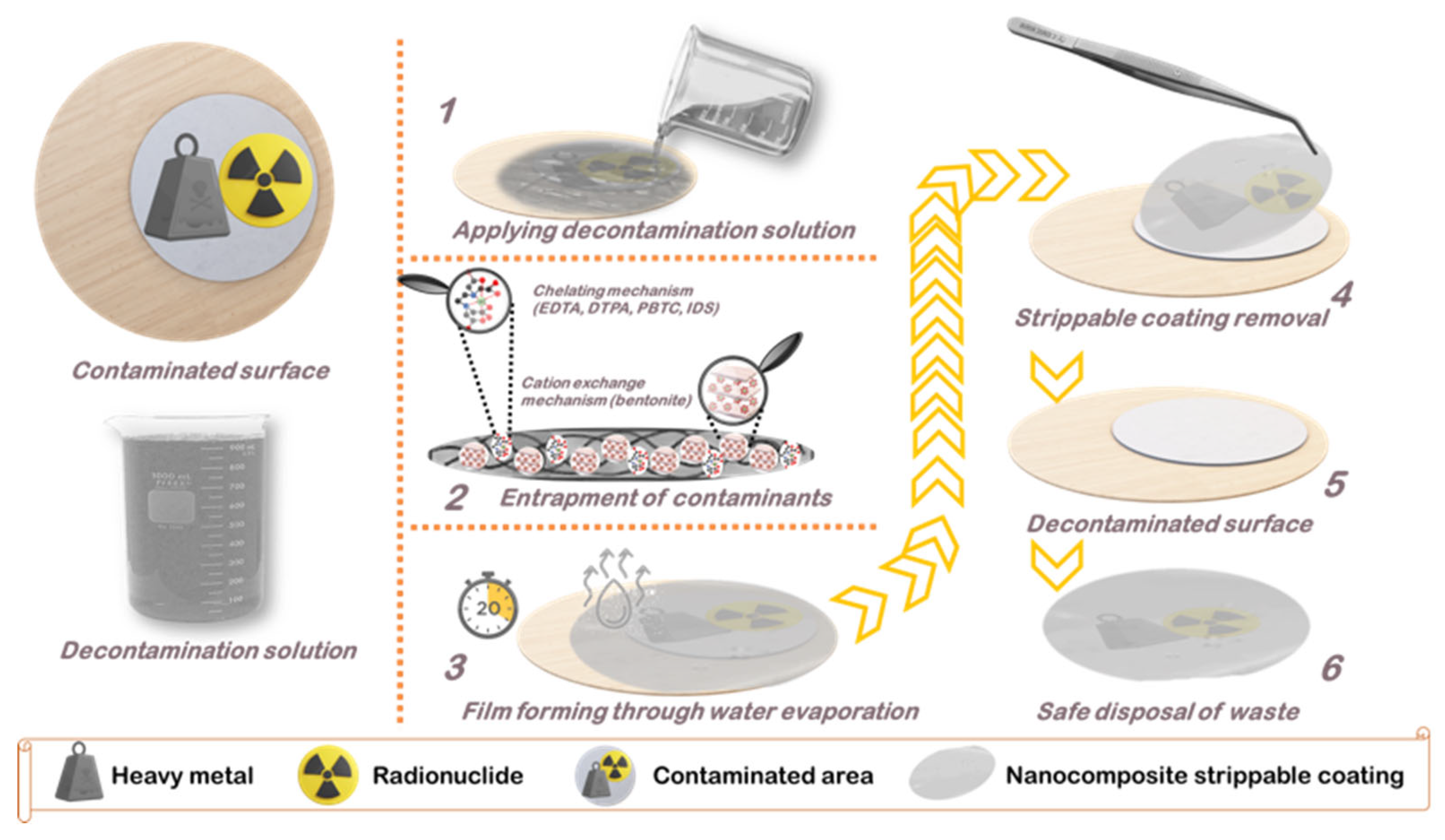
3.1. Basic Principles of HM/RN Decontamination with Nanocomposite Peelable Coatings
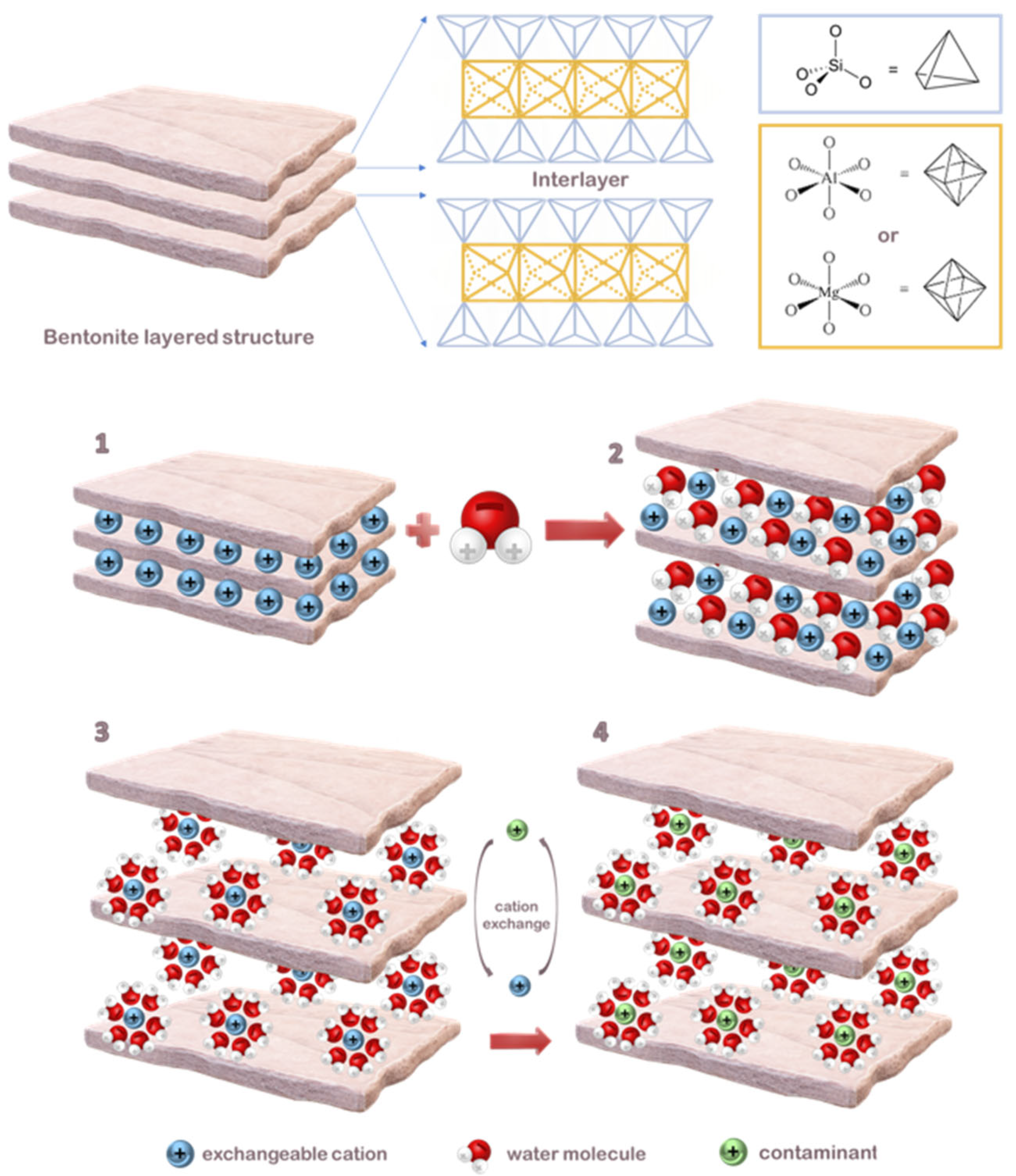
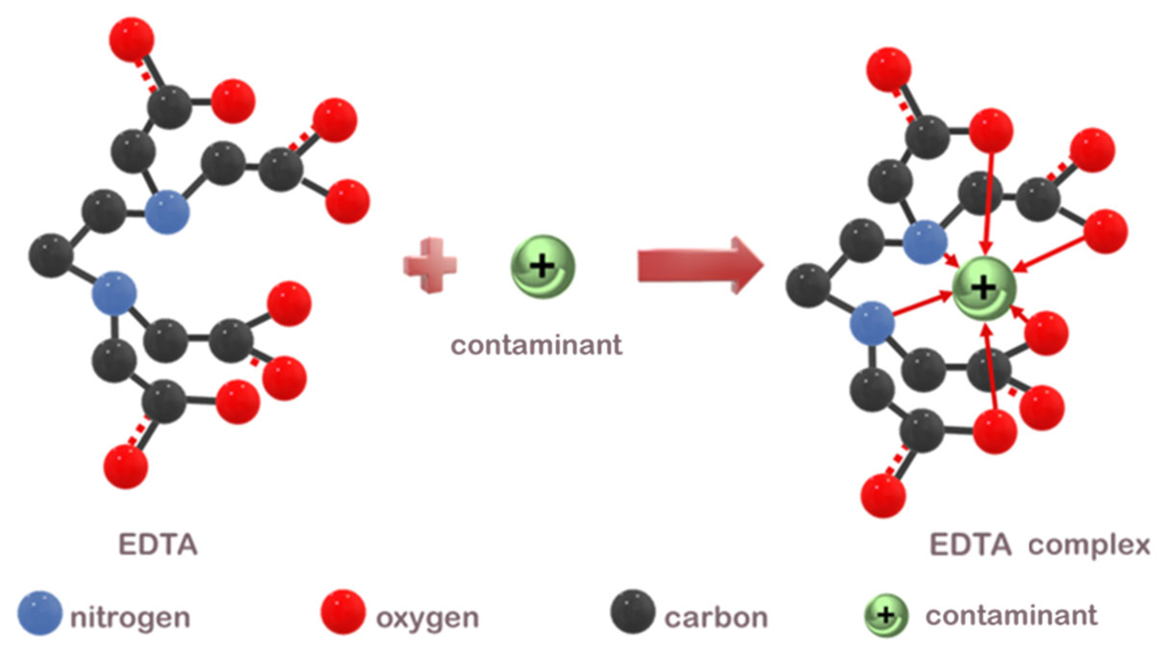
3.2. Decontamination Mechanism Analysis
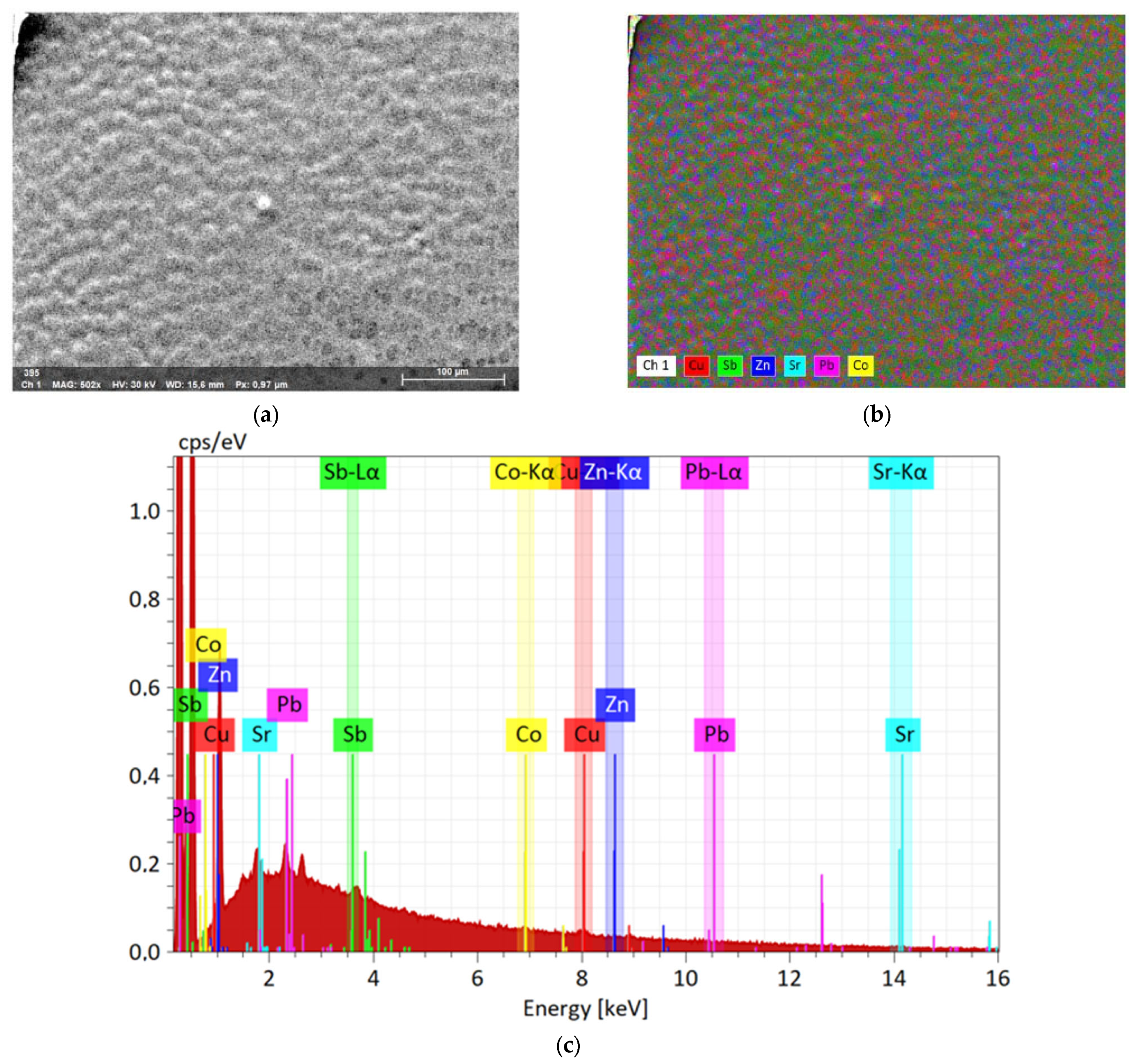
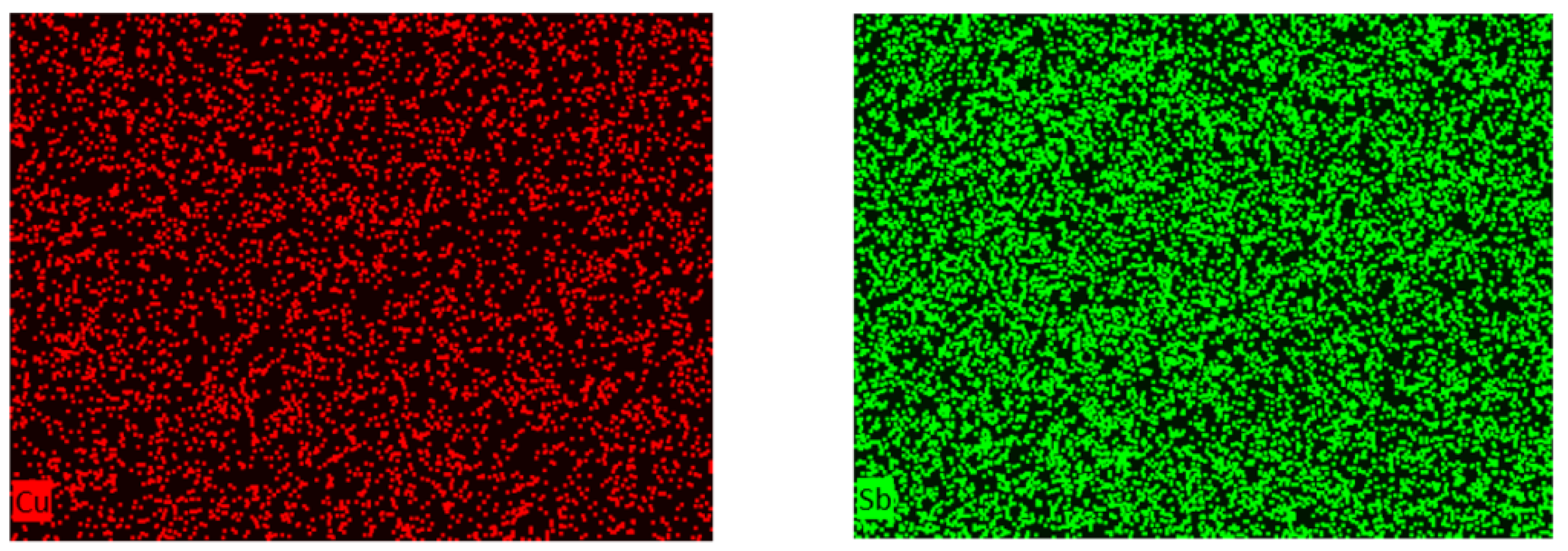
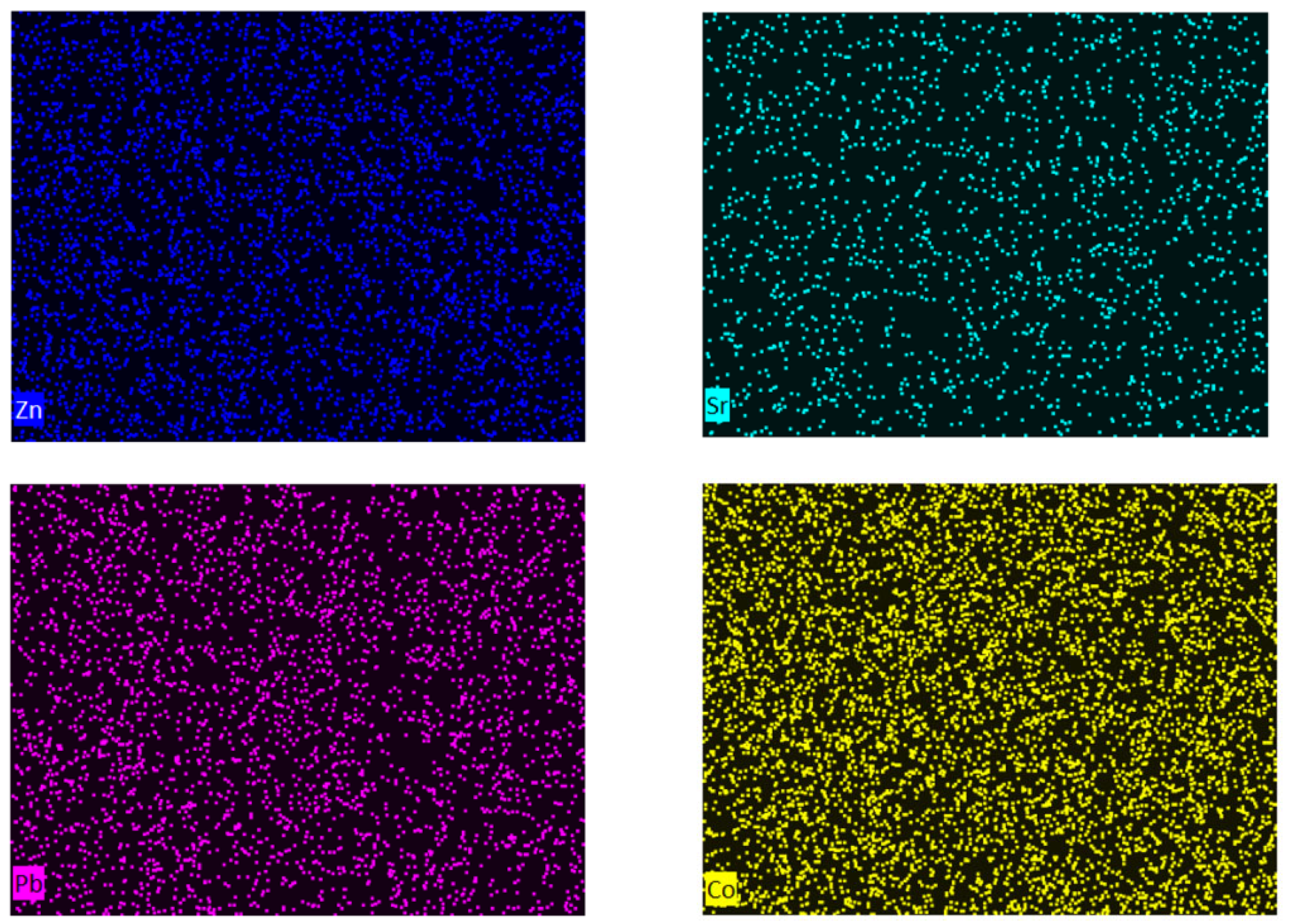
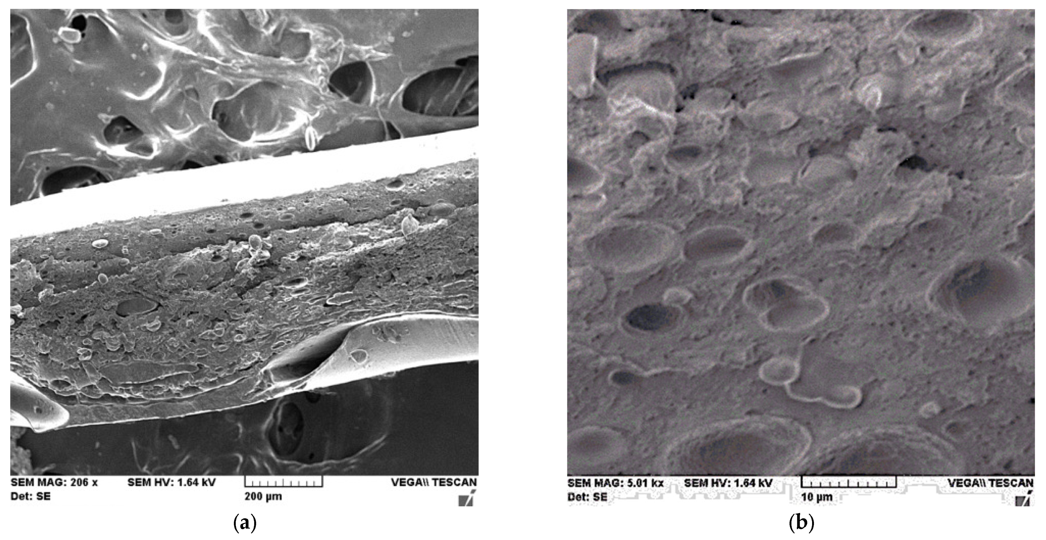
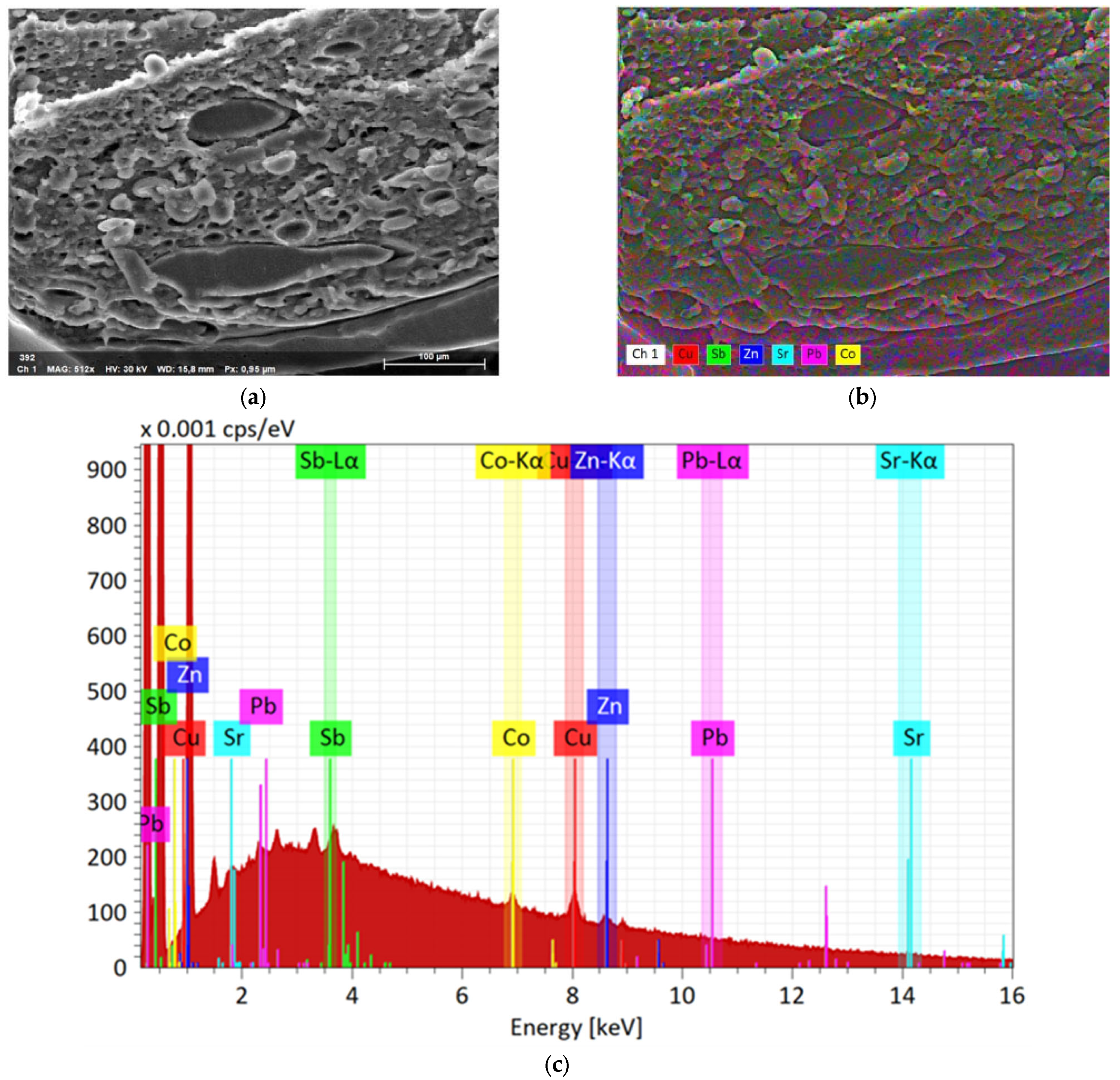
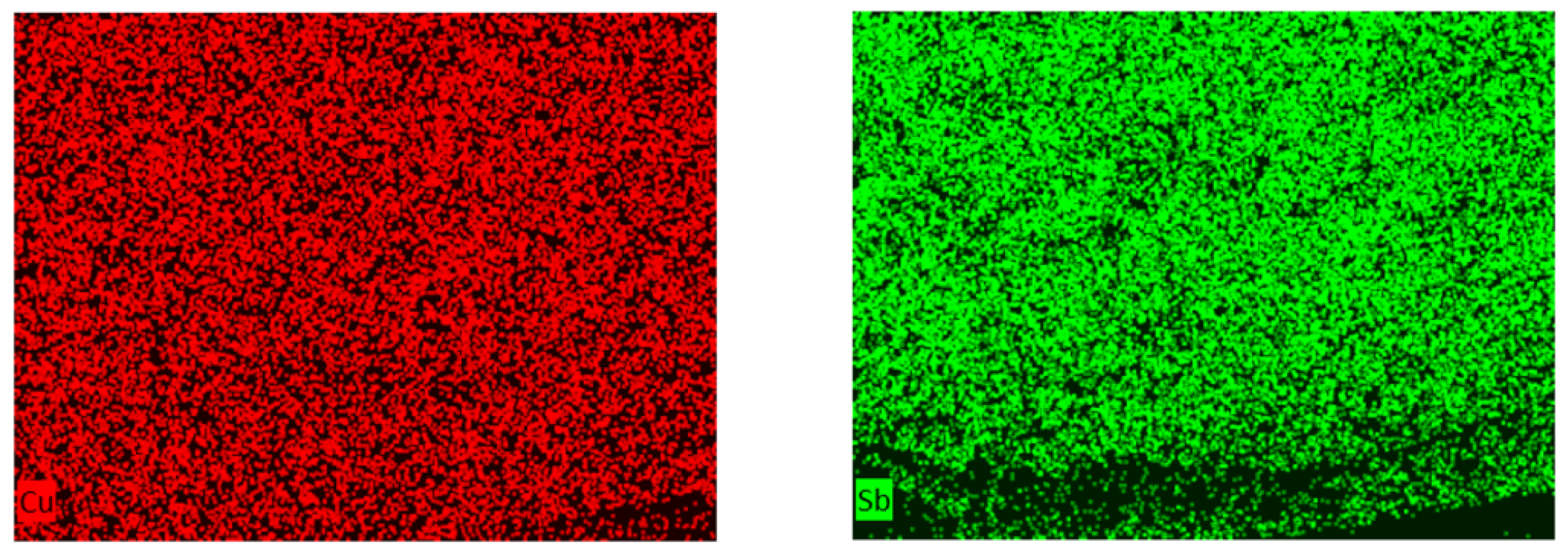
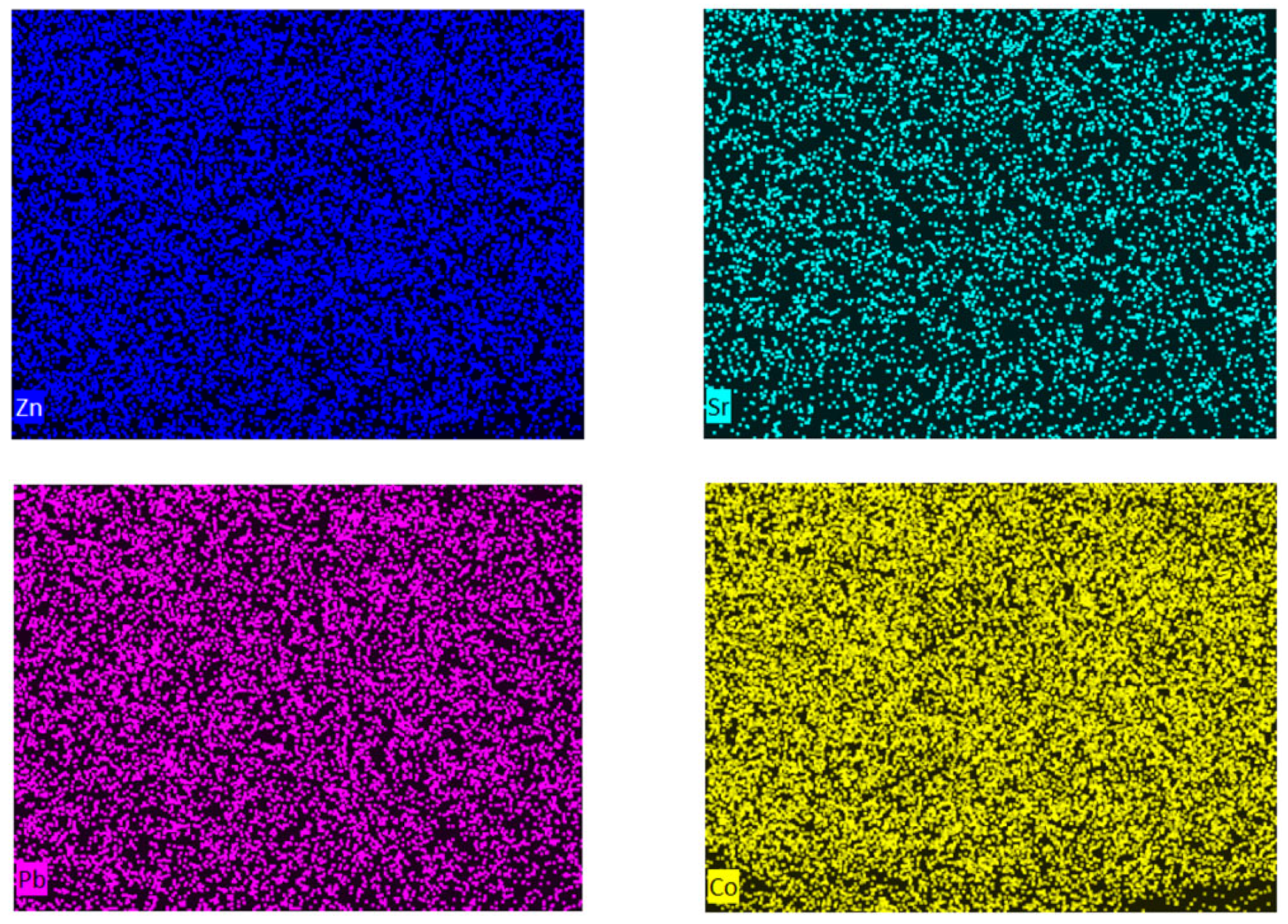
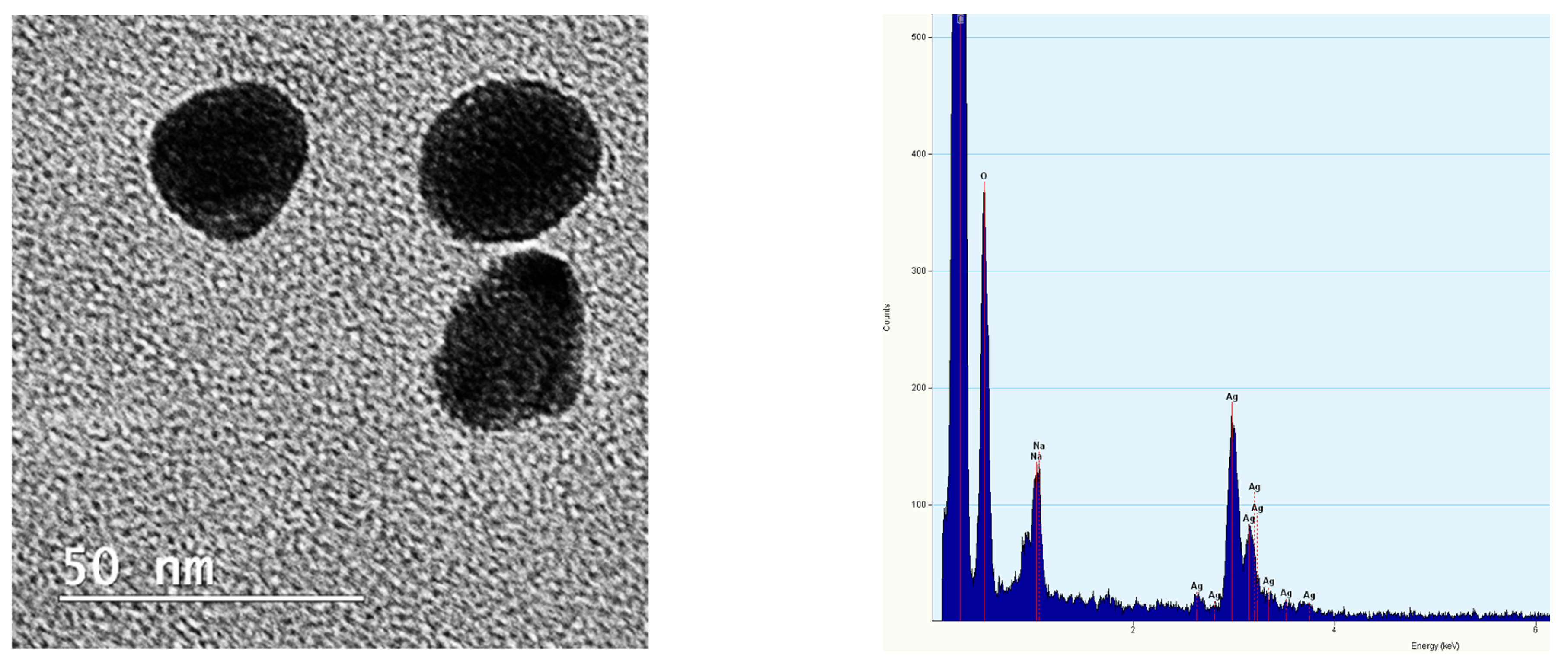
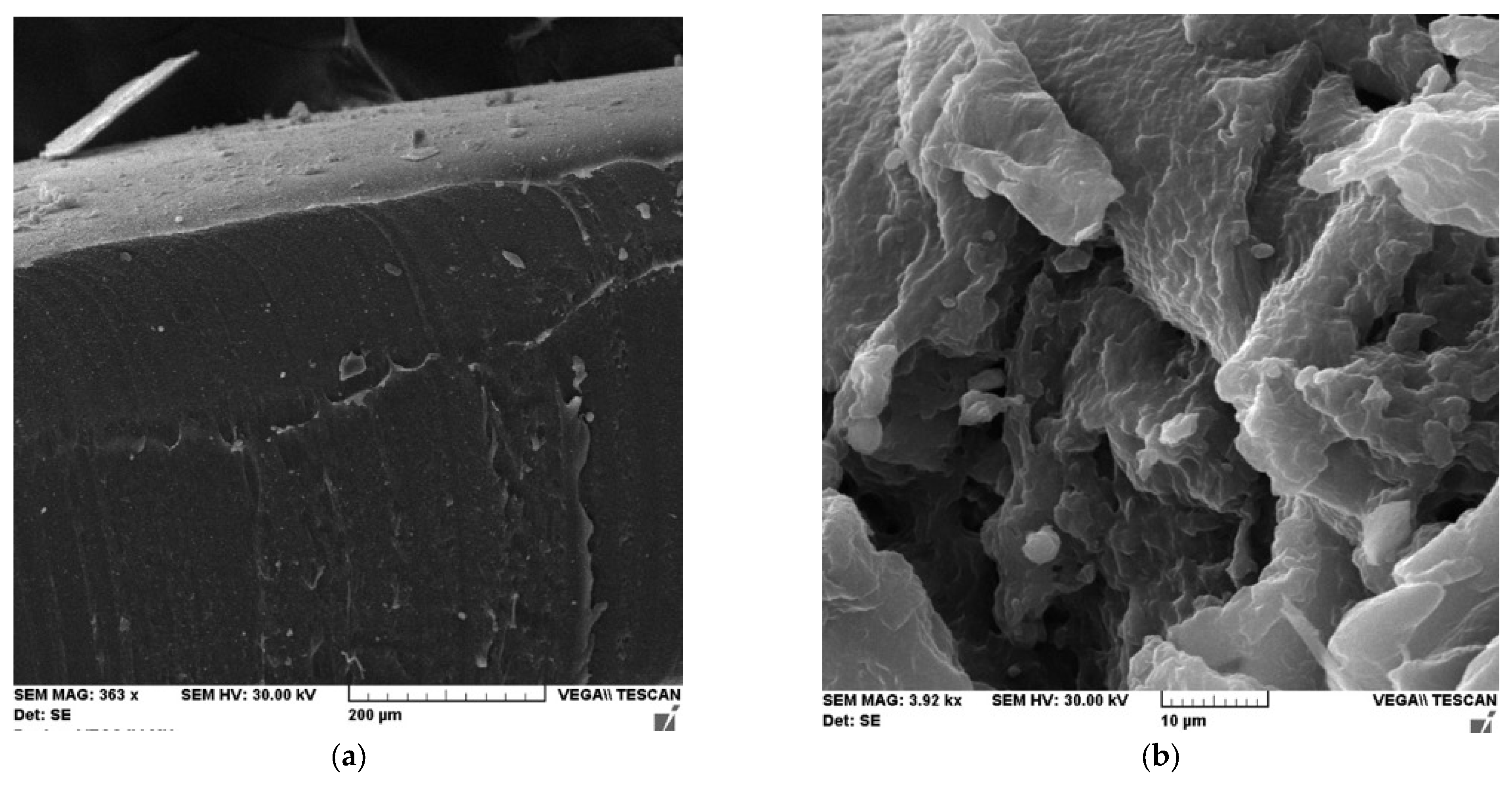
3.3. SEM—EDX
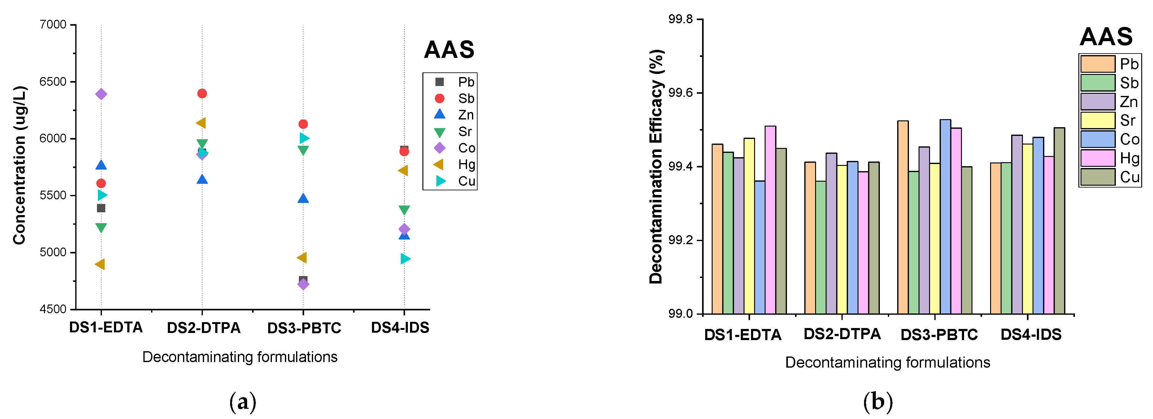
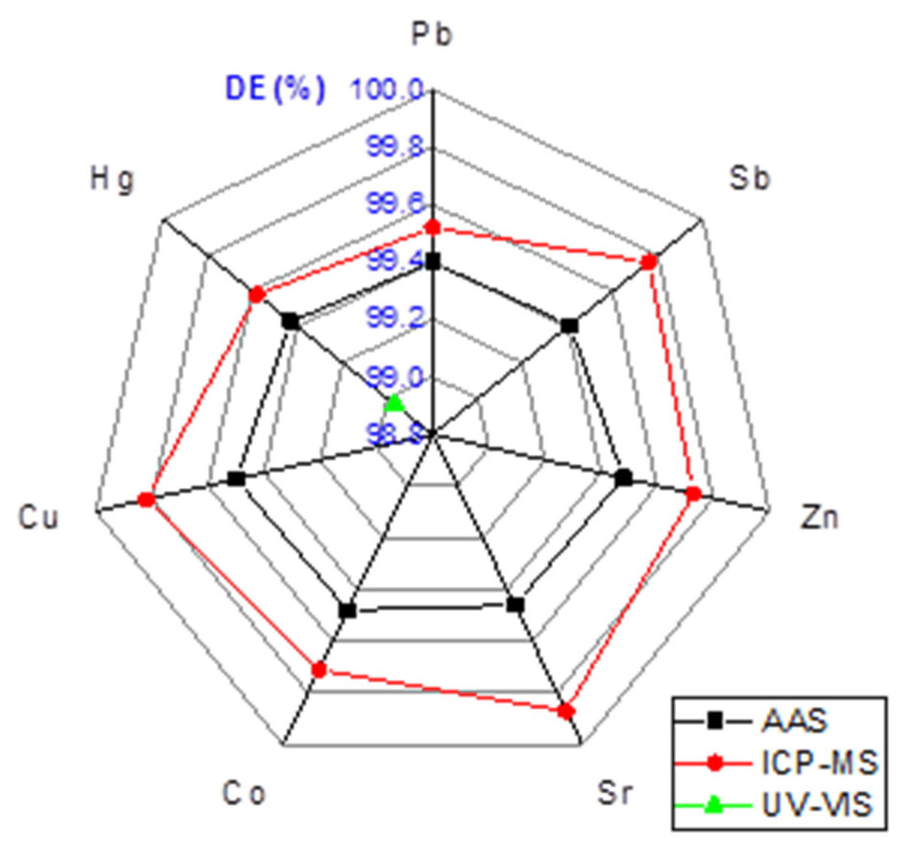
3.4. AAS
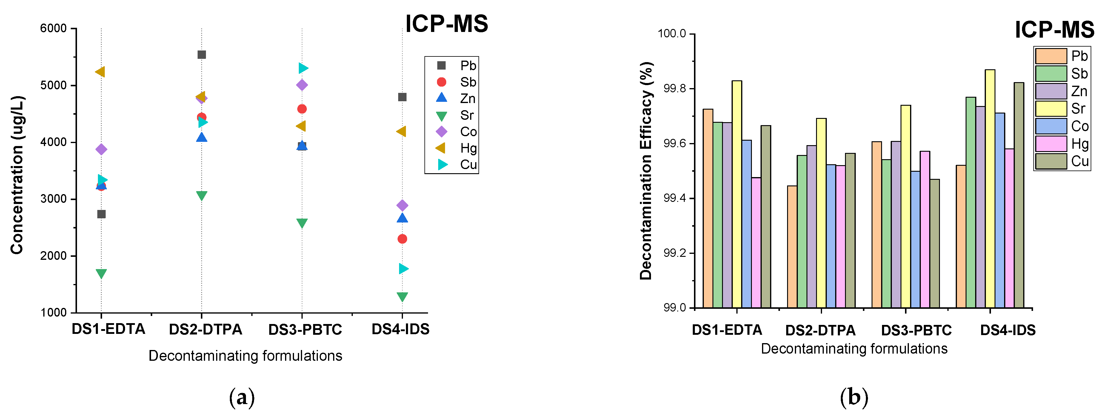
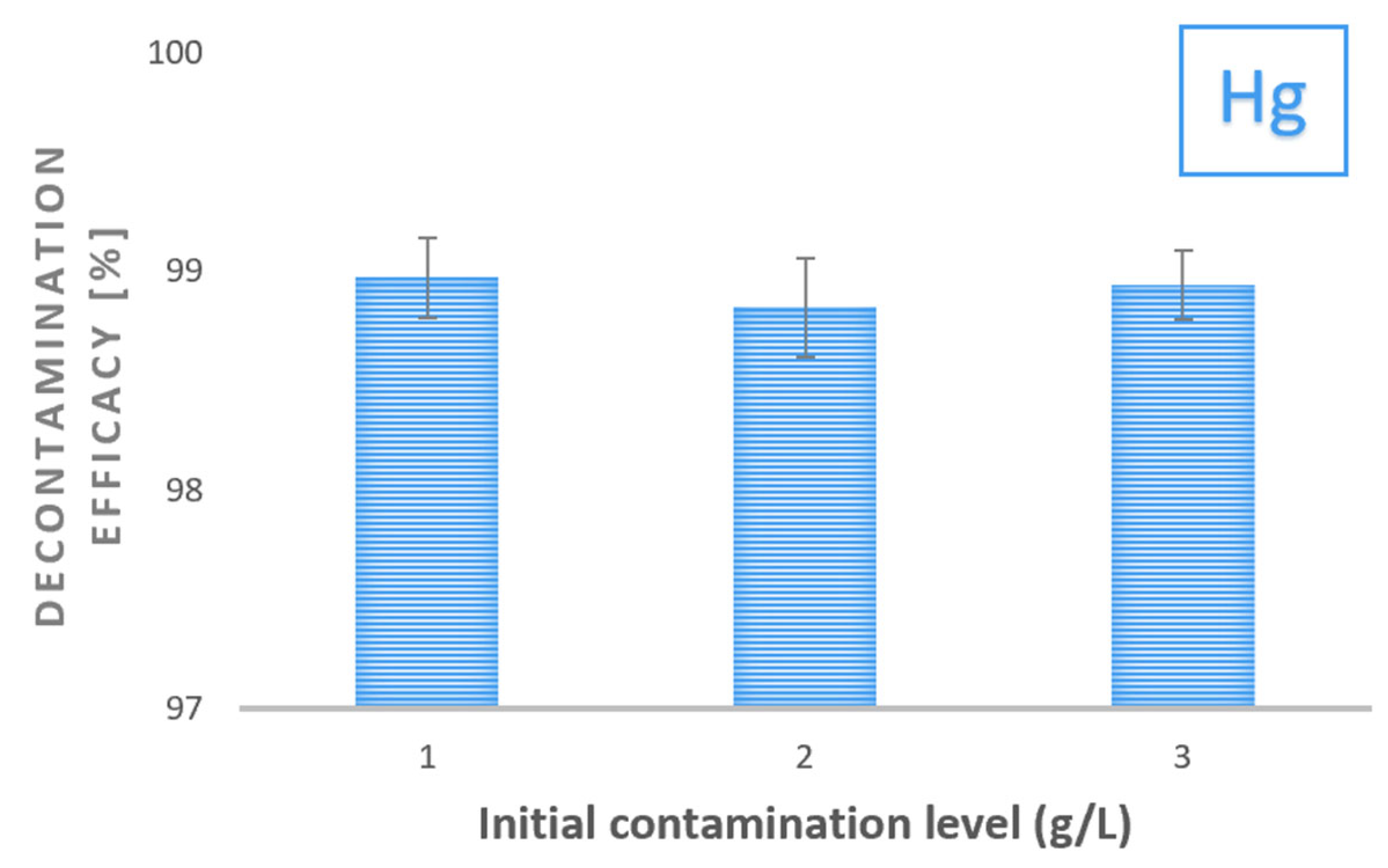
3.5. ICP-MS
3.6. Silver Nanoparticles Characterization
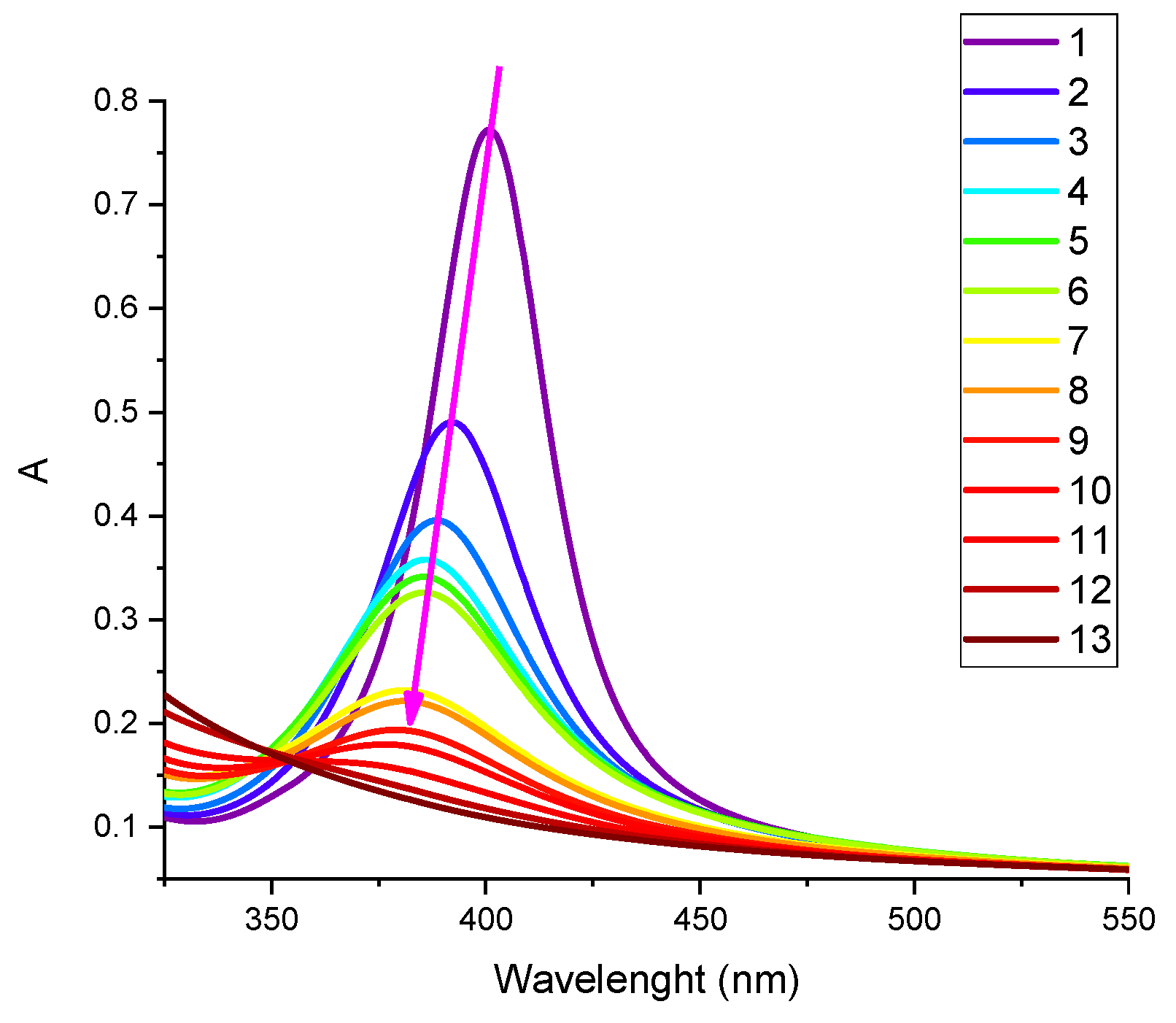
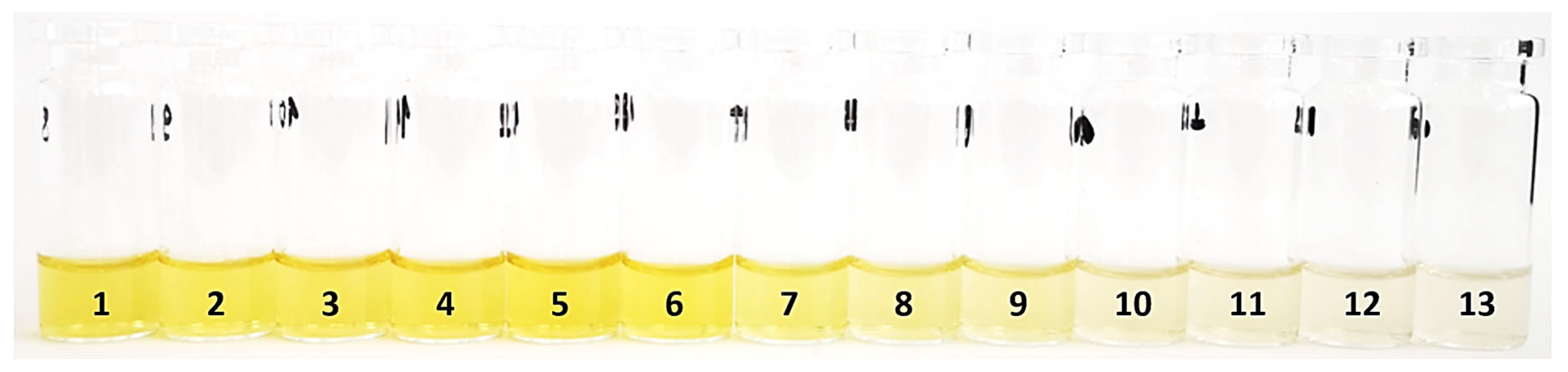
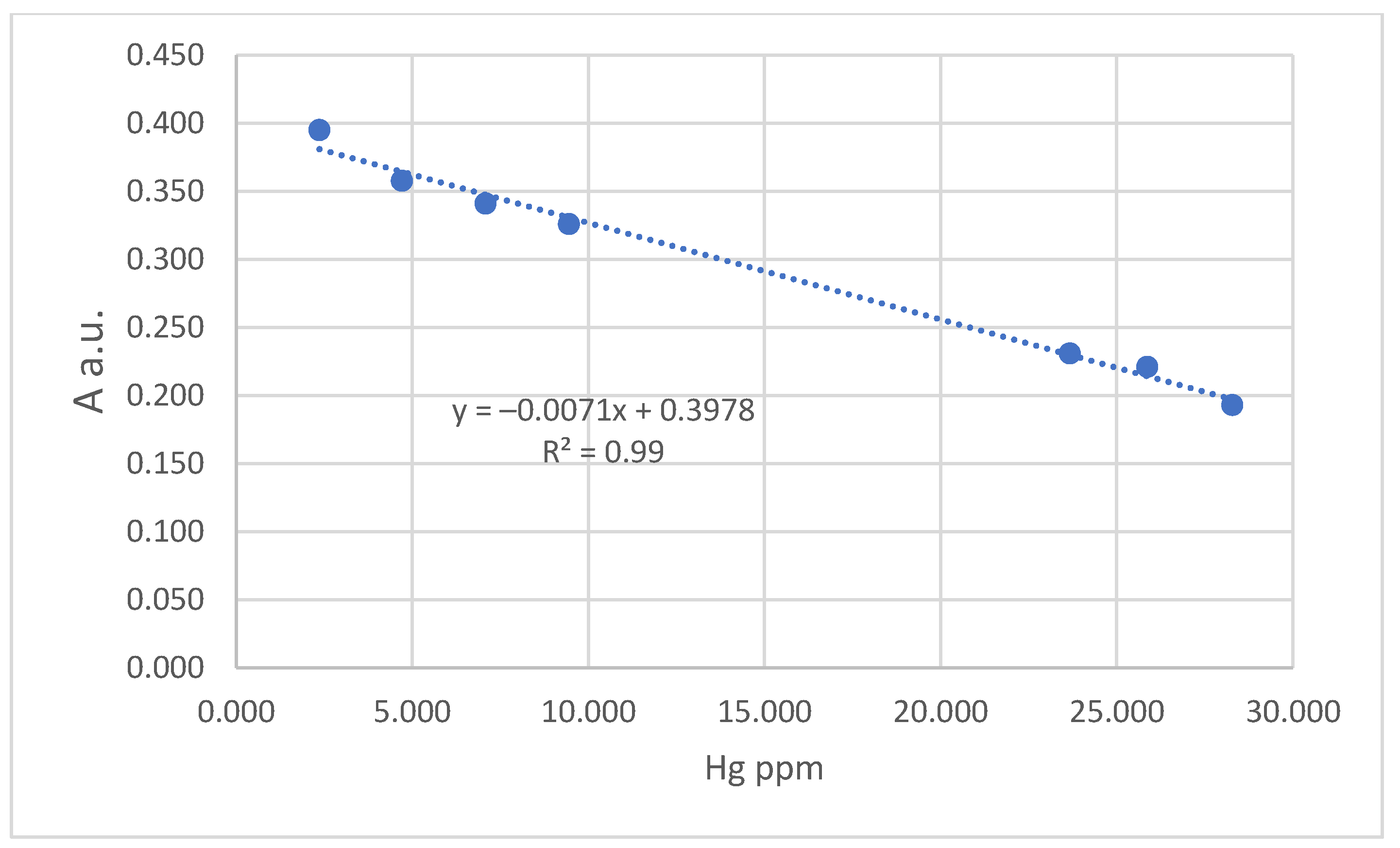
3.7. UV–Vis
4. Conclusions
Supplementary Materials
Author Contributions
Funding
Institutional Review Board Statement
Data Availability Statement
Acknowledgments
Conflicts of Interest
References
- Das, S.; Das, S.; Ghangrekar, M.M. Efficacious bioremediation of heavy metals and radionuclides from wastewater employing aquatic macro- and microphytes. J. Basic Microbiol. 2022, 62, 260–278. [Google Scholar] [CrossRef] [PubMed]
- Leonardi, N.M.; Tesán, F.C.; Zubillaga, M.B.; Salgueiro, M.J. Radioactivity Decontamination of Materials Commonly Used as Surfaces in General-Purpose Radioisotope Laboratories. J. Nucl. Med. Technol. 2014, 42, 292. [Google Scholar] [CrossRef] [PubMed] [Green Version]
- Liu, S.; He, Y.; Xie, H.; Ge, Y.; Lin, Y.; Yao, Z.; Jin, M.; Liu, J.; Chen, X.; Sun, Y.; et al. A State-of-the-Art Review of Radioactive Decontamination Technologies: Facing the Upcoming Wave of Decommissioning and Dismantling of Nuclear Facilities. Sustainability 2022, 14, 4021. [Google Scholar] [CrossRef]
- Toader, G.; Stănescu, P.-O.; Zecheru, T.; Rotariu, T.; El-Ghayoury, A.; Teodorescu, M. Water-based strippable coatings containing bentonite clay for heavy metal surface decontamination. Arab. J. Chem. 2019, 12, 4026–4034. [Google Scholar] [CrossRef] [Green Version]
- Vandebroek, E.; Haufroid, V.; Smolders, E.; Hons, L.; Nemery, B. Occupational Exposure to Metals in Shooting Ranges: A Biomonitoring Study. Saf. Health Work. 2019, 10, 87–94. [Google Scholar] [CrossRef]
- Laidlaw, M.A.; Filippelli, G.; Mielke, H.; Gulson, B.; Ball, A.S. Lead exposure at firing ranges—A review. Environ. Health 2017, 16, 1–15. [Google Scholar] [CrossRef] [Green Version]
- Diaz, E.; Souza Sarkis, J.E.; Viebig, S.; Saldiva, P. Measurement of airborne gunshot particles in a ballistics laboratory by sector field inductively coupled plasma mass spectrometry. Forensic Sci. Int. 2012, 214, 44–47. [Google Scholar] [CrossRef]
- Bai, J.; Zhao, X. Ecological and Human Health Risks of Heavy Metals in Shooting Range Soils: A Meta Assessment from China. Toxics 2020, 8, 32. [Google Scholar] [CrossRef]
- Wojtkowska, M.; Wojtkowski, K.; Długosz-Lisiecka, M. Assessment of Heavy Metals and Radionuclides Concentration in Selected Mineral Waters Available on the Polish Market. Appl. Sci. 2022, 12, 11401. [Google Scholar] [CrossRef]
- Custodio, M.; Fow, A.; Chanamé, F.; Orellana-Mendoza, E.; Peñaloza, R.; Alvarado, J.C.; Cano, D.; Pizarro, S. Ecological Risk Due to Heavy Metal Contamination in Sediment and Water of Natural Wetlands with Tourist Influence in the Central Region of Peru. Water 2021, 13, 2256. [Google Scholar] [CrossRef]
- Shirkhanloo, H.; Golbabaei, F.; Hassani, H.; Eftekhar, F.; Kian, M.J. Occupational Exposure to Mercury: Air Exposure Assessment and Biological Monitoring based on Dispersive Ionic Liquid-Liquid Microextraction. Iran. J. Public Health 2014, 43, 793–799. [Google Scholar] [PubMed]
- Ahmed, M.A.; Hasan, N.; Mohiuddin, S. Silver Nanoparticles: Green Synthesis, Characterization, and Their Usage in Determination of Mercury Contamination in Seafoods. ISRN Nanotechnol. 2014, 2014, 148184. [Google Scholar] [CrossRef] [Green Version]
- Wu, L.P.; Zhao, H.W.; Qin, Z.H.; Zhao, X.Y.; Pu, W.D. Highly Selective Hg (II) Ion Detection Based on Linear Blue-Shift of the Maximum Absorption Wavelength of Silver Nanoparticles. J. Anal. Methods Chem. 2012, 2012, 856947. [Google Scholar] [CrossRef] [PubMed] [Green Version]
- Velgosova, O.; Mačák, L.; Čižmárová, E.; Mára, V. Influence of Reagents on the Synthesis Process and Shape of Silver Nanoparticles. Materials 2022, 15, 6829. [Google Scholar] [CrossRef] [PubMed]
- Johansson, I.; Somasundaran, P. Handbook for Cleaning/Decontamination of Surfaces; Elsevier Science: Amsterdam, The Netherlands, 2007. [Google Scholar]
- Pulpea, D.; Rotariu, T.; Toader, G.; Pulpea, G.B.; Neculae, V.; Teodorescu, M. Decontamination of radioactive hazardous materials by using novel biodegradable strippable coatings and new generation complexing agents. Chemosphere 2020, 258, 127227. [Google Scholar] [CrossRef]
- Lewandowski, K.; Krepski, L.R.; Mickus, D.E. Dry-peelable temporary protective coatings from waterborne self-crosslinkable sulfourethane–silanol dispersions. J. Appl. Polym. Sci. 2004, 91, 1443–1449. [Google Scholar] [CrossRef]
- Wagle, P.G.; Tamboli, S.S.; More, A.P. Peelable coatings: A review. Prog. Org. Coat. 2021, 150, 106005. [Google Scholar] [CrossRef]
- Xue, Y.; Yang, W.; Yue, R.; Chen, Y. Aqueous Strippable Polymer Coating for Highly Efficient Primary Radioactive Uranium Decontamination with Versatility on Diversified Surface. Polymers 2022, 14, 1656. [Google Scholar] [CrossRef]
- Chiellini, E.; Corti, A.; D’Antone, S.; Solaro, R. Biodegradation of poly (vinyl alcohol) based materials. Prog. Polym. Sci. 2003, 28, 963–1014. [Google Scholar] [CrossRef]
- Alonso-López, O.; López-Ibáñez, S.; Beiras, R. Assessment of Toxicity and Biodegradability of Poly(vinyl alcohol)-Based Materials in Marine Water. Polymers 2021, 13, 3742. [Google Scholar] [CrossRef]
- Ahmad, A.; Mubarak, N.M.; Jannat, F.T.; Ashfaq, T.; Santulli, C.; Rizwan, M.; Najda, A.; Bin-Jumah, M.; Abdel-Daim, M.M.; Hussain, S.; et al. A Critical Review on the Synthesis of Natural Sodium Alginate Based Composite Materials: An Innovative Biological Polymer for Biomedical Delivery Applications. Processes 2021, 9, 137. [Google Scholar] [CrossRef]
- Sun, J.; Tan, H. Alginate-Based Biomaterials for Regenerative Medicine Applications. Materials 2013, 6, 1285–1309. [Google Scholar] [CrossRef]
- Azar, A.T. Modeling and Control of Drug Delivery Systems; Elsevier Science: Amsterdam, The Netherlands, 2021. [Google Scholar]
- Additives, E.P.O.; Bampidis, V.; Azimonti, G.; Bastos, M.D.L.; Christensen, H.; Dusemund, B.; Fašmon Durjava, M.; Kouba, M.; López-Alonso, M.; Marcon, m.; et al. Safety and efficacy of a feed additive consisting of sodium alginate for all animal species (ALGAIA). EFSA J. 2022, 20, e07164. [Google Scholar] [CrossRef]
- Wang, M.; Chen, L.; Zhang, Z. Potential applications of alginate oligosaccharides for biomedicine–A mini review. Carbohydr. Polym. 2021, 271, 118408. [Google Scholar] [CrossRef]
- Gaggero, G.; Delucchi, M.; Di Tanna, G.; Lagazzo, A.; Vicini, S.; Botter, R. Effect of different alginate salts on the rheological and tensile properties of waterborne paints. Prog. Org. Coat. 2022, 163, 106676. [Google Scholar] [CrossRef]
- Toader, G.; Pulpea, D.; Rotariu, T.; Diacon, A.; Rusen, E.; Moldovan, A.; Podaru, A.; Ginghină, R.; Alexe, F.; Iorga, O.; et al. Strippable Polymeric Nanocomposites Comprising “Green”; Chelates, for the Removal of Heavy Metals and Radionuclides. Polymers 2021, 13, 4194. [Google Scholar] [CrossRef]
- Toader, G.; Diacon, A.; Rotariu, T.; Alexandru, M.; Rusen, E.; Ginghină, R.E.; Alexe, F.; Oncioiu, R.; Zorila, F.L.; Podaru, A.; et al. Eco-Friendly Peelable Active Nanocomposite Films Designed for Biological and Chemical Warfare Agents Decontamination. Polymers 2021, 13, 3999. [Google Scholar] [CrossRef]
- NATO. AEP-58-Combined Operational Characteristics, Technical Specifications, Test Procedures, and Evaluation Criteria for Chemical, Biological, Radiological, and Nuclear Decontamination Equipment; NATO. 2014. Available online: https://standards.globalspec.com/std/14358280/aep-58-vol-1 (accessed on 15 December 2022).
- Chilcott, R.P. Managing mass casualties and decontamination. Environ. Int. 2014, 72, 37–45. [Google Scholar] [CrossRef]
- Gossard, A.; Lilin, A.; Faure, S. Gels, coatings and foams for radioactive surface decontamination: State of the art and challenges for the nuclear industry. Prog. Nucl. Energy 2022, 149, 104255. [Google Scholar] [CrossRef]
- Alexander, J.A.; Ahmad Zaini, M.A.; Surajudeen, A.; Aliyu, E.-N.U.; Omeiza, A.U. Surface modification of low-cost bentonite adsorbents—A review. Part. Sci. Technol. 2019, 37, 538–549. [Google Scholar] [CrossRef]
- Rotariu, T.; Pulpea, D.; Toader, G.; Rusen, E.; Diacon, A.; Neculae, V.; Liggat, J. Peelable Nanocomposite Coatings: “Eco-Friendly” Tools for the Safe Removal of Radiopharmaceutical Spills or Accidental Contamination of Surfaces in General-Purpose Radioisotope Laboratories. Pharmaceutics 2022, 14, 2360. [Google Scholar] [CrossRef] [PubMed]
- Carter, J.A.; Barros, A.I.; Nóbrega, J.A.; Donati, G.L. Traditional Calibration Methods in Atomic Spectrometry and New Calibration Strategies for Inductively Coupled Plasma Mass Spectrometry. Front. Chem. 2018, 6, 504. [Google Scholar] [CrossRef] [PubMed]
- NATO, AEP-7 - Chemical, Biological, Radiological and Nuclear (CBRN) Contamination Survivability Factors in the Design, Testing and Acceptance of Military Equipment; NATO. 2012. Available online: https://standards.globalspec.com/std/9857648/AEP-7 (accessed on 15 December 2022).


| Sample Code | Chelating Agent [wt.%] | PVA [wt.%] | Alg [wt.%] | BT [wt.%] | Gly [wt.%] |
|---|---|---|---|---|---|
| DS1—EDTA | 1 | 5 | 0.7 | 1 | 3 |
| DS2—DTPA | 1 | 5 | 0.7 | 1 | 3 |
| DS3—PBTC | 1 | 5 | 0.7 | 1 | 3 |
| DS4—IDS | 1 | 5 | 0.7 | 1 | 3 |
Disclaimer/Publisher’s Note: The statements, opinions and data contained in all publications are solely those of the individual author(s) and contributor(s) and not of MDPI and/or the editor(s). MDPI and/or the editor(s) disclaim responsibility for any injury to people or property resulting from any ideas, methods, instructions or products referred to in the content. |
© 2023 by the authors. Licensee MDPI, Basel, Switzerland. This article is an open access article distributed under the terms and conditions of the Creative Commons Attribution (CC BY) license (https://creativecommons.org/licenses/by/4.0/).
Share and Cite
Toader, G.; Pulpea, D.; Diacon, A.; Rusen, E.; Ginghina, R.E.; Rotariu, T.; Podaru, A.I.; Moldovan, A.E.; Gavrilescu, M.; Gavrila, A.-M.; et al. Comparative Study on the Decontamination Efficacy of Peelable Coatings for Heavy Metals Removal. Water 2023, 15, 982. https://doi.org/10.3390/w15050982
Toader G, Pulpea D, Diacon A, Rusen E, Ginghina RE, Rotariu T, Podaru AI, Moldovan AE, Gavrilescu M, Gavrila A-M, et al. Comparative Study on the Decontamination Efficacy of Peelable Coatings for Heavy Metals Removal. Water. 2023; 15(5):982. https://doi.org/10.3390/w15050982
Chicago/Turabian StyleToader, Gabriela, Daniela Pulpea, Aurel Diacon, Edina Rusen, Raluca Elena Ginghina, Traian Rotariu, Alice Ionela Podaru, Andreea Elena Moldovan, Maria Gavrilescu, Ana-Mihaela Gavrila, and et al. 2023. "Comparative Study on the Decontamination Efficacy of Peelable Coatings for Heavy Metals Removal" Water 15, no. 5: 982. https://doi.org/10.3390/w15050982