Article

Beneath the Aegean Sun: Investigating Dunaliella Strains’ Diversity from Greek Saltworks

by
Urania Lortou
1,†,
Manthos Panou
1,†,
Georgia Papapanagiotou
1,2,†,
Georgia Florokapi
1,
Christos Giannakopoulos
1,
Savvas Kavoukis
3,
Georgios Iakovou
3,
Giorgos Zalidis
2,
Konstantinos Triantafyllidis
3 and
Spyros Gkelis
1,*
1
School of Biology, Aristotle University of Thessaloniki, GR-541 24 Thessaloniki, Greece
2
School of Agriculture, Aristotle University of Thessaloniki, GR-541 24 Thessaloniki, Greece
3
Department of Chemistry, Aristotle University of Thessaloniki, GR-541 24 Thessaloniki, Greece
*
Author to whom correspondence should be addressed.
The authors have equally contributed to this work.
Water 2023, 15(6), 1037; https://doi.org/10.3390/w15061037
Submission received: 19 February 2023 / Revised: 6 March 2023 / Accepted: 7 March 2023 / Published: 9 March 2023

Abstract

:
The genus Dunaliella belongs to the division Chlorophyta and is known for its ability to survive in highly saline environments. Dunaliella is an important source of carotenoids, especially beta-carotene and has a wide range of applications. In this study, we aimed to isolate and identify Dunaliella strains from active and abandoned saltworks in Greece. Four seasonal samplings were carried out in seven active saltworks and two samplings were performed in an abandoned saltwork throughout the year 2020. Strains were characterized based on their morphological and phylogenetic traits, whilst their beta-carotene potential were evaluated. Fifteen (15) Dunaliella strains were isolated and classified into nine species based on morphological and morphometrical features. The isolated strains were assigned to different species such as D. parva, D. granulata, D. minuta, D. terricola, D. asymmetrica, D. bioculata, D. viridis, D. minutissima, and D. polymorpha. The results of the phylogenetic analysis indicate the formation of distinct clades among different Dunaliella species and suggest that morphological and morphometrical features may not always align with the phylogenetic position of species in the Dunaliella clade. Strains were found to produce a low amount of beta-carotene under default laboratory conditions. This study comprises the first phylogenetic inference of several Dunaliella species and highlights a gap on molecular data for Dunaliella spp. We provide valuable information on the diversity of Dunaliella strains in the saltworks of Greece, which can be used for further research and biotechnological applications.

1. Introduction

Dunaliella is a cosmopolitan genus of green algae (Chlorophyta) that is widely distributed in various saline environments with a prominent presence in hypersaline habitats, including saltworks [1]. Dunaliella cells can be ellipsoid, ovoid to almost spherical, motile, biflagellate, and may appear green and/or orange and from ovoid to pyriform (5 to 15 μm), characterized by the lack of a rigid cell wall [2]. Dunaliella has received significant attention due to its ability to produce high levels of carotenoids, particularly beta-carotene, which has potential applications in the food, cosmetic, and pharmaceutical industries [3]. The taxonomy of the green algal genus Dunaliella is often seen as confusing and the names associated with species in culture collections can be problematic [4]. The molecular phylogeny of Dunaliella has helped to establish the evolutionary relationships between different Dunaliella species and to identify the major lineages within the genus [5,6,7]. The use of molecular markers such as the ribosomal DNA and the internal transcribed spacer region has been critical in providing a better understanding of the diversity, evolution, and distribution of Dunaliella species in different environments, including saltworks [8].
Saltworks are unique ecosystems characterized by high salinity levels and fluctuations in temperature, light, and water availability [9]. The presence of Dunaliella in saltworks has been reported in various parts of the world, including Greece [2,4,10]. In these environments, Dunaliella has been shown to be capable of adapting to extreme conditions and having the ability to produce high levels of carotenoids. Carotenogenesis in Dunaliella may serve to protect cells from oxidative stress, as a result of environmental stressors such as increased salinity and nutrient limitations; beta-carotene, which has strong antioxidant properties, is produced in higher amounts under these conditions [11,12]. The production of beta-carotene by Dunaliella species has been extensively studied, and various factors that influence its production have been identified. These include light intensity, salinity, temperature, and nutrient availability [13]. Different Dunaliella species have been shown to have varying capacities to produce beta-carotene, and the selection of the most appropriate species for carotenoid production depends on the specific application and the environmental conditions in which the algae are cultivated [14,15]. The presence of Dunaliella in saltworks provides a suitable environment for the isolation, cultivation, and characterization of different Dunaliella species [4].
In recent years, the isolation and characterization of the Dunaliella species from saltworks in Greece has become an area of interest for researchers due to the high diversity of algae species, the varying salinities and other environmental conditions found in these ecosystems [16,17,18]. This study investigates the diversity of Dunaliella strains isolated from saltworks in Greece using a polyphasic approach including morphology, 18S rRNA, ITS, and rbcL phylogeny, as well as their growth rates and pigment production.

2. Materials and Methods

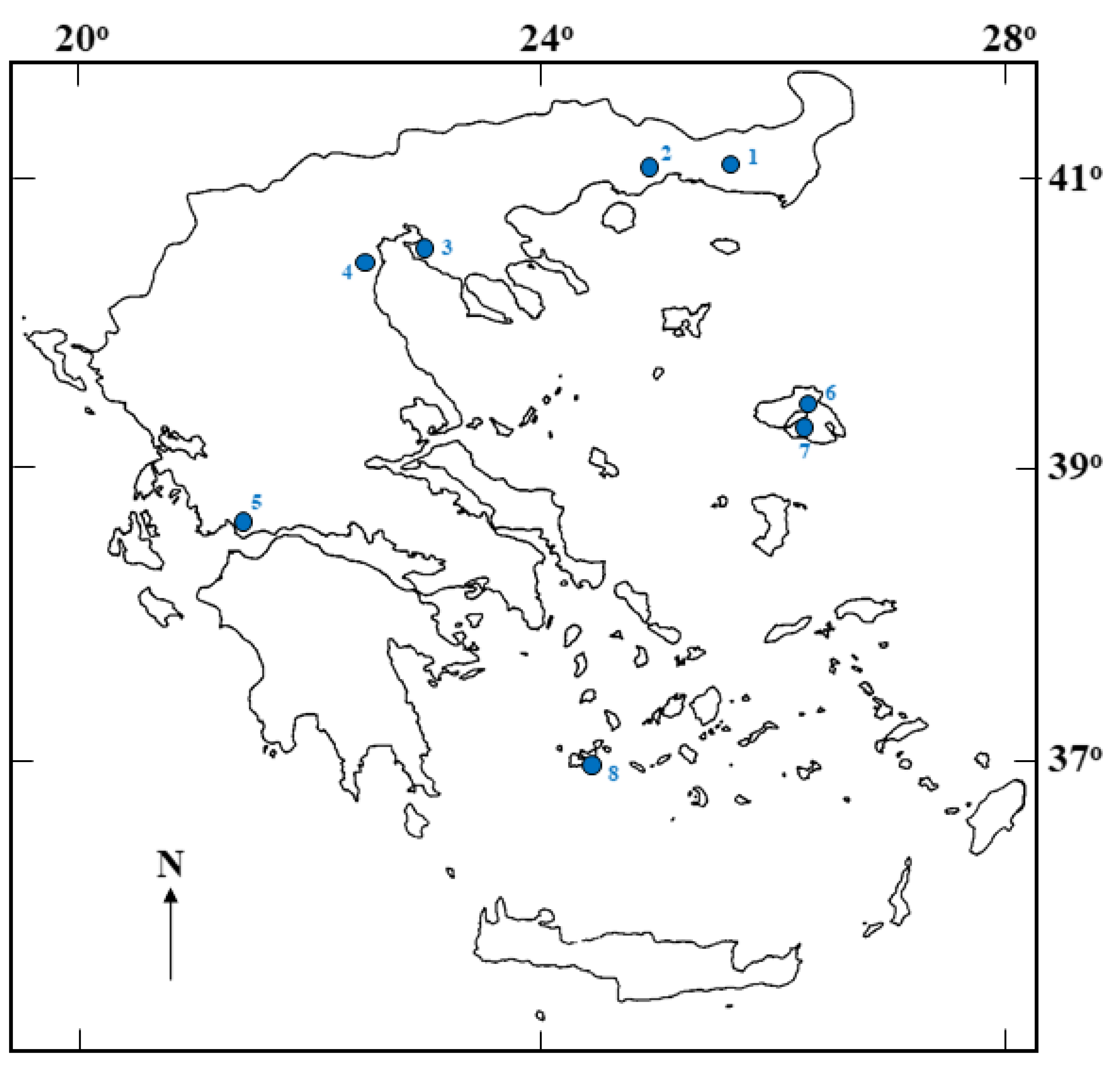
2.1. Site Description, Sampling and Strain Isolation

A series of four seasonal samplings was carried out throughout the year 2020 in seven active saltworks of Greece (Figure 1). Moreover, two samplings were performed in the abandoned saltwork of Adamas in Milos island during the winter and summer period of the same year (Table 1).
Strains were isolated from water samples placed in sterile 15 mL falcon tubes and transferred to the laboratory in the dark at 4 °C within 4–16 h upon collection. For the isolation of strains, four media were used, namely Zarrouk [19], MN [20], Johnson’s [21], and CP [22], as liquid or solid in agar (1.4% w/v) plates (agar No. A-7002 (Sigma Usa)) [23].
Isolation was performed according to Gkelis et al. [24] and Lortou and Gkelis [25]. Briefly, 100 μL of sample material was transferred to Eppendorf tubes containing 1 mL of liquid medium. After three weeks of incubation, the cultures were checked under microscopy and Dunaliella cells were transferred in agar plates or in liquid subcultures after serial dilutions. Strains were grown as unialgal cultures, and purified with Carbendazim (70 μg mL−1) (BCM, Methyl 2-benzimidazolecarbamate, Methyl benzimidazol-2-ylcarbamate, 378,674 Sigma-Aldrich) when necessary. All strains are deposited in the Thessaloniki Aristotle University Microalgae and Cyanobacteria (TAU-MAC) culture collection [26] and are maintained as liquid batch cultures at a photosynthetic photon flux density of 20 μmol m−2s−1 using cool white light fluorescent tubes (Sylvania Standard F36W/154-T8, SLI) at 22 ± 2 °C in a culture room, in a 16:8 h light:dark cycle.

2.2. Polyphasic Characterization

The morphological traits of the strains were examined using a Zeiss Axio Imager.Z2 microscope and micrographs were captured with an AxioCam MRc5 digital camera (Carl Zeiss, Jena, Germany). Cell dimensions were calculated after measuring the length and width of at least 50 individuals (100–160 cells) of each strain. Morphological descriptions were produced based on the taxonomic key by Borowitzka and Siva [2]. Microphotographs of all 15 isolates from this study are available in the TAU-MAC collection database [http://cyanobacteria.myspecies.info/ (accessed on 10 January 2023)]. Information and microphotographs of the strains are available under a Creative Commons Attribution-NonCommercial (CC BY-NC) license.
Culture material from the strain (1.5 mL) was harvested during exponential growth and the DNA was extracted using the protocol described in Atashpaz et al. [27]. Thermal cycling was carried out using an Eppendorf MasterCycler Pro (Eppendorf, Hamburg, Germany). PCR was carried out using the primer pairs and under the conditions described in Table 2. PCR products were separated by 1.5% (w/v) agarose gel in 1X TAE buffer. The gels were stained with Midori Green Advanced (NIPPON Genetics Europe GmbH, Düren, Germany) and photographed under UV transillumination.
The 18S rRNA gene, the 18S-28S rRNA internal transcribed spacer (ITS), and the ribulose-1,5-bisphosphate carboxylase/oxygenase large subunit (rbcL) gene, were used to assess the molecular phylogeny of the strains. PCR was carried out using the primer pairs shown in Table 2 and PCR conditions described in detail in Lortou and Gkelis [25]. Sequence data were obtained by capillary electrophoresis (GENEWIZ, Takeley, UK). The obtained nucleotide sequences were edited with Unipro UGENE 1.29.0 [32]. Nucleotide sequences were deposited in GenBank—National Center for Biotechnology Information (NCBI) (Table S1). Sequences were blasted and the closest relative(s) for each sequence were included in the phylogenetic trees. For the phylogenetic analyses, we selected sequences (>1500 bp, >500 bp, and 1050 bp, for 18S rRNA gene, the 18S-28S rRNA, and rbcL, respectively) in order to examine the phylogenetic position of our strains. The phylogenetic analyses were conducted with Mega (V7.0) software [33]. MUSCLE was used for the alignment; all missing data and gaps were excluded from the analysis by choosing the complete deletion option. A consensus phylogenetic tree was constructed using maximum likelihood (ML). The best fitting evolutionary models for the ML analyses were the Kimura 2-parameter + G model [18S rRNA gene and 18S-28S rRNA internal transcribed spacer (ITS)] or Tamura 3-parameter + G + I model (rbcL gene). Bootstrap replicates (n = 1000) were performed.

2.3. Growth Rates and Pigments

For each Dunaliella strain that was isolated, cell growth was estimated every 48 h by measuring the optical density (OD) of the sample at two wavelengths, 750 and 680 nm, using a UV–visible spectrophotometer (UV-2401PC, Shimadzu, Kyoto, Japan). The wavelength of 750 nm was used for excluding absorption interferences from photosynthetic pigments, while the wavelength of 680 nm was chosen to represent the maximum pigment absorbance, as an additional information of the physiological status of each Dunaliella strain [34].
Pigment accumulation (chlorophylls and carotenoids) was quantified by applying the protocol of [35]; at the end of the experimental procedure, the total biomass of each strain was collected, centrifuged, and the supernatant was stored at −80 °C and freeze-dried (ALPHA 1-4, Martin Christ, Gefriertrockungsan-langen GmbH, Osterode am Harz, Germany). Samples of 1 mg of dry biomass were obtained in triplicate for each strain. One mL of pure methanol (99.8%) was injected into the freshly freeze-dried biomass, followed by 20 min of periodical stirring in the dark. After pigment extraction, the sample was centrifugated again to sediment the discolored cell pellet and receive the extract. The absorbance (A) at 666 nm (A666), 653 nm (A653), and 470 nm (A470) of the extraction solution was measured using a UV–visible spectrophotometer (UV-2401PC, Shimadzu, Kyoto, Japan) and pigment concentration (in μg mg−1) was calculated using the Wellburn [35] equations:
chla concentration = (15.65 × A666) − (7.34 × A653)
chlb concentration = (27.05 × A653) − (11.21 × A666)
carotenoids concentration = (1000 × A470 − 2.86chla − 129.2chlb)/221

3. Results

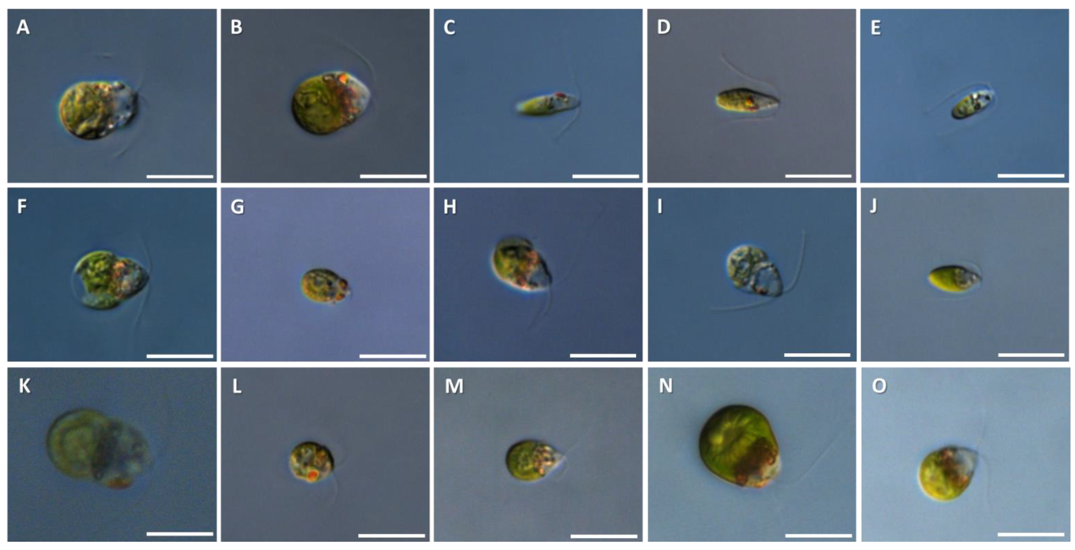
3.1. Morphological Analysis

Fifteen Dunaliella strains were isolated from eight saltworks in Greece (Table 1). Based on the morphological and morphometrical features of the isolated Dunaliella strains, they were classified into nine different species: strain TAU-MAC 0120, D. parva; strains TAU-MAC 0220, 1120, 1420, D. granulata; strains TAU-MAC 0320, 0420, D. minuta; strain TAU-MAC 0520, D. terricola; strains TAU-MAC 0620, 0820, D. asymmetrica; strain TAU-MAC 0720, D. bioculata; strains TAU-MAC 0920, 1020, D. viridis; strain TAU-MAC1220, D. minutissima; strains TAU-MAC 1320, 1520, D. polymorpha (Table 3, Figure 2).

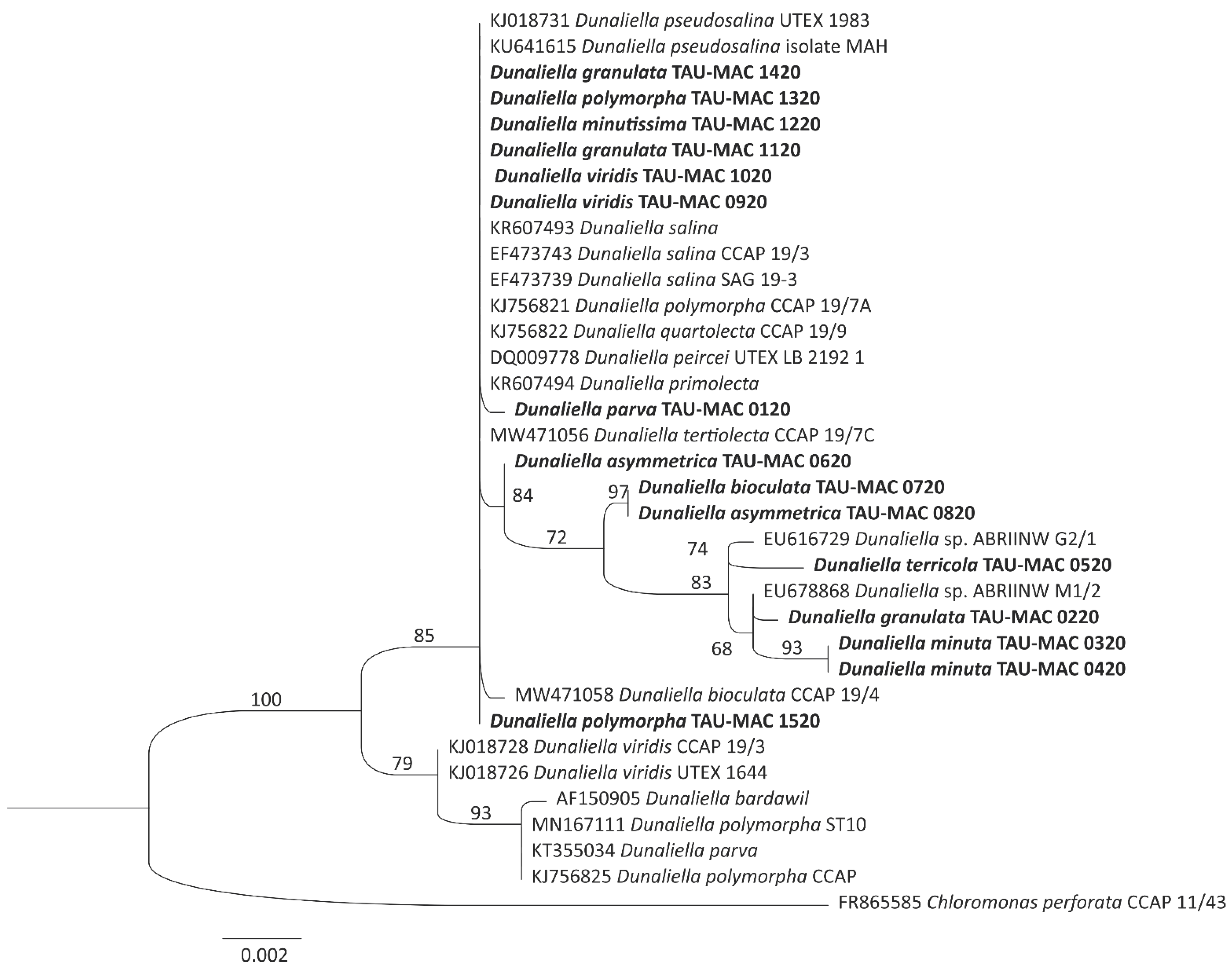
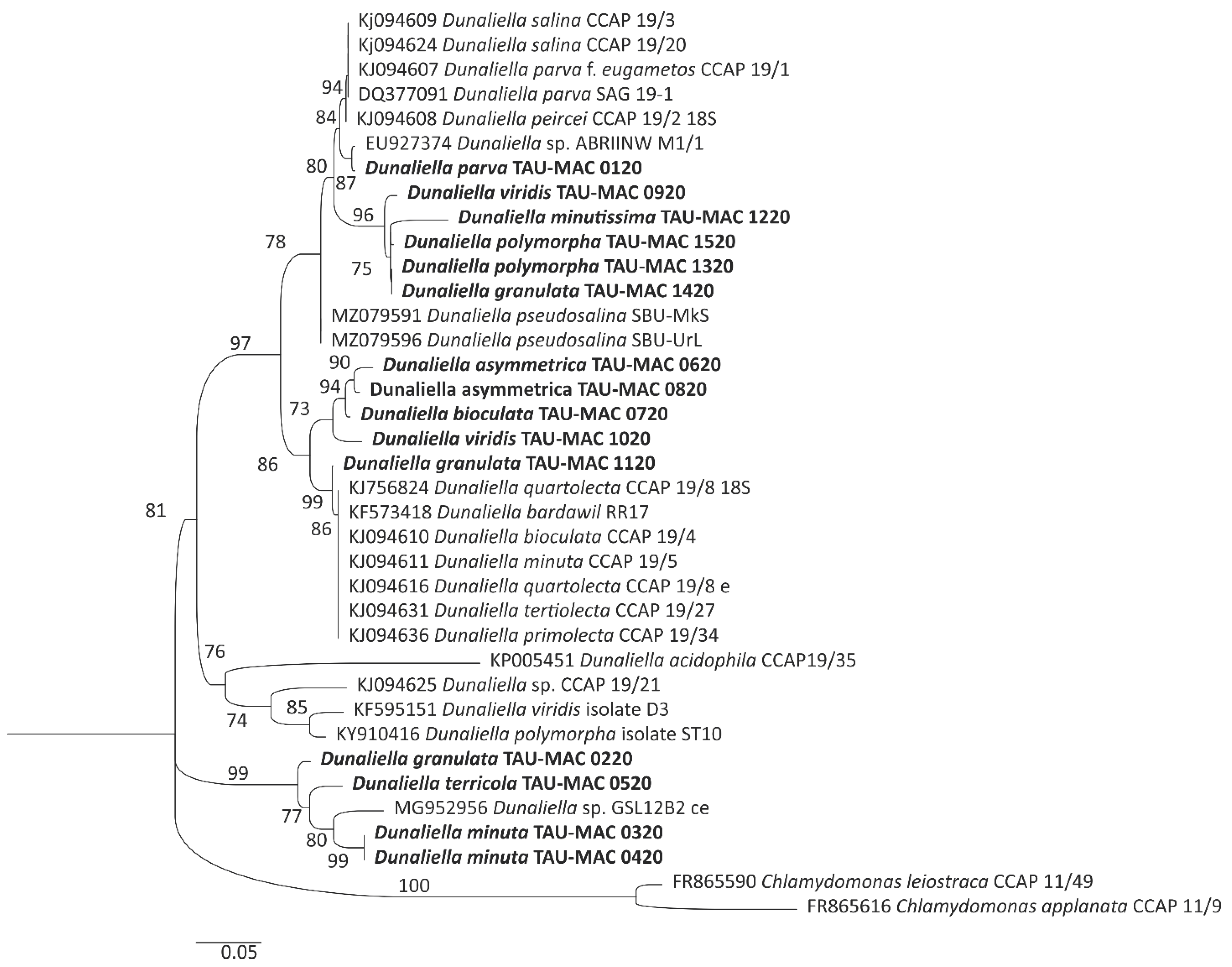
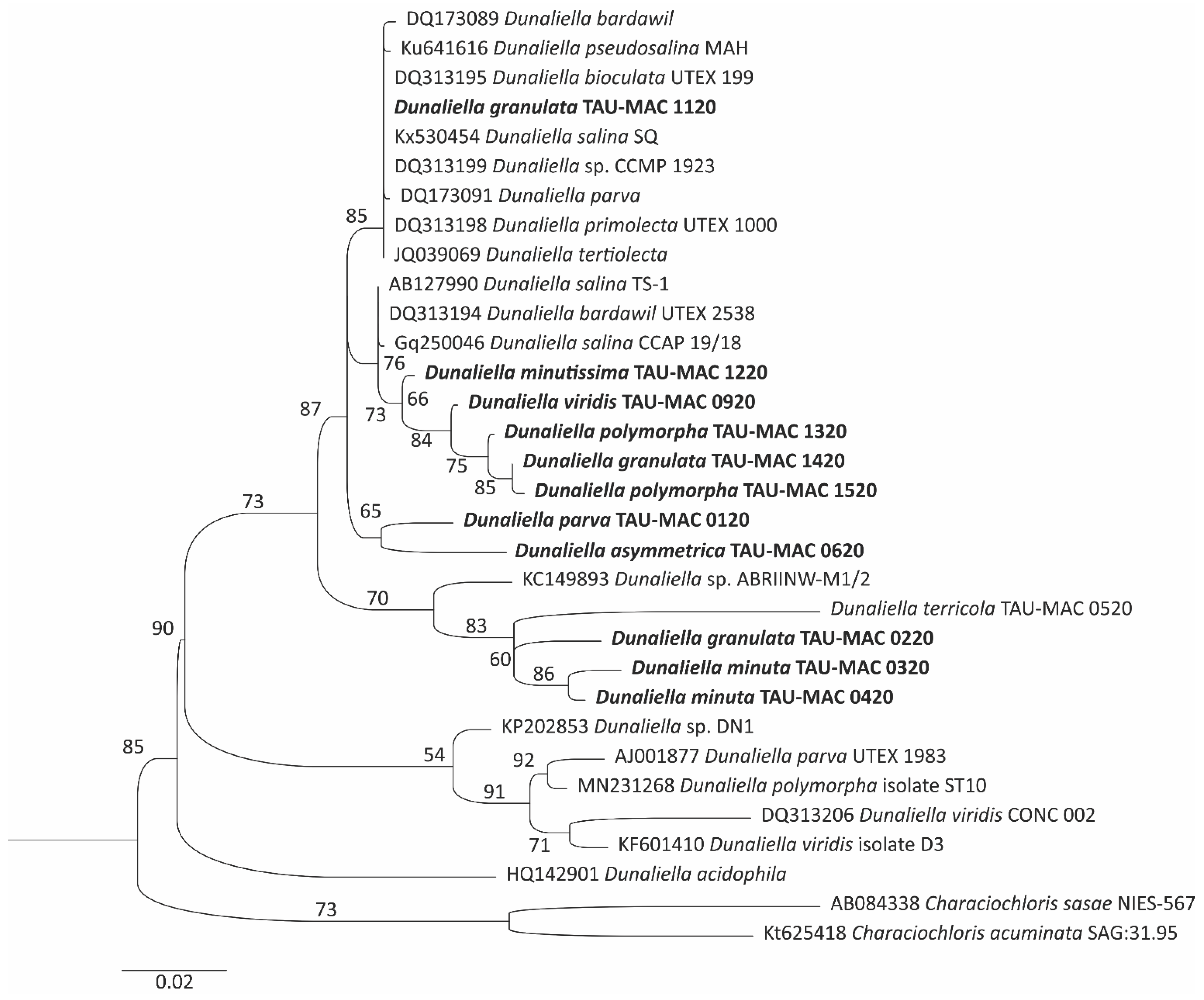
3.2. Phylogenetic Analysis

The phylogenetic relationships of the Dunaliella strains TAU-MAC 0120-1520 were analyzed and presented in Figure 3, Figure 4 and Figure 5. The results showed that all strains were grouped within the Dunaliella clade in all three analyses. The 18S rRNA phylogeny (Figure 3) showed that strains D. parva TAU-MAC 0120, D. viridis 0920 and 1020, D. granulata 1120 and 1420, and D. minutissima 1220, D. polymorpha 1320, were closely related and formed a clade along with several other species of the genus (D. salina, D. pseudosalina, D. quartolecta, D. peircei, D. primolecta), indicating the high degree of conservation of the genetic region. However, D. asymmetrica TAU-MAC 0620 was placed into a separate branch with high bootstrap support, and D. bioculata TAU-MAC 0720 and D. asymmetrica 0820 formed a distinct clade. D. minuta TAU-MAC 0320 and 0420, D. granulata 0220, and D. terricola TAU-MAC 0520 were grouped in a well-supported clade with several unidentified species of the genus.
The ITS region phylogeny (Figure 4) reinforced the results of the 18S rRNA phylogeny but provided a better resolution of relationships within the clades. A clade comprising the strains D. minutissima TAU-MAC 1220, D. granulata TAU-MAC 1120 and 1420, and D. polymorpha TAU-MAC 1320 and 1520 was positioned between the clades of D. salina and D. pseudosalina. D. parva TAU-MAC 0120 was placed in a clade with an unidentified strain of the genus, while in the rbcL phylogeny, it was positioned alone in a branch. The ITS phylogeny revealed a unique lineage distinct from other Dunaliella representatives, which included D. asummetrica TAU-MAC 0620 and 0820, D. bioculata TAU-MAC 0720, and D. viridis TAU-MAC 1020. The presence of multiple species in a distinct clade may be due to the lack of deposited sequences, as only 13 out of 30 described species of Dunaliella have deposited sequences (Table S2).
The molecular data based on the rbcL gene (Figure 5) confirmed the results obtained from the ITS phylogeny. The main difference between the ITS and rbcL phylogenetic trees was the genetic distance between the Dunaliella species. The rbcL gene appeared to be more divergent than the other two markers, resulting in better resolution in clade formation. However, the overall grouping pattern was similar to the ITS phylogeny, with a large clade containing multiple Dunaliella species (D. bardawil, D. pseudosalina, D. bioculata, D. salina, D. parva, D. primolecta, D. tertiolecta, D. salina) and smaller subclades composed of different species. It is important to note that the species forming the smaller subclades did not have any representatives in the genetically conserved clade in the rbcL phylogeny.

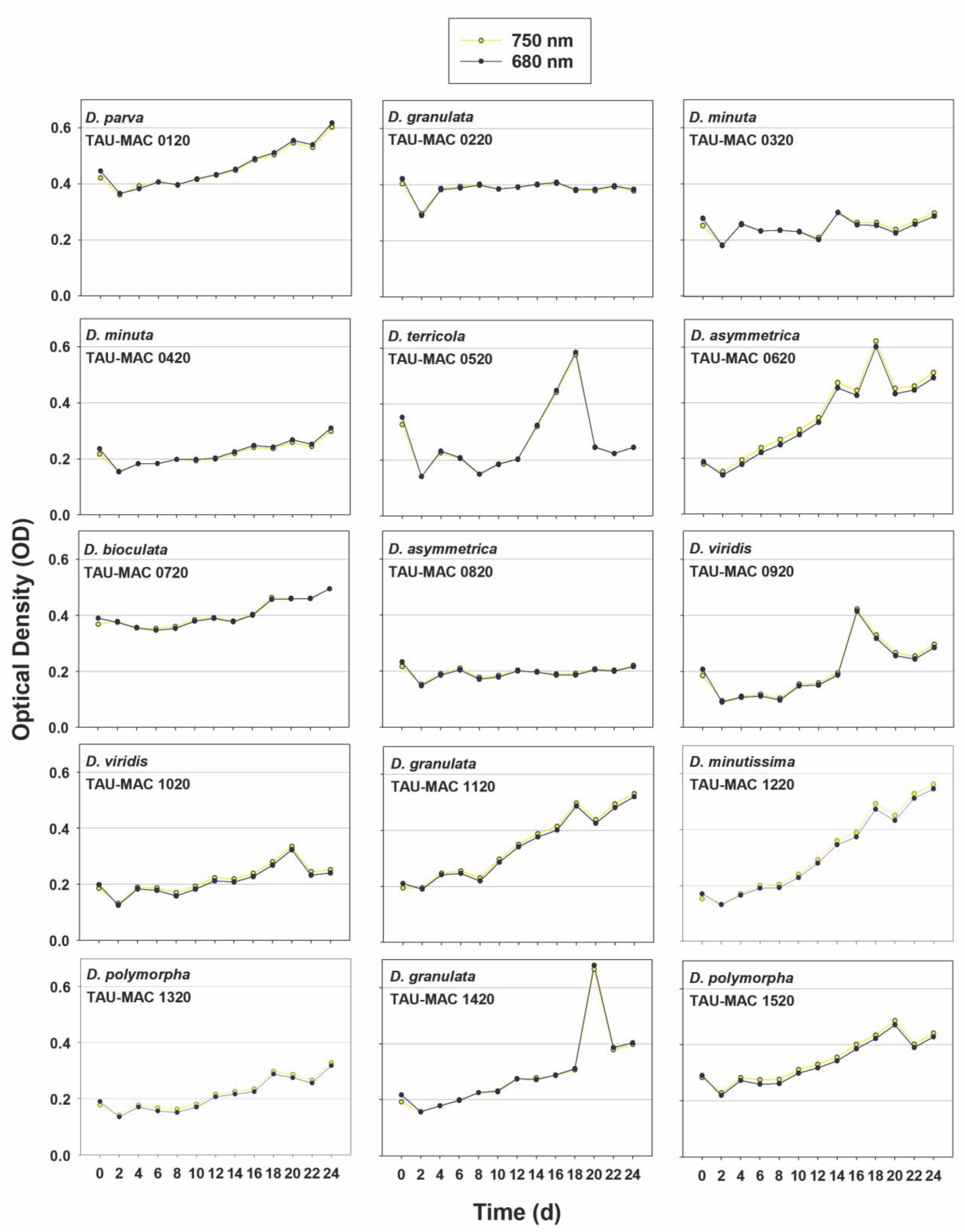
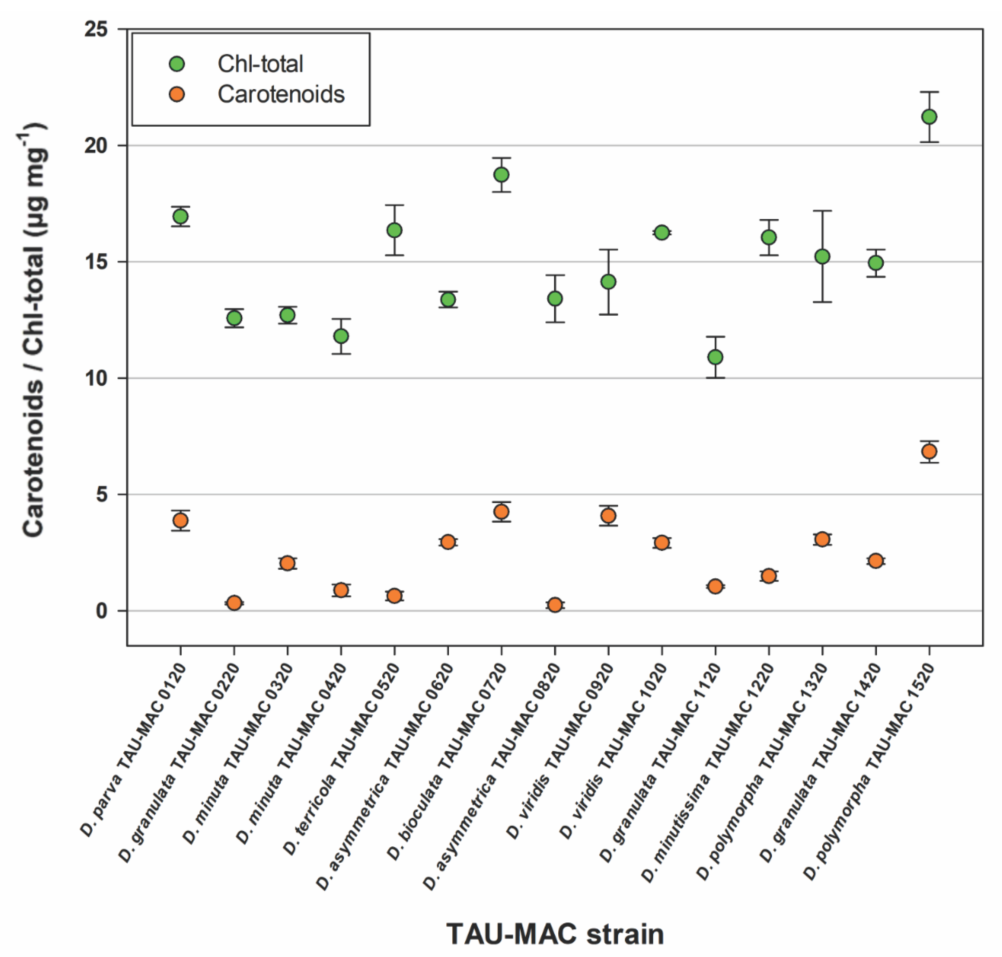
3.3. Growth Rates and Pigments

The results obtained from 15 biomass measurements within 24 days demonstrated a slow growth (Figure 6). No significant biomass accumulation was observed in the 15 Dunaliella strains according to the OD measurements, and the highest value was measured in the D. granulata TAU-MAC 1420 strain at 0.67 OD. The results at two wavelengths, 680 nm (corresponding to chla) and 750 nm (turbidity), were similar and overlapped.
Concerning the amount of the pigments produced during the growth experiments in the 15 TAU-MAC Dunaliella strains, total chlorophyll content was higher than total carotenoid content (Figure 7). The strain with the highest accumulation of both pigments was D. polymorpha TAU-MAC 1520. All other strains showed pigment accumulation on a scale of 0 to 5 and 10 to 20 μg mg−1 for total carotenoids and chlorophylls, respectively. Strains that produced either none or a very small amount of beta-carotene were the lowest concerning the chla production too.

4. Discussion

In this study, 15 strains of Dunaliella were isolated from eight saltworks in Greece and were classified into nine different species (D. parva, D. granulata, D. minuta, D. terricola, D. asymmetrica, D. bioculata, D. viridis, D. minutissima, and D. polymorpha) based on diacritical morphological and morphometrical features [2]. Our findings seem to be in congruence with other studies that examined the morphology of Dunaliella species [2,36]. Morphological discrepancies among strains are depicted mainly in the cell size, as well as in the size of Dunaliella’s distinct stigma. For instance, our strains (TAU-MAC 0320, 0420) classified as D. minuta demonstrate smaller cell size than the one described by Borowitzka and Siva [2], even though the rest of the morphological features are identical. Similarly, TAU-MAC strains classified as D. parva and D. minuta seem to have smaller cells compared with the ones decribed by Borowitzka and Siva [2]; however, our cell sizes are in range with the morphometric measurements performed by Beuzenberg et al. [37]. Different studies [5,37] suggest that morphological features used in Dunaliella’s classification could be affected by either nutrient concentration or even the growth medium itself. This could lead to misidentifications, as the cell size is a diacritical feature for some species. Preetha et al. [36] reported that D. salina had a cell size signficantly larger than other common strains such as D. parva, D. viridis, and D. tertiolecta, when tested under high salinity experiments. Borowitzka and Siva [2] reported that differences in cell or stigma dimensions could be explained from the existence of different subforms of Dunaliella species. Recent research [5,6,13,17,36] mainly focuses on the characterization and morphology of industrial cultivated species (e.g., D. salina, D. parva), whilst there is scarce information regarding the morphology of other species. This classification is crucial as it provides insight into the genetic relationships and metabolic capacities of different Dunaliella strains, which could have potential applications in the production of carotenoids for the food and pharmaceutical industries.
While morphological and morphometrical features are widely used in the classification and identification of microalgae, they may not always align with the phylogenetic position of species in the Dunaliella clade. Several studies [5,6,7,8,36,37,38,39] have attempted to validate the morphological features of Dunaliella spp. with phylogenetic traits, but these studies mainly focus on the intraspecies phylogenetic position within the Dunaliella lineage. To the best of our knowledge, this study comprises the first phylogenetic inference of several Dunaliella species. Based on 18S rRNA phylogeny, TAU-MAC strains, classified as different Dunaliella species (D. granulata, D. polymorpha, D. minutissima, D. viridis), formed a distinct clade with other well-studied species, such as D. salina and D. pseudosalina. González et al. [5] examined the phylogenetic position of D. salina and demonstrated the existence of different distinct clades within the D. salina lineage; however, they did not include sequences from other Dunaliella species. D. salina is mainly investigated based on its ability to produce beta-carotene and the classification of strains might be incorrect as it might fail in its ability to turn red [36,39]. This might explain the clustering of TAU-MAC species and representatives from other species with D. salina in our 18S rRNA analysis, indicating an unknown high degree of conservation of the specific genetic region. Other molecular markers, such as tufa also failed to distinguish the phylogenetic position of different Dunaliella species [8]. Additionally, species that did not have any representatives in GenBank (D. asymmetrica, D. granulata, D. minutissima, D. terricola) seem to cluster in distinct clades with different Dunaliella morphospecies. One study [36] used a combination of molecular and morphological data to investigate the phylogenetic relationships between different species of Dunaliella. The authors found that D. salina and D. bardawil were closely related, and that D. parva was a distinct lineage that was more closely related to other species such as D. viridis. The study also revealed that there was significant genetic diversity within the genus, with some species being more closely related to each other than to other species. This is in congruence with our results as some Dunaliella species, such as D. minuta, D. parva, and D. polymorpha formed distinct clades in our 18S phylogenetic analysis.
In our study, TAU-MAC strains presented a better resolution in the ITS analysis, compared to the 18S, as almost all the TAU-MAC strains formed distinct subclades from other Dunaliella species, which were clustered together. Additionally, species that did not have any representatives in GenBank (D. asymmetrica, D. granulata, D. minutissima, D. terricola) seem to cluster in distinct clades with different Dunaliella morphospecies. The utilization of ITS markers for Dunaliella species is a popular choice due to their abundance in the genome, which facilitates their amplification, and their tendency for frequent insertions and deletions within the sequence, leading to notable variation between different species [8]. However, ITS did not corroborate with the morphological analysis, as the clades formed contained several species. This could be resolved if more strains, classified in these species, were characterized and included in further phylogenetic studies [2,5,39,40]. For example, a clade comprising different species was placed between the D. salina and D. pseudosalina groups and distinguished them, in contrast to a previous study [5] reporting that the ITS failed to discriminate the position of D. salina and D. pseudosalina, whose classification was based on ecophysiological characteristics (ability to turn red). Another distinct clade formed by TAU-MAC strains grouped near a large clade of different Dunaliella species, which was revised by Assuncao et al. [40] as the Dunaliella tertiolecta clade. Dunaliella minuta was firstly characterized as a segregation of D. viridis [40]. However, its phylogenetic position was always problematic, mainly due to either a lack of sequences or identical sequences representing the same species. TAU-MAC strains classified as D. minuta formed a well-defined clade at the basis of our ITS tree (also in 18S rRNA and rbcL), and this clade could be considered as D. minuta, which could partly resolve the problematic status of Dunaliella taxonomy. TAU-MAC strains 0920 and 1020 classified as Dunaliella viridis based on morphology formed different single clades in ITS phylogeny; this phylogenetic discrepancy could indicate that D. viridis is a polyphyletic lineage. In fact, Assuncao et al. 2013 [40] examined the phylogenetic position of different D. viridis strains, using ITS, and proposed four different subclades of the D. viridis species. The authors suggest that the different clades should be considered as different biological species. Such a case has already been made for D. salina [5,39,41]. Similarly, TAU-MAC strains 0220, 1120, and 1420 classified as D. granulata fell into different clades in our ITS phylogeny. However, there is still no sufficient information in the scientific literature concerning the placement of D. granulata inside Dunaliella.
More recent studies [6,8,36] have used the 18S rRNA gene, ITS, and rbcL genetic regions in combination to infer the relationships between species of Dunaliella. For example, Preetha et al. [36] used these three genetic markers to investigate the relationships between different species of Dunaliella. This study confirmed the presence of two main clades within the genus, but also identified several other subclades within the halophilic clade. In our study, TAU-MAC strains presented a better resolution comparing the rbcL analysis, as almost all the TAU-MAC strains formed distinct subclades from other Dunaliella species, that clustered together. Highfield et al. [8] reported that the rbcL region played a key role in the evolution of Dunaliella. These results are in congruence with our phylogenetic analysis, as the genetic distances in our phylogenetic tree are higher than the other two tested markers, thus rcbL could be used for a better demonstration of the “true” number of representatives from the Dunaliella genus. Highfield et al. [8] used rbcL in a consensus phylogenetic analysis with other markers and they were able to distinguish different evolutionary patters in D. salina.
In addition to the potential of the rbcL (or the combination with other markers) to be an adequate marker for resolving Dunaliella’s phylogeny, there is a need expressed among the scientific community that a “type strain” should be established for each Dunaliella taxon (including subspecies, forms, or varieties), thereby greatly facilitating comparison with new isolates, and avoiding misleading information and/or false conclusions. Actually, the only Dunaliella strains that are described as “type strains” in the different Official Culture Collections are Dunaliella bioculata CCAP19/4 (UTEX199, SAG19-4) and Dunaliella primolecta CCAP11/34 (UTEX1000, SAG183-80), surprisingly both are included in the Tertioleta-clade  [38]. We also identified that among the 30 morphologically differentiated species [2,5], molecular aspects of only a few ones have been extensively studied and reported, and there is an important gap on molecular taxonomic markers (Table S2).
In the Mediterranean region, a number of studies have been conducted to determine the presence and diversity of Dunaliella species in various habitats, including saltworks and hypersaline lakes [2,4,8,10,17,42]. Some of the species that have been reported in this region include D. salina, D. parva, D. granulata, and D. terricola. The results of the present study are consistent with the findings of previous studies reporting the presence of different Dunaliella species in the Mediterranean region and other salt-rich habitats around the world [4,7,10,17,18]. However only the occurrence of D. salina and D. viridis has been reported from Greek saltworks [17,18,43,44]. Thus, to the best of our knowledge, this study is the first report of the presence of several other Dunaliella species, demonstrating the high diversity and adaptability of these microalgae, which can survive and thrive in a range of extreme environments [1]. In our phylogenetic analysis, a number of Dunaliella strains from Greek saltworks formed subclades, which could depict evidence of endemism inside the genus. However, further studies are required to determine the distribution, the prevalence, and the preference of Dunaliella species in saltworks around the world.
In terms of biomass accumulation, the results of the study demonstrated a slow growth for the strains, with no significant accumulation observed. The highest value measured was in D. granulata TAU-MAC 1420, at 0.67 OD. The total chlorophyll and carotenoid concentrations of the 15 Dunaliella strains were analyzed, with the highest accumulation of both pigments observed in D. polymorpha TAU-MAC 1520. One possible explanation for the low biomass accumulation and pigment concentrations observed in our strains is suboptimal growth conditions. If any of the tested growth conditions were not optimal, it could affect the growth rate and metabolic activity of Dunaliella [45]. Therefore, it is possible that the conditions used in this experiment were not optimal for the growth and pigment production of the Dunaliella strains. Another possible explanation is genetic variability among the strains. Different strains of Dunaliella can exhibit significant genetic diversity [3], which can result in variations in growth rates, pigment production, and stress tolerance, hence the low rates observed in our study. One of the most studied Dunaliella species is D. salina, which is known to produce high levels of beta-carotene under conditions of high salinity and exposure to light [11,13,15,18]. Studies have shown that under optimal growth conditions, D. salina can produce up to 20% of its dry weight in beta-carotene. This makes it one of the most efficient beta-carotene-producing microalgae known to date. Even though we report the isolation of Dunaliella species from Greek saltworks, our strains did not produce such amounts of beta-carotene reported in the scientific literature under normal laboratory conditions. D. parva is another species that is known to produce beta-carotene, although the exact levels produced by this species have not been well documented [46,47,48,49]. Some studies have shown that D. parva can produce beta-carotene levels of up to 5% of its dry weight under optimal growth conditions, while others have reported much lower levels [5]. TAU-MAC 0120 strain was classified as D. parva and was one of the highest producers of beta-carotene among the Dunaliella strains analyzed in this work. The production of beta-carotene could be hindered by different parameters, such as high salinity, temperature, and light. González et al. [5] demonstrated that in D. salina strains isolated from Chile, low temperature seems to be the trigger point of beta-carotene production. In fact, the amounts of Car/Chla reported from González et al. [5] in the highest temperature tested are in accordance or even lower with the ones reported from this study. Further investigation is required in order to identify the strains with the ability to produce high amounts of beta-carotene under a wide range of environmental conditions, including high salinity, temperature, and light.

5. Conclusions

In conclusion, the study of the Dunaliella species highlights their potential for biotechnological applications. In our study, TAU-MAC strains did not produce significant amounts of beta-carotene under normal laboratory conditions. The presence of various Dunaliella species in Greece was classified based on morphological and morphometrical features, in line with previous studies showing the diversity and adaptability of these microalgae in salt-rich environments around the world. The study also includes the first examination of the phylogenetic position of a large number of different Dunaliella species. The results indicate the formation of distinct clades among different Dunaliella species and suggest that morphological and morphometrical features may not always align with the phylogenetic position of species in the Dunaliella clade. Additionally, this study bolsters the lack of correlation between molecular data and morphological attributes used so far for taxonomic circumscription within the genus Dunaliella. In light of this information, we are still far from solving the taxonomic problem of some of the above species. Some of the existing problems in the proper analysis of phylogenetic data for taxa originate from the uncertainty about the original type of material from culture collections. In order to gain better knowledge on the phylogenetic relationship among the taxa within the genus, which should lead to a better definition of the species, more focused work is required on biochemical (pigment content analysis), physiological (growth rates under different salt concentration), and molecular attributes.

Supplementary Materials

The following supporting information can be downloaded at: https://www.mdpi.com/article/10.3390/w15061037/s1, Table S1: NCBI accession numbers of the TAU-MAC Dunaliella strains for the molecular markers 18S rRNA gene, the 18S-28S rRNA internal transcribed spacer (ITS), and the ribulose-1,5-bisphosphate carboxylase/oxygenase large subunit (rbcL) gene; Table S2: List with the accepted Dunaliella species from algae base and their representation in GenBank concerning the phylogenetic markers 18S rRNA gene, the 18S-28S rRNA internal transcribed spacer (ITS), and the ribulose-1,5-bisphosphate carboxylase/oxygenase large subunit (rbcL) gene.

Author Contributions

Conceptualization, S.G., G.Z. and K.T.; study design, S.G., G.P., U.L. and M.P.; formal analysis, M.P., U.L., G.P., G.F., C.G., S.K. and G.I.; validation, S.G., K.T. and G.Z.; data curation, S.G., M.P. and G.P.; writing-original draft preparation, S.G., M.P., G.P., U.L., G.F. and C.G.; writing-review and editing, S.G. and M.P.; visualization, S.G. and M.P.; supervision, S.G., G.Z. and K.T.; project administration, S.G. All authors have read and agreed to the published version of the manuscript.

Funding

This research is co-financed by the European Union (European Regional Development Fund) and Greek resources through the National Action “Special Actions” AQUACULTURE INDUSTRIAL MATERIALS and OPENNESS Innovation (EPANEK), GSRT, NSRF 2014–2020 (PILOUS project, MIS 5045805).

Data Availability Statement

Data is contained within the article or supplementary material.

Acknowledgments

We thank N. Korovesis as well as all the board of directors of Hellenic Saltworks SA for the provision of sampling permits.

Conflicts of Interest

The authors declare no conflict of interest.

References

  1. Oren, A. The microbiology of red brines. In Advances in Applied Microbiology; Academic Press Inc.: Cambridge, MA, USA, 2020; Volume 113, pp. 57–110. ISBN 9780128207093. [Google Scholar]
  2. Borowitzka, M.A.; Siva, C.J. The taxonomy of the genus Dunaliella (Chlorophyta, Dunaliellales) with emphasis on the marine and halophilic species. J. Appl. Phycol. 2007, 19, 567–590. [Google Scholar] [CrossRef]
  3. da Silva, M.R.O.B.; Moura, Y.A.S.; Converti, A.; Porto, A.L.F.; Viana Marques, D.d.A.; Bezerra, R.P. Assessment of the potential of Dunaliella microalgae for different biotechnological applications: A systematic review. Algal Res. 2021, 58, 102396. [Google Scholar] [CrossRef]
  4. Oren, A. The ecology of Dunaliella in high-salt environments. J. Biol. Res. 2014, 21, 23. [Google Scholar] [CrossRef] [Green Version]
  5. González, M.A.; Gómez, P.I.; Polle, J.E.W. Taxonomy and Phylogeny of the Genus Dunaliella. In The Alga Dunaliella; CRC Press: Boca Raton, FL, USA, 2019; pp. 15–44. [Google Scholar]
  6. Henley, W.J.; Cobbs, M.; Novoveská, L.; Buchheim, M.A. Phylogenetic analysis of Dunaliella (Chlorophyta) emphasizing new benthic and supralittoral isolates from Great Salt Lake. J. Phycol. 2018, 54, 483–493. [Google Scholar] [CrossRef]
  7. Gao, F.; Nan, F.; Feng, J.; Lv, J.; Liu, Q.; Liu, X.; Xie, S. Comparative morphological, physiological, biochemical and genomic studies reveal novel genes of Dunaliella bioculata and D. quartolecta in response to salt stress. Mol. Biol. Rep. 2022, 49, 1749–1761. [Google Scholar] [CrossRef] [PubMed]
  8. Highfield, A.; Ward, A.; Pipe, R.; Schroeder, D.C. Molecular and phylogenetic analysis reveals new diversity of Dunaliella salina from hypersaline environments. J. Mar. Biol. Assoc. UK 2021, 101, 27–37. [Google Scholar] [CrossRef]
  9. Rodrigues, C.M.; Bio, A.; Amat, F.; Vieira, N. Artisanal salt production in Aveiro/Portugal—An ecofriendly process. Saline Syst. 2011, 7, 3. [Google Scholar] [CrossRef] [PubMed] [Green Version]
  10. Chekanov, K. Diversity and Distribution of Carotenogenic Algae in Europe: A Review. Mar. Drugs 2023, 21, 108. [Google Scholar] [CrossRef]
  11. Zhang, X.; Tang, X.; Wang, M.; Zhang, W.; Zhou, B.; Wang, Y. ROS and calcium signaling mediated pathways involved in stress responses of the marine microalgae Dunaliella salina to enhanced UV-B radiation. J. Photochem. Photobiol. B Biol. 2017, 173, 360–367. [Google Scholar] [CrossRef] [PubMed]
  12. Xi, Y.; Kong, F.; Chi, Z. ROS Induce β-Carotene Biosynthesis Caused by Changes of Photosynthesis Efficiency and Energy Metabolism in Dunaliella salina Under Stress Conditions. Front. Bioeng. Biotechnol. 2021, 8, 1447. [Google Scholar] [CrossRef]
  13. Pourkarimi, S.; Hallajisani, A.; Nouralishahi, A.; Alizadehdakhel, A.; Golzary, A. Factors affecting production of beta-carotene from Dunaliella salina microalgae. Biocatal. Agric. Biotechnol. 2020, 29, 101771. [Google Scholar] [CrossRef]
  14. Gallego-Cartagena, E.; Castillo-Ramírez, M.; Martínez-Burgos, W. Effect of stressful conditions on the carotenogenic activity of a Colombian strain of Dunaliella salina. Saudi J. Biol. Sci. 2019, 26, 1325–1330. [Google Scholar] [CrossRef]
  15. Xi, Y.; Bian, J.; Luo, G.; Kong, F.; Chi, Z. Enhanced β-carotene production in Dunaliella salina under relative high flashing light. Algal Res. 2022, 67, 102857. [Google Scholar] [CrossRef]
  16. Hotos, G.N. A Preliminary Survey on the Planktonic Biota in a Hypersaline Pond of Messolonghi Saltworks (W. Greece). Diversity 2021, 13, 270. [Google Scholar] [CrossRef]
  17. Hotos, G.; Avramidou, D.; Mastropetros, S.G.; Tsigkou, K.; Kouvara, K.; Makridis, P.; Kornaros, M. Isolation, identification, and chemical composition analysis of nine microalgal and cyanobacterial species isolated in lagoons of Western Greece. Algal Res. 2023, 69, 102935. [Google Scholar] [CrossRef]
  18. Chantzistrountsiou, X.; Ntzouvaras, A.; Papadaki, S.; Tsirigoti, A.; Tzovenis, I.; Economou-Amilli, A. Carotenogenic Activity of Two Hypersaline Greek Dunaliella salina Strains under Nitrogen Deprivation and Salinity Stress. Water 2023, 15, 241. [Google Scholar] [CrossRef]
  19. Sili, C.; Torzillo, G.; Vonshak, A. Arthrospira (Spirulina). In Ecology of Cyanobacteria II: Their Diversity in Space and Time; Springer: Berlin/Heidelberg, Germany, 2012; Volume 9789400738, pp. 677–705. ISBN 9789400738553. [Google Scholar]
  20. Waterbury, J.B.; Stanier, R.Y. Patterns of growth and development in pleurocapsalean cyanobacteria. Microbiol. Rev. 1978, 42, 2–44. [Google Scholar] [CrossRef] [PubMed]
  21. Johnson, M.K.; Johnson, E.J.; MacElroy, R.D.; Speer, H.L.; Bruff, B.S. Effects of salts on the halophilic alga Dunaliella viridis. J. Bacteriol. 1968, 95, 1461–1468. [Google Scholar] [CrossRef] [PubMed] [Green Version]
  22. Chitlaru, E.; Pick, U.; Physiology, S.P.; Oct, N.; Chitlaru, E.; Pick, U. Selection and Characterization of Dunaliella salina Mutants Defective in Haloadaptation Linked references are available on JSTOR for this article: Selection and Characterization of Dunaliella salina Mutants Defective in Haloadaptation. Plant Physiol. 1989, 91, 788–794. [Google Scholar] [CrossRef] [Green Version]
  23. De Chazal, N.M.; Smaglinski, S.; Smith, G.D. Methods involving light variation for isolation of cyanobacteria: Characterization of isolates from central Australia. Appl. Environ. Microbiol. 1992, 58, 3561–3566. [Google Scholar] [CrossRef] [Green Version]
  24. Gkelis, S.; Fernández Tussy, P.; Zaoutsos, N. Isolation and preliminary characterization of cyanobacteria strains from freshwaters of Greece. Open Life Sci. 2015, 10, 52–60. [Google Scholar] [CrossRef]
  25. Lortou, U.; Gkelis, S. Polyphasic taxonomy of green algae strains isolated from Mediterranean freshwaters. J. Biol. Res. 2019, 26, 11. [Google Scholar] [CrossRef] [Green Version]
  26. Gkelis, S.; Panou, M. Capturing biodiversity: Linking a cyanobacteria culture collection to the “scratchpads” virtual research environment enhances biodiversity knowledge. Biodivers. Data J. 2016, 4, e7965-1. [Google Scholar] [CrossRef] [PubMed] [Green Version]
  27. Atashpaz, S.; Khani, S.; Barzegari, A.; Barar, J.; Vahed, S.Z.; Azarbaijani, R.; Omidi, Y. A robust universal method for extraction of genomic DNA from bacterial species. Microbiology 2010, 79, 538–542. [Google Scholar] [CrossRef]
  28. Medlin, L.; Elwood, H.J.; Stickel, S.; Sogin, M.L. The characterization of enzymatically amplified eukaryotic 16S-like rRNA-coding regions. Gene 1988, 71, 491–499. [Google Scholar] [CrossRef] [Green Version]
  29. Lane, D. 16S/23S rRNA Sequencing. In Nucleic Acid Techniques in Bacterial Systematic; Stackebrandt, E., Goodfellow, M., Eds.; John Wiley and Sons: New York, NY, USA, 1991; pp. 115–175. [Google Scholar]
  30. Johnson, J.L.; Fawley, M.W.; Fawley, K.P. The diversity of Scenedesmus and Desmodesmus (Chlorophyceae) in Itasca State Park, Minnesota, USA. Phycologia 2007, 46, 214–229. [Google Scholar] [CrossRef]
  31. Nozaki, H.; Itoh, M.; Sano, R.; Uchida, H.; Watanabe, M.M.; Kuroiwa, T. Phylogenetic relationships within the colonial Volvocales (Chlorophyta) inferred from rbcL gene sequence data. J. Phycol. 1995, 31, 970–979. [Google Scholar] [CrossRef]
  32. Okonechnikov, K.; Golosova, O.; Fursov, M. UGENE team Unipro UGENE: A unified bioinformatics toolkit. Bioinformatics 2012, 28, 1166–1167. [Google Scholar] [CrossRef] [PubMed] [Green Version]
  33. Kumar, S.; Stecher, G.; Tamura, K. MEGA7: Molecular Evolutionary Genetics Analysis Version 7.0 for Bigger Datasets. Mol. Biol. Evol. 2016, 33, 1870–1874. [Google Scholar] [CrossRef] [Green Version]
  34. Griffiths, M.J.; Garcin, C.; van Hille, R.P.; Harrison, S.T.L. Interference by pigment in the estimation of microalgal biomass concentration by optical density. J. Microbiol. Methods 2011, 85, 119–123. [Google Scholar] [CrossRef]
  35. Wellburn, A.R. The Spectral Determination of Chlorophylls a and b, as well as Total Carotenoids, Using Various Solvents with Spectrophotometers of Different Resolution. J. Plant Physiol. 1994, 144, 307–313. [Google Scholar] [CrossRef]
  36. Preetha, K.; John, L.; Subin, C.S.; Vijayan, K.K. Phenotypic and genetic characterization of Dunaliella (Chlorophyta) from Indian salinas and their diversity. Aquat. Biosyst. 2012, 8, 27. [Google Scholar] [CrossRef] [Green Version]
  37. Beuzenberg, V.; Smith, K.F.; Packer, M.A. Isolation and characterisation of halo-tolerant Dunaliella strains from Lake Grassmere/Kapara Te Hau, New Zealand. N. Z. J. Bot. 2014, 52, 136–152. [Google Scholar] [CrossRef]
  38. Assunção, P.; Jaén-Molina, R.; Caujapé-Castells, J.; de la Jara, A.; Carmona, L.; Freijanes, K.; Mendoza, H. Phylogenetic position of Dunaliella acidophila (Chlorophyceae) based on ITS and rbcL sequences. J. Appl. Phycol. 2012, 24, 635–639. [Google Scholar] [CrossRef]
  39. González, M.A.; Coleman, A.W.; Gómez, P.I.; Montoya, R. Phylogenetic relationship among various strains of Dunaliella (Chlorophyceae) based on nuclear its rDNA sequences. J. Phycol. 2001, 37, 604–611. [Google Scholar] [CrossRef]
  40. Assunção, P.; Jaén-Molina, R.; Caujapé-Castells, J.; Wolf, M.; Buchheim, M.A.; de la Jara, A.; Freijanes, K.; Carmona, L.; Mendoza, H. Phylogenetic analysis of ITS2 sequences suggests the taxonomic re-structuring of Dunaliella viridis (Chlorophyceae, Dunaliellales). Phycol. Res. 2013, 61, 81–88. [Google Scholar] [CrossRef]
  41. Polle, J.E.W.; Struwe, L.; Jin, E. Identification and characterization of a new strain of the unicellular green alga Dunaliella salina (Teod.) from Korea. J. Microbiol. Biotechnol. 2008, 18, 821–827. [Google Scholar] [PubMed]
  42. Hejazi, M.A.; Barzegari, A.; Gharajeh, N.H.; Hejazi, M.S. Introduction of a novel 18S rDNA gene arrangement along with distinct ITS region in the saline water microalga Dunaliella. Saline Syst. 2010, 6, 4. [Google Scholar] [CrossRef] [Green Version]
  43. Dolapsakis, N.P.; Tafas, T.; Abatzopoulos, T.J.; Ziller, S.; Economou-Amilli, A. Abundance and growth response of microalgae at Megalon Embolon solar saltworks in northern Greece: An aquaculture prospect. J. Appl. Phycol. 2005, 17, 39–49. [Google Scholar] [CrossRef]
  44. Hotos, G.N.; Avramidou, D. The Effect of Various Salinities and Light Intensities on the Growth Performance of Five Locally Isolated Microalgae [Amphidinium carterae, Nephroselmis sp., Tetraselmis sp. (var. red pappas), Asteromonas gracilis and Dunaliella sp.] in Laboratory Batch Cultures. J. Mar. Sci. Eng. 2021, 9, 1275. [Google Scholar]
  45. Al-Mhanna, N.; Pistorius, M.; Al Sammarraie, L. Optimization of the Cultivation Conditions of the Green Algae Dunaliella salina by Using Simplex Method. Processes 2023, 11, 292. [Google Scholar] [CrossRef]
  46. Gan, S.; Liang, S.; Zou, Q.; Shang, C. Optimization of carotenoid extraction of a halophilic microalgae. PLoS ONE 2022, 17, e0270650. [Google Scholar] [CrossRef] [PubMed]
  47. Li, Y.; Huang, X.; Luo, L.; Shang, C. Optimization of Extraction Conditions of Carotenoids from Dunaliella parva by Response Surface Methodology. Molecules 2022, 27, 1444. [Google Scholar] [CrossRef] [PubMed]
  48. Ismaiel, M.M.S.; El-Ayouty, Y.M.; Fathey, H.A. Disparity of the carotenoids antioxidant properties of wild-type and D-PSY-transgenic Dunaliella parva strains under three environmental stresses. Physiol. Mol. Biol. Plants 2021, 27, 2151. [Google Scholar] [CrossRef] [PubMed]
  49. Ismaiel, M.M.S.; El-Ayouty, Y.M.; Said, A.A.; Fathey, H.A. Transformation of Dunaliella parva with PSY gene: Carotenoids show enhanced antioxidant activity under polyethylene glycol and calcium treatments. Biocatal. Agric. Biotechnol. 2018, 16, 378–384. [Google Scholar] [CrossRef]
Figure 1. Map of Greece showing the locations of the eight salt marshes, sampled for the isolation of Dunaliella strains. The numbers 1 to 8 identify the water bodies: (1) Mesi, (2) Nea Kessani, (3) Angelochori, (4) Kitros, (5) Messolonghi, (6) Kalloni, (7) Polichnitos, (8) Adamas.
Water 15 01037 g001
Figure 2. Light micrographs of Dunaliella strains isolated in this study. (A) D. parva TAU-MAC 0120, (B) D. granulata TAU-MAC 0220, (C) D. minuta TAU-MAC 0320, (D) D. minuta TAU-MAC 0420, (E) D. terricola TAU-MAC 0520, (F) D. asymmetrica TAU-MAC 0620, (G) D. bioculata TAU-MAC 0720, (H) D. asymmetrica TAU-MAC 0820, (I) D. viridis TAU-MAC 0920, (J) D. viridis TAU-MAC 1020, (K) D. granulata TAU-MAC 1120, (L) D. minutissima TAU-MAC 1220, (M) D. polymorpha TAU-MAC 1320, (N) D. granulata TAU-MAC 1420, (O) D. polymorpha TAU-MAC 1520. Scale bar 10 μm.
Water 15 01037 g002
Figure 3. Phylogenetic tree of Dunaliella species relationships based on 18S rRNA (c. 1500 bp), including TAU-MAC Dunaliella strains isolated from Greece (bold). Support values are indicated as bootstrap support for maximum likelihood (ML) analysis. GenBank accession numbers for the studied strains are given in Table S1 (Supplementary Materials).
Water 15 01037 g003
Figure 4. Phylogenetic tree of Dunaliella species relationships based on ITS region (c. 500 bp), including TAU-MAC Dunaliella strains isolated from Greece (bold). Support values are indicated as bootstrap support for maximum likelihood (ML) analysis. GenBank accession numbers for the studied strains are given in Table S1 (Supplementary Materials).
Water 15 01037 g004
Figure 5. Phylogenetic tree of Dunaliella species based on rbcL gene (c. 1050 bp), including TAU-MAC Dunaliella strains isolated from Greece (bold). Support values are indicated as bootstrap support for maximum likelihood (ML) analysis. GenBank accession numbers for the studied strains are given in Table S1 (Supplementary Materials).
Water 15 01037 g005
Figure 6. Growth curves of 15 TAU-MAC Dunaliella strains from 12 biomass measurements within 24 days. The absorbance at 680 nm (blue line) was used to measure the amount of chlorophyll α absorbed, while a wavelength of 750 nm (yellow line) was used to measure the apparent turbidity of the sample. At 750 nm, there was no light absorption by the pigment, and the measurement corresponded to the light scattering.
Water 15 01037 g006
Figure 7. Total carotenoid (orange) and chlorophyll (green) concentrations (μg m−1) of 15 Dunaliella strains. Dots correspond to the average of the triplicate measurements; bars refer to standard errors.
Water 15 01037 g007
Table 1. Dunaliella strains isolated from solar saltworks of Greece.
Strain
(TAU-MAC)
Isolation SiteGeographic Coordinates (N, E)Collection Date
0720Nea Kessani41.019039, 25.07718123 January 2020
082026 May 2020
152010 September 2020
0120Mesi40.973328, 25.22087923 January 2020
022023 January 2020
132010 September 2020
1220Angelochori40.491461, 22.8219339 September 2020
0520Kitros40.374210, 22.63491117 January 2020
14209 September 2020
0920Kalloni39.213665, 26.25267523 June 2020
0620Polichnitos39.114962, 26.17666723 February 2020
0320Messolonghi38.399854, 21.41590127 January 2020
042027 January 2020
102012 June 2020
1120Adamas, Milos36.711771, 24.46783026 August 2020
Table 2. PCR primers used in the analyses of strains isolated from freshwaters of Greece.
PrimerTarget Gene-RegionSequence (5′-3′)Size (bp)ReferenceConditions
EukA18S rRNAAACCTGGTTGATCCTGCCAGT1750[28]Initial denaturation step at 95 °C for 5 min, 35 cycles consisting of denaturation at 95 °C for 60 s, annealing at 55 °C for 60 s and elongation at 72 °C for 90 s; a final 7-min elongation step at 72 °C was included.
EukBTGATCCTTCTGCAGGTTCACCTAC
U1391RGGGCGGTGTGTACAARGR *[29]
ITS-AFITSCGTTTCCGTAGGTGAACCTGC700[30]Initial denaturation step at 94 °C for 4 min, 35 cycles consisting of denaturation at 95 °C for 60 s, annealing at 58 °C for 2 min and elongation at 72 °C for 2 min; a final 7 min elongation step at 72 °C was included.
ITS-BRCATATGCTTAAGTTCAGCGGG T
rbcL1-20rbcLATGGTTCCACAAACAGAAAC1100[31]
rbcL1181-1160AAGATTTCAACTAAAGCTGGCA
*: The letter R is for unspecified purine nucleoside (A, G) based on the International Union of Biochemistry (IUB).
Table 3. Taxonomic assignment of 15 Dunaliella strains isolated from solar saltworks of Greece, based on morphological and morphometrical features.
Strain
(TAU-MAC)
Taxonomic Assignment Cells Length and Width (μm)Morphology
0120D. parval: 10–13
w: 6–9
Cells: green (faint or pale), oval to cylindrical, radially symmetric with widely rounded posterior and slightly narrowed anterior. Flagella: length slightly longer than the cell. Stigma small, red and distinctive. Chloroplast: slightly cup-shaped, with poorly developed lateral lobes that do not reach the anterior end of the cell. Figure 2A
0220
1120
1420
D. granulatal: 10–13
w: 6–9
Cells: intense green, broad oval, ovoid or slightly pyriform, wide and rounded at the posterior end and gradually narrowed and rounded at the anterior end. Chloroplast: cup-shaped with poorly developed lateral lobes, always granulated on the edge. Constant presence of a ring of dark granules at the anterior edge of the chloroplast. Stigma: large, very distinct, protuberant. Figure 2B,K,N
0320
0420
D. minutal: 8–11
w: 2.5–4.5
Cells: green, narrow-cylindrical, oval or elliptical, usually with rounded anterior and posterior ends. Flagella: length slightly longer than cell length, often with the one of the two flagellums to be shorter than the other. Anterior large elongated rod-shaped, narrow, bright red stigma. Vegetative cysts 7–9 μm diameter. Figure 2C,D
0520D. terricolal: 5.5–9
w: 2–3.5
Cells: green, wide and rounded at the anterior end, narrow towards the posterior end, elongate spindle shaped, sometimes slightly bent. Flagella: 1.5–2 times cell length. Cup-shaped chloroplast with thin lateral lobes reaching nearly to the cell apex. Pyrenoid axial and central, usually spherical. Apical stigma, usually small. Figure 2E
0620
0820
D. asymmetrical: 8–12
w: 6–9
Cells: green, asymmetrical, irregularly dorsiventral. They are irregularly ovoid, pyriform from the dorsal side, oval or ellipsoid with widely rounded posterior end and slightly narrowed anterior end. Flagella are slightly longer than the cell, oriented forward or slightly to the side during motion. The cup-shaped chloroplast is usually fragmented at the anterior edge. Pyrenoid is basal, large and is sometimes slightly asymmetrical in form and position. Large stigma, usually located on the ventral side of the cell. Figure 2F,H
0720D. bioculatal: 7–10
w: 3.5–6.5
Cells: green, oval, ovoid, cylindrical or nearly spherical, wide from the posterior end and gradually narrowing towards the anterior end, with both ends rounded. Flagella (not obvious in the Figure 2G) is 1.5–2 times the cell length. Cup-shaped chloroplast with wide lateral lobes which do not reach the anterior end of the cell. Pyrenoid is axial or basal, spherical or elongated with a distinct amylosphere. Two distinct stigmata at the anterior end of the cell. Figure 2G
0920
1020
D. viridisl: 7–11
w: 3.5–7
Cells: green, pyriform, ellipsoid, oval, ovoid or spindle shaped. Flagella: about 1.3–1.5 times cell length. Chloroplast: cup-shaped with lateral lobes not reaching anterior end. Anterior end of cell is usually transparent. Stigma large, usually elongated and ellipsoidal. Figure 2I,J
1220D. minutissimad: 5–7.0Cells: green, spherical, 5–7.0 μm in diameter. Flagella: longer than the cell. Stigma large and distinct. Figure 2L
1320
1520
D. polymorphal: 8–10
w: 5–7
Cells: green, radially symmetrical, mostly ellipsoidal or pyriform. Flagella: length about 1.5 times the cell length. Stigma: small and medial. Linear or U-shaped zone of irregular refractile granules at anterior sinus of chloroplast. Figure 2M,O
Disclaimer/Publisher’s Note: The statements, opinions and data contained in all publications are solely those of the individual author(s) and contributor(s) and not of MDPI and/or the editor(s). MDPI and/or the editor(s) disclaim responsibility for any injury to people or property resulting from any ideas, methods, instructions or products referred to in the content.

