Article

Decreased Efficiency of Free Naked DNA Transformation by Chlorine and UV Disinfection and Its Detection Limitations

1
Graduate School of Science and Technology, University of Tsukuba, 1-1-1 Tennodai, Tsukuba 305-8572, Japan
2
Faculty of Life and Environmental Sciences, University of Tsukuba, 1-1-1 Tennodai, Tsukuba 305-8572, Japan
3
Department of Agricultural Engineering and Socio-Economics, Kobe University, 1-1 Rokkodai-cho, Nadaku, Kobe 657-8501, Japan
4
Biological Research Institute, National Institute of Advanced Industrial Science and Technology, 1-8-31 Midorigaoka, Osaka 563-8577, Japan
5
Faculty of Life Sciences, Toyo University, 1-1-1 Izumino, Oura-Gun, Itakura 374-0193, Japan
*
Author to whom correspondence should be addressed.
Water 2023, 15(6), 1232; https://doi.org/10.3390/w15061232
Submission received: 30 December 2022 / Revised: 17 February 2023 / Accepted: 15 March 2023 / Published: 21 March 2023
(This article belongs to the Section Wastewater Treatment and Reuse)

Abstract

:
Antibiotic resistance genes can be spread via gene horizontal transfer (GHT). Chlorination and UV irradiation are common disinfection methods used in wastewater treatment plants before the discharge of treated wastewater. This study aimed to elucidate the effects of disinfection on the transformation of naked DNA in the aquatic environment. The pUC19 plasmid possessing ampicillin-resistant beta-lactamase and subjected to different dosages of chlorine or UV irradiation was used for transformation in Escherichia coli to estimate the transformation efficiency and GHT in the environment after disinfection. The results showed that doses > 0.5 mg-Cl2/L can effectively decrease transformation efficiency (1.21 to 8.83-log10) based on pUC19 as the positive control. UV irradiation can decrease the efficiency (2.37 to 3.39-log10) following 10–60 min of treatment. PCR and qPCR detection have limitations for determining transformation efficiency because they provide approximate estimates damaged DNAs. Overall, these results indicate that proper disinfection management using chlorine and/or UV for treated wastewater before discharge from wastewater treatment plants can prevent the spread of antibiotic resistant bacteria and genes, by decreasing the efficiency of naturally occurring bacterial transformations in wastewater treatment plants.

1. Introduction

Problems associated with antibiotic-resistant bacteria (ARB) are becoming widespread in recent years owing to the increasing use of antibiotics in livestock, hospitals, and domestic wastewater [1,2]. If bacterial strains gain resistance to an antibiotic during clinical therapy, higher dosages or other types of antibiotics are needed to suppress or kill them, which increases medical costs and public health risks. The antibiotic resistance mechanisms include membrane permeability changes, target modification by mutation, drug efflux pumps (a molecular pump to eliminate antibiotics and heavy metals from cells), and degradation [3]. Wastewater treatment plants (WWTPs) receive antibiotics from many sources, including livestock, household, and pharmaceutical plants. The per capita/total consumption of antibiotics varies among countries. For example, the concentrations of ceftazidime detected in sewage from Hong Kong and Shenzhen differ by as much as 70-fold [4,5]. The microbes in biological treatment facilities at WWTPs are affected by the antibiotics contained in wastewater. Microbes gather at high densities and bacterial cells easily come in contact with each other during the biological treatment process at WWTPs. The concentration of microorganisms in activated sludge during wastewater treatment is higher than that observed in nature. Environmental DNA including antibiotic-resistant genes (ARGs) is detected at levels of 5–20 µg/mL as cell associates in biological treatment tanks and at levels of 1.1–15.9 ng/mL in the effluent at WWTPs [6].
Municipal WWTPs use chlorine and/or ultraviolet (UV) irradiation to disinfect treated wastewater before discharge to eliminate bacterial human pathogens from biological treatment facilities [7,8,9,10]. Chlorine and UV combination treatments are reported to be highly efficient at inactivating human pathogens and genetic materials [9]. Another concern is that the release of genetic material from bacterial cells broken during the disinfection process increases the secondary contamination risk. Disinfection by chlorination in this study is based on the hydrolysis of sodium hypochlorite to hypochlorous acid and its ionization to hypochlorite ions, as described below.
NaClO + H2O → HClO + NaOH
HClO ↔ H+ + ClO
HClO and ClO are strongly oxidizing and can react with ammonia, humic acids, protein, polysaccharides, and DNA. Although these soluble microbial products can be removed by chlorine, they simultaneously generate various disinfection by-products (DBPs). Some studies have shown the reaction of pyrimidine bases (cytosine and thymine) with chlorine in producing haloacetic acids (HAAs) and haloacetonitriles (HANs) [11]. Chlorination of binary pyrimidine bases can also produce carbonous DBPs (C-DBPs) and nitrogenous DBPs (N-DBPs), which are harmful and included under controlled substances in America, the EU, and Canada. The chlorination process can also produce other toxic substances, such as chlorinated polycyclic aromatic hydrocarbons (Cl-PAHs) [12]. These substances raise environmental concerns owing to their toxicity and potentially harmful effects on human health. UV is a physical disinfection method. UV light penetrates the cell wall and can cause damage to DNA, thus inhibiting cellular replication. Use of UV treatment alone is seldom reported to generate DBPs, whereas post-UV chlorination can generate trihalomethanes and brominated trihalomethanes [13]. This indicates that UV disinfection alone has fewer by-products than those of chlorination. UV disinfection does not require the addition of corrosive chemicals; therefore, it does not produce DBPs, and it has low operating costs, safe operation, simple management, and broad-spectrum disinfection. However, in practice, UV disinfection has been found to be ineffective because some cells can repair UV damage.
Horizontal gene transfer, indicating the spread of resistance genes between different bacterial cells and from environmental DNA to bacteria, plays an important role in the spread of antibiotic resistance. Bacteria can acquire resistance genes via transformation, transduction, and conjugation. Transformation is the direct uptake of genetic materials by bacteria from the environment. Previous research indicates that the plasmid-quinolone resistance gene, qnr, can be transferred via a plasmid [14]. Therefore, the role of plasmids needs to be clarified, especially with respect to treated plasmids as mediators in resistance transfer. Evaluating the efficacy of commonly used disinfection methods in reducing antibiotic resistance transfer between WWTPs and the environment is also necessary.
In this study, the transformation efficiency of pUC19, a double-stranded circular DNA plasmid commonly used as a cloning vector in Escherichia coli, was evaluated upon treatment with different concentrations of chlorine and UV treatments. After treatment, the plasmid DNA quantity was evaluated by conventional PCR and quantitative PCR qPCR). This research contributes to increasing our understanding of the effect of the common disinfection process on plasmid transformation under univariate conditions (different concentrations of chlorine treatment or UV irradiation, applied individually) and the possibility of monitoring DNA quality and quantity after disinfection.

2. Materials and Methods

2.1. Plasmid Preparation

E. coli DH5α competent cells transformed with pUC19 (Takara Bio Inc., Shiga, Japan) were cultured in a Luria–Bertani (LB) medium with ampicillin (50 µg/L) at 37 °C. In total, a 50 mL overnight culture of recombinant E. coli was harvested by centrifugation; pUC19 was then extracted using the GenElute HP Plasmid Miniprep Kit (Sigma-Aldrich, Taufkirchen, Germany). The quality and purity of pUC19 DNA were verified by electrophoresis using 1.5% agarose gel and by NanoVue Plus (GE Healthcare, Tokyo, Japan).

2.2. Disinfection of Plasmid DNA Using Chlorine and UV Irradiation

Sodium hypochlorite (NaClO, Nacalai Tesque Inc., Kyoto, Japan) was mixed with sterilized Milli-Q water to make 0.1, 0.5, 1.0, and 2.0 mg-Cl2/L of chlorine solutions. The concentration of free residual chlorine was determined using a DPD (N, N-diethyl-p-phenylenediamine) comparator (Sibata Scientific Technology Ltd., Saitama, Japan).
In total, 500 ng of pUC19-plasmid DNA dissolved in 50 µL of TE buffer (10 mM Tris-HCl, 0.1 mM EDTA, pH 8.0) were added to each chlorine solution (49.950 mL) to obtain a DNA concentration of 10 ng/mL. Another 500 ng of pUC19-plasmid DNA dissolved in 50 µL of TE buffer were added to 49.950 mL sterilized Milli-Q water (equal to 10 ng/mL) and then exposed to UV illumination (40 µW/cm2) in a biosafety cabinet (Thermo Scientific 1300 Series A2, Tokyo, Japan). Simultaneously, pUC19 was suspended in sterilized Milli-Q water to prepare a 10 ng/mL plasmid DNA solution under the same conditions; the control group was prepared in the dark. The above solution containing pUC19 was shaken at room temperature at 30 rpm to facilitate the distribution of plasmid DNA. Then, 10 µL samples were taken at 10 min, 30 min, and 60 min for the next step of transformation and PCR experiments. The original plasmid DNA concentration was determined using a Qubit fluorometer (Qubit 4.0, Thermo Fisher Scientific, Tokyo, Japan).

2.3. pUC19 Transformation into E. coli and Plate Colony-Forming Unit Counting

At 10 min, 30 min, and 60 min, 5 µL samples of the treated plasmid DNA samples were added to 50 µL of E. coli DH5α competent cells (Takara Bio Inc., Shiga, Japan) for plasmid transformation using electroporation. Immediately after electroporation, 1 mL of SOC medium was added to the transformed cells followed by culture for 1 h at 37 °C. Then, 100 µL of 10-fold gradient dilution samples was taken and inoculated onto a 1.5% Luria–Bertani (LB) agar medium containing 50 ng/mL ampicillin. All plates were then incubated at 37 °C for 14–16 h to count the colony-forming units (CFU) of E. coli. Only successfully transformed E. coli could grow on the medium as pUC19 contains beta-lactamase, an ampicillin-resistant gene (ampR). All heterotrophic growth samples were analyzed in triplicate. The formula below was used to calculate the transformation efficiency of E. coli in each experimental group.
Transformation efficiency = CFU on plate/(volume plated (µL) × dilution factor × plasmid concentration (ng/µL))

2.4. PCR and qPCR Detection of the Treated pUC19

Both conventional PCR and qPCR were performed to determine the quantity of the treated plasmid DNA. The primers, pUC19amp121F and pUC19amp646R, shown in Table 1, were used to amplify longer fragments (526 bp) by conventional PCR using MightyAmp DNA polymerase ver. 3 kit (Takara Bio Inc., Shiga, Japan). The thermal conditions were 30 cycles of denaturation at 98 °C for 10 s, annealing at 60 °C for 15 s, and elongation at 68 °C, for 32 s. In total, 2 µL of PCR products were used for electrophoresis to compare band intensity under UV light. ImageJ 1.53t software was used to compare the band brightness. For qPCR, the primers of pUC19amp587F and pUC19amp646R, as shown in Table 1, were used for amplifying the shorter fragment (60 bp) using MightyAmp™ for Real Time (TB Green® Plus) (Takara Bio Inc., Shiga, Japan). The same thermal conditions as described above were used for qPCR with 40 cycles on the Applied Biosystems 7500 Real-Time PCR System (Thermo Fisher Scientific, Tokyo, Japan).

3. Results

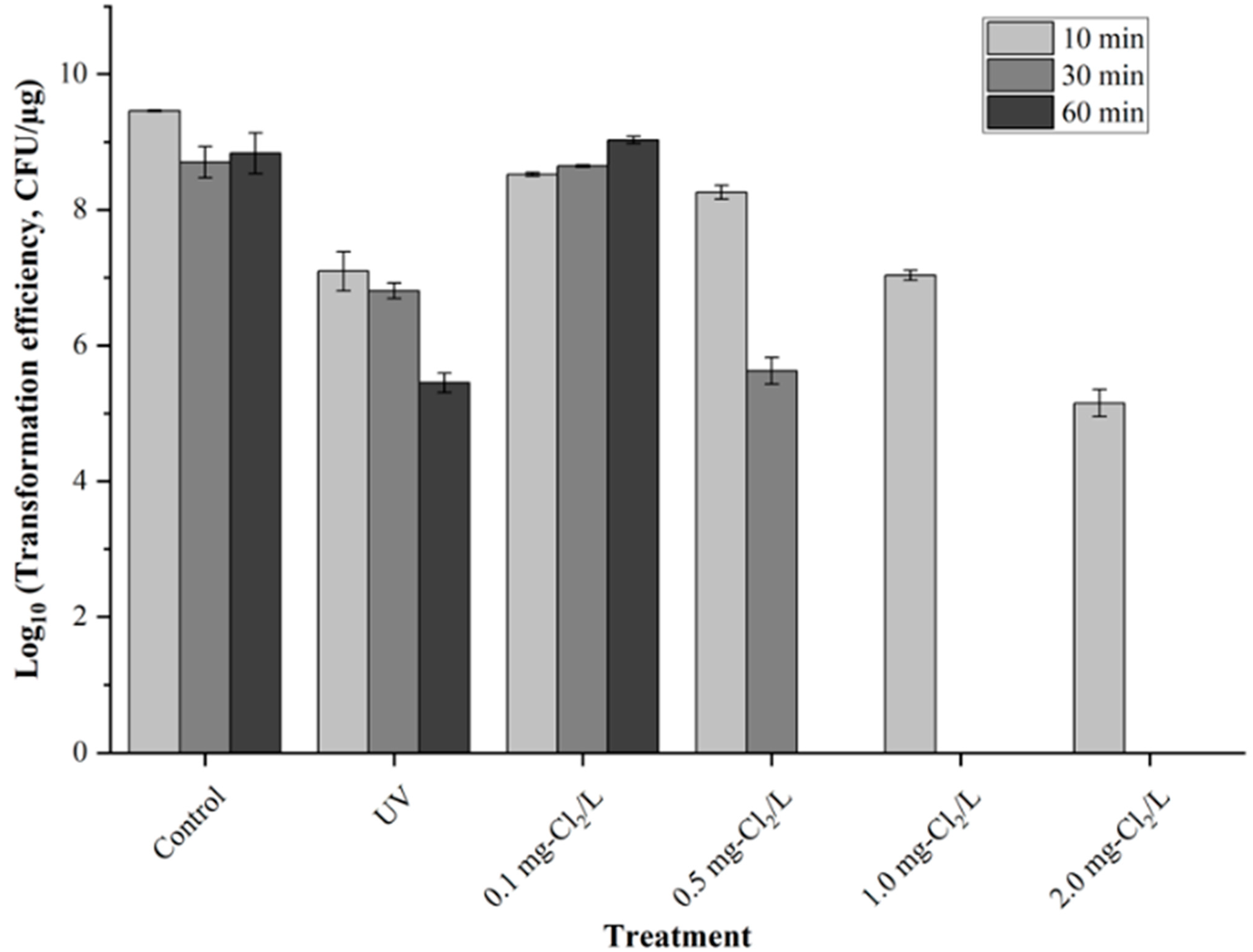
3.1. Reduction in Transformation Efficiency by Chlorine and UV Irradiation

The results show that the different concentrations of free chlorine could affect the transformation efficiency of pUC19 in E. coli. Treatment using 0.1 mg-Cl2/L could not significantly decrease the transformation efficiency (Figure 1). With the chlorine concentration increased from 0.5 to 2.0 mg-Cl2/L, the CFU of successfully transformed E. coli decreased from 1.20 to 4.74 log10/µg-plasmid DNA after 10 min of treatment. After 30 min of treatment, the 1.0 and 2.0 mg-Cl2/L treatments decreased the plasmid DNA transformation efficiency by over 8.71 log10, and no transformed E. coli was counted. When the treatment duration was extended to 60 min, even 0.5 mg-Cl2/L could decrease the transformed E. coli by more than 8.83 log10 compared to the 10 min and 30 min treatments.
UV irradiation also resulted in a decreasing trend with respect to pUC19 transformation efficiency from 2.48 to 3.78 log10.

3.2. Detection of Treated pUC19 Using PCR and qPCR

As a universally used plasmid, pUC19 was used for evaluating the transformation efficiency of plasmid DNA and for PCR and qPCR processes to determine the quantity of treated plasmid DNA in this study. Based on the brightness of PCR-amplified products, the 0.1 mg-Cl2/L treatment oxidized pUC19 to the extent that the relative brightness decreased by 11.3%, 8.8%, and 76.5%, respectively, over 10, 30, and 60 min. The 10-fold serial dilution of the PCR template also showed a similar trend (Figure 2). However, the 0.5–2.0 mg-Cl2/L treatment did not show any bands after the 10 to 60 min treatment. The qPCR results of the pUC19 log10 copy numbers are presented in Figure 3. The pUC19 log10 copy numbers decreased by −0.7%, −2.8%, and 12.9% after the 10-, 30-, and 60-minute treatments with 0.1 mg-Cl2/L, respectively. The copy numbers were out of the range of the standard curve of qPCR and could not be detected correctly when the chlorine doses increased to 0.5 mg-Cl2/L.
UV irradiation decreased the brightness of PCR-amplified products from 4.4% to 19.2% over the course of treatment from 10 to 60 min compared with those in the control group. The log10 copy numbers of pUC19 decreased by −2.2%, −2.4%, and 1.8% after UV irradiation for 10-, 30-, and 60-minute treatments, respectively.

4. Discussion

WWTPs can spread ARB and ARG to the environment, even though wastewater is typically treated by chlorination and/or UV irradiation prior to discharge. The oxidative properties of hypochlorous acid (HOCl) as free chlorine in the disinfection process are considered to significantly affect nucleic acids, including plasmid and genomic DNA [8,15]. Some researchers have suggested that plasmid DNA is more persistent than genomic DNA because of its circular structure [15]. One disadvantage of chlorine treatment is that it can produce potentially harmful by-products. In contrast, UV disinfection is a chemical-free method. However, the effectiveness of UV disinfection can be reduced by the presence of particles in water, whereas chlorine is not affected by these [16,17]. Other studies have revealed that higher UV dosages are required to achieve reduced ARGs (200–400 mJ/cm2 for 3- to 4-log reduction) than for ARB (10–20 mJ/cm2 for 4- to 5-log reduction) [18]. UV irradiation can also induce bacterial cells into a viable but nonculturable (VBNC) state, which explains the practical importance of this increased risk relative to public health [19]. In this study, UV treatment for 10 min had a similar effect that of the 1.0 mg-Cl2/L treatment on decreasing the plasmid DNA transformations into E. coli but did not show better efficiency than 0.5–2.0 mg-Cl2/L treatments for 30 and 60 min. The results of the UV treatment results are consistent with those of previous studies in which 40 mJ/cm2 of 265 nm UV-LED could decrease the artificial transformation efficiency by approximately 3.3-log10 [20]. Previous research indicates that free DNA concentrations in WWTP usually range from 1.1 to 15.9 ng/mL [6]. In this study, the 10 ng/mL plasmid DNA solution was disinfected using different concentrations of chlorine and UV irradiation. Application of chlorine doses greater than 0.5 mg-Cl2/L for treatment durations of 60 min or of doses greater than 1.0 mg-Cl2/L for treatment durations of over 30 min significantly reduced the plasmid DNA transformation efficiency into E. coli, as evidenced by a decrease of greater than 9.46 log10 in CFUs. These results indicate that under these experimental conditions, no viable transformed E. coli colonies were detected. However, we used E. coli and pUC19, which are common and easy recipients/vectors for artificial transformation. As the natural occurrence of transformation is much lower [21,22], so if chlorination/UV can significantly decrease artificial transformation efficiency, the risk of ARG/ARB spread from WWTPs is considered to be controlled using chlorine or UV treatments.
The decrease in transformation efficiency can be calculated using the equation below.
KT = log10 (transformation efficiencyt) − log10 (transformation efficiency0)
  • transformation efficiencyt: the transformation efficiency after t min of treatment
  • transformation efficiency0: the no treatment transformation efficiency
The kinetics of transformation efficiency affected by UV and 0.5, 1.0, and 2.0 mg-Cl2/L treatments are shown in Figure 4. The processing time required to reduce the 2-log10 transformation efficiency based on the kinetics diagram is calculated, as shown in Table 2.
Degradation of pUC19 treated with low dosages of chlorine can be detected using PCR or qPCR. At chlorine concentrations over 0.5 mg-Cl2/L for 10 min no bright bands were detected in the electrophoresed PCR products; further, the results were outside the standard curve of qPCR. The high chlorine concentration caused all plasmid DNAs to be oxidatively damaged, and none of the templates could be amplified by PCR. Moreover, the high concentration of chlorine could have inhibited the PCR, even though the template was diluted 10 to 1000 times in the PCR amplification (Figure 2).
After the 2.0 mg-Cl2/L treatment for 10 min, E. coli bacteria amounting to 1.41 × 10⁵ CFU/µg-pUC19 were still successfully transformed, but no plasmid DNA could be detected by PCR and qPCR (Figure 1, Figure 2 and Figure 3). The linear fit results between the transformation efficiency of E. coli with the relative brightness of the 0.1 mg-Cl2/L chlorine-treated PCR product (R2 = 0.935) and the copy numbers (R2 = 0.815) are shown in Figure 3. The 0.5–2.0 mg-Cl2/L treatment made all plasmid DNA below the detection limit, which could not be analyzed by a spectrophotometer (NanoVue Plus, GE Healthcare, Tokyo, Japan). As the amount of DNA could not be analyzed by qPCR, the DNA concentration could be less than 10–4 ng/µL, which is the minimum DNA concentration in the standard curve of the qPCR used in this study. However, it is only a crude fitting result and is insufficient because we only tested three data sets for the linear fit. A potential risk of persistent mobile genetic elements possessing the ability to transfer resistant genes cannot be denied, but these cannot be detected using PCR and/or qPCR after high-concentration chlorine disinfection by chlorination. However, the incidence of natural transformation is much lower than that of artificial transformation, which may alleviate these concerns.
The linear fit between the transformation efficiency of E. coli with the UV-treated DNA and the PCR band brightness has better results (R2 = 0.981) than that of the copy numbers determined using qPCR (R2 = 0.681) (Figure 5). The adjacent pyrimidine, especially adjacent TT (thymine and thymine) in DNA, is considered a UV-active site [23]. A previous study evaluating the efficacy of UV irradiation, 1 mg/L chlorine, and a combination of both for the disinfection of Pseudomonas aeruginosa found that the intracellular opr gene copy numbers and relative brightness did not significantly decrease following treatments with chlorine or UV alone for 1 to 30 min. This lack of efficacy was attributed to the protective effect of the cell membrane and the lower percentage of adjacent thymine in the bacterial genome [9]. The lengths of the target amplicons and the percentage of adjacent pyrimidine in this and other studies are shown in Table 3. UV irradiation can cause thymine dimer formation in DNA, inducing DNA damage, which can lead to the inability of DNA replication and the death; UV irradiation results in fewer DNA transformations relative to other bacteria (Figure 1). Methods to identify the formation of thymine dimers are already established [24]. The adjacent pyrimidine of qPCR amplicons is 30.0%, and that of pUC19 is 22.7%, which is much lower than that reported in previous studies. This may be one of the reasons for the poor fit of copy numbers in qPCR and transformation efficiency. Consequently, these results suggest that the percentage of adjacent pyrimidine is an indicator for estimating the removal capacity by UV. However, the relationship between the percentage of adjacent pyrimidine and transformation efficiency should be examined in the future.
WWTPs also use other technologies used for disinfection. Besides the widely used chlorination and UV irradiation processes, the other methods include combined UV/chlorination, ozone [25], silver ions, solar photocatalysis [26], boiling water [27], and so on. Silver ions are especially efficient at damaging extracellular resistance or recombinant genes [28]. The biggest advantage of chlorine disinfection is that it is cheap and easy to use, especially in developing regions [29]. UV irradiation is considered a safe physical disinfection method with no by-products but has disadvantages in monitoring the disinfection effects compared to those of chlorine treatments, and the cost is higher. However, UV LEDs can overcome some of the disadvantages of traditional UV [30].
Both the relative brightness of the bands in PCR and the copy numbers in qPCR can be representative parameters of the transformation efficiency of UV disinfected DNA samples (Figure 5). With chlorine treatments, some plasmids can still be transformed into E. coli at high chlorine concentrations, but these are not detected by relative brightness in PCR or by copy numbers in qPCR. In addition, other research indicated that qPCR detection could lead to an overestimation of ARG risk using shorter qPCR amplicons [20]. However, in this study, combining the results of UV or chlorine treatments, PCR and qPCR detection was found to underestimate the risk of ARG transmissions. The actual disinfection efficiency and suitable treatment for the disinfection against ARB as well as ARGs thus needs to be investigated. The present study suggests that one should not rely on only one method (only PCR or only qPCR of molecular biology method) when estimating the spread risk of ARB and ARG. Other research also suggests that bacterial isolation and culture-dependent risk assessment methods can be applied to different samples and regions [31]. Kim et al. [31] reported a clear difference between environmental (4% of strains showed antibiotic resistance) and clinical strains (35.7% of strains showed antibiotic resistance) using antibiotic susceptibility tests. F, the percentage of antibiotic tolerance, which is an indicator of potential resistance risks for strains that have not become resistant, was 78.8% for clinical strains and 90% for environmental strains. Thus, it is necessary to develop a method combining both culture-independent methods (molecular methods such as PCR and qPCR) and culture-dependent methods (plate counting such as transformation efficiency) to estimate the risk of spreading ARB/ARG and to assess the actual situation in the environment.
The processing time required for 2-log10 reduction in transformation efficiency by using UV irradiation is 18.04 min, as the UV lamp is 40 mW/cm2; thus, the energy consumed is 43.296 mJ/cm2. This is substantially lower than the energy required for a 2-log10 reduction in pUC19 by qPCR in this study. This study is consistent with a previous study showing that more UV irradiation power is needed to cause a 2-log10 copy number reduction in 16S rDNA compared to the same reduction in transformation efficiency [28]. This can be explained by the fact that even a little damage to pUC19 can affect transformation efficiency, whereas more DNA damage is needed to be reflected in the qPCR results.
Chlorine oxidation treatment is more advantageous than UV treatment in damaging plasmid DNA, and previous studies have shown that only 1.980 mg/L treatment for one minute can reduce 16S rDNA by 2-log10. This study also demonstrates that pUC19 is easily oxidized by chlorine and cannot be detected by PCR and qPCR, but plasmids that are still transformed into E. coli after chlorine treatment cannot be ignored to reduce the spread of resistance genes.
Some combined technologies indicate that energy costs can be reduced. For example, the energy consumption of the UV/electrolysis hybrid reactor was reported as 0.17 kWh/m3 [32,33]. Some studies even indicated that boiling, especially at water temperatures higher than 90 °C, can significantly remove the ARGs; however, this is uneconomical with respect to energy use [27]. Furthermore, it is necessary to develop a system that provides easy access to real-time ARB and ARG data via web-based tools and platforms, such as interactive maps, as reported by Stedtfeld et al. [34]. As medical doctors share information about the clinical strains of ARB in the region when diagnosing patients for chemical therapy, citizens are also required to know their situation and act in preventing environmental damage to wildlife, livestock, and human health [35].

5. Conclusions

The effectiveness of chlorine or UV irradiation treatment in reducing the transformation efficiency of free naked pUC19 into E. coli reminds us that commonly used technologies play an important role in reducing the spread of antibiotic resistance at WWTP if sufficient doses of chlorine (>0.5 mg-Cl2/L) and sufficient treatment times (>60 min) are used. This study conducted a dynamics analysis of naked plasmid DNA disinfected using chlorine or UV and analyzed the relationship between transformation efficiency and PCR/qPCR detection, which helps understand disinfection efficiencies in actual complex conditions.

Author Contributions

Conceptualization, K.S. and C.Z.; methodology and experiments, C.Z., H.M. (Hanchen Miao) and K.S.; data analysis, C.Z., K.S., Z.L. and T.Y.; writing—original draft preparation, C.Z. and K.S.; writing—review and editing, C.Z., K.S., I.I., H.M. (Hideaki Maseda), Z.L. and Z.Z. All authors have read and agreed to the published version of the manuscript.

Funding

This study was supported by JSPS KAKENHI Grant Number JP20H03107 to I. I. and China Scholarship Council (CSC) (No. 202008050305) to Z. C.

Data Availability Statement

Not applicable.

Conflicts of Interest

The authors declare no conflict of interest.

References

  1. van Boeckel, T.P.; Gandra, S.; Ashok, A.; Caudron, Q.; Grenfell, B.T.; Levin, S.A.; Laxminarayan, R. Global antibiotic Consumption 2000–2010: An Analysis of National Pharmaceutical Sales Data. Lancet Infect. Dis. 2014, 14, 742–750. [Google Scholar] [CrossRef] [PubMed]
  2. Terahara, F.; Nishiura, H. Fluoroquinolone consumption and Escherichia coli resistance in Japan: An ecological study. BMC Public Health 2019, 19, 426. [Google Scholar] [CrossRef] [PubMed] [Green Version]
  3. Blair, J.M.A.; Webber, M.A.; Baylay, A.J.; Ogbolu, D.O.; Piddock, L.J.V. Molecular mechanisms of antibiotic resistance. Nat. Rev. Microbiol. 2015, 13, 42–51. [Google Scholar] [CrossRef]
  4. Pazda, M.; Kumirska, J.; Stepnowski, P.; Mulkiewicz, E. Antibiotic resistance genes identified in wastewater treatment plant systems—A review. Sci. Total Environ. 2019, 697, 134023. [Google Scholar] [CrossRef] [PubMed]
  5. Zhang, T.; Li, B. Occurrence, transformation, and fate of antibiotics in municipal wastewater treatment plants. Crit. Rev. Environ. Sci. Technol. 2011, 41, 951–998. [Google Scholar] [CrossRef]
  6. Zhang, Y.; Li, A.; Dai, T.; Li, F.; Xie, H.; Chen, L.; Wen, D. Cell-free DNA: A neglected source for antibiotic resistance genes spreading from WWTPs. Environ. Sci. Technol. 2018, 52, 248–257. [Google Scholar] [CrossRef]
  7. Collivignarelli, M.C.; Abbà, A.; Benigna, I.; Sorlini, S.; Torretta, V. Overview of the Main Disinfection Processes for Wastewater and Drinking Water Treatment Plants. Sustainability 2018, 10, 86. [Google Scholar] [CrossRef] [Green Version]
  8. Furukawa, T.; Jikumaru, A.; Ueno, T.; Sei, K. Inactivation effect of antibiotic-resistant gene using chlorine disinfection. Water 2017, 9, 547. [Google Scholar] [CrossRef]
  9. Wang, L.; Ye, C.; Guo, L.; Chen, C.; Kong, X.; Chen, Y.; Shu, L.; Wang, P.; Yu, X.; Fang, J. Assessment of the UV/chlorine process in the disinfection of Pseudomonas aeruginosa: Efficiency and mechanism. Environ. Sci. Technol. 2021, 55, 9221–9230. [Google Scholar] [CrossRef]
  10. Liu, L.; Xing, X.; Hu, C.; Wang, H.; Lyu, L. Effect of sequential UV/free chlorine disinfection on opportunistic pathogens and microbial community structure in simulated drinking water distribution systems. Chemosphere 2019, 219, 971–980. [Google Scholar] [CrossRef]
  11. Zhang, S.; Lin, T.; Chen, H.; Chen, W.; Xu, H.; Tao, H. DNA pyrimidine bases in water: Insights into relative reactivity, by-products formation and combined toxicity during chlorination. Sci. Total Environ. 2020, 717, 137205. [Google Scholar] [CrossRef] [PubMed]
  12. Xu, X.; Xiao, R.; Dionysiou, D.D.; Spinney, R.; Fu, T.; Li, Q.; Wang, Z.; Wang, D.; Wei, Z. Kinetics and mechanisms of the formation of chlorinated and oxygenated polycyclic aromatic hydrocarbons during chlorination. Chem. Eng. J. 2018, 351, 248–257. [Google Scholar] [CrossRef]
  13. Spiliotopoulou, A.; Hansen, K.M.S.; Andersen, H.R. Secondary formation of disinfection by-products by UV treatment of swimming pool water. Sci. Total Environ. 2015, 520, 96–105. [Google Scholar] [CrossRef]
  14. Tran, J.H.; Jacoby, G.A. Mechanism of plasmid-mediated quinolone resistance. Proc. Natl. Acad. Sci. USA 2002, 99, 5638–5642. [Google Scholar] [CrossRef] [PubMed] [Green Version]
  15. Zhang, M.; Chen, S.; Yu, X.; Vikesland, P.; Pruden, A. Degradation of extracellular genomic, plasmid DNA and specific antibiotic resistance genes by chlorination. Front. Environ. Sci. Eng. 2019, 13, 38. [Google Scholar] [CrossRef]
  16. Gopal, K.; Tripathy, S.S.; Bersillon, J.L.; Dubey, S.P. Chlorination byproducts, their toxicodynamics and removal from drinking water. J. Hazard. Mater. 2007, 140, 1–6. [Google Scholar] [CrossRef]
  17. Gibson, J.; Drake, J.; Karney, B. UV disinfection of wastewater and combined sewer overflows. Adv. Exp. Med. Biol. 2017, 996, 267–275. [Google Scholar] [CrossRef]
  18. McKinney, C.W.; Pruden, A. Ultraviolet disinfection of antibiotic resistant bacteria and their antibiotic resistance genes in water and wastewater. Environ. Sci. Technol. 2012, 46, 13393–13400. [Google Scholar] [CrossRef]
  19. Zhang, S.; Ye, C.; Lin, H.; Lv, L.; Yu, X. UV disinfection induces a Vbnc State in Escherichia coli and Pseudomonas aeruginosa. Environ. Sci. Technol. 2015, 49, 1721–1728. [Google Scholar] [CrossRef] [PubMed]
  20. Umar, M.; Anglès D’auriac, M.; Wennberg, A.C. Application of UV-LEDs for antibiotic resistance genes inactivation—Efficiency monitoring with QPCR and transformation. J. Environ. Chem. Eng. 2021, 9, 105260. [Google Scholar] [CrossRef]
  21. Komiyama, Y.; Maeda, S. Natural transformation in Escherichia coli. In Horizontal Gene Transfer: Methods and Protocols; de la Cruz, F., Ed.; Springer: New York, NY, USA, 2020; pp. 179–187. ISBN 978-1-4939-9877-7. [Google Scholar] [CrossRef]
  22. Sun, D. Two different routes for double-stranded DNA transfer in natural and artificial transformation of Escherichia coli. Biochem. Biophys. Res. Commun. 2016, 471, 213–218. [Google Scholar] [CrossRef]
  23. Sorinolu, A.J.; Munir, M. Degradation and horizontal gene transfer analysis of plasmid-encoded antibiotic resistance genes during UV254, hydroxyl radical and sulphate radical treatments. Chem. Eng. J. 2022, 450, 138380. [Google Scholar] [CrossRef]
  24. Yadav, V.K.; Awasthi, P.; Kumar, A. Detection of UV-induced thymine dimers. In Genotoxicity Assessment: Methods and Protocols; Dhawan, A., Bajpayee, M., Eds.; Springer: New York, NY, USA, 2019; pp. 313–322. ISBN 978-1-4939-9646-9. [Google Scholar] [CrossRef]
  25. Ding, W.; Jin, W.; Cao, S.; Zhou, X.; Wang, C.; Jiang, Q.; Huang, H.; Tu, R.; Han, S.F.; Wang, Q. Ozone disinfection of chlorine-resistant bacteria in drinking water. Water Res. 2019, 160, 339–349. [Google Scholar] [CrossRef] [PubMed]
  26. Venieri, D.; Mantzavinos, D. Disinfection of waters/wastewaters by solar photocatalysis. In Advances in Photocatalytic Disinfection; An, T., Zhao, H., Wong, P.K., Eds.; Springer: Berlin/Heidelberg, Germany, 2017; pp. 177–198. ISBN 978-3-662-53496-0. [Google Scholar] [CrossRef]
  27. Wan, K.; Zheng, S.; Ye, C.; Hu, D.; Zhang, Y.; Dao, H.; Chen, S.; Yu, X. Ancient oriental wisdom still works: Removing ARGs in drinking water by boiling as compared to chlorination. Water Res. 2021, 209, 117902. [Google Scholar] [CrossRef]
  28. Van Aken, B.; Lin, L.S. Effect of the disinfection agents chlorine, UV irradiation, silver ions, and TiO2 nanoparticles/near-UV on DNA molecules. Water Sci. Technol. 2011, 64, 1226–1232. [Google Scholar] [CrossRef] [PubMed]
  29. Nielsen, A.M.; Garcia, L.A.T.; Silva, K.J.S.; Sabogal-Paz, L.P.; Hincapié, M.M.; Montoya, L.J.; Galeano, L.; Galdos-Balzategui, A.; Reygadas, F.; Herrera, C.; et al. Chlorination for low-cost household water disinfection—A critical review and status in three Latin American countries. Int. J. Hyg. Environ. Health 2022, 244, 114004. [Google Scholar] [CrossRef]
  30. Ibrahim, M.A.S.; MacAdam, J.; Autin, O.; Jefferson, B. Evaluating the impact of LED bulb development on the economic viability of ultraviolet technology for disinfection. Environ. Technol. 2014, 35, 400–406. [Google Scholar] [CrossRef]
  31. Kim, S.; Masai, S.; Murakami, K.; Azuma, M.; Kataoka, K.; Sebe, M.; Shimizu, K.; Itayama, T.; Whangchai, N.; Whangchai, K.; et al. Characteristics of antibiotic resistance and tolerance of environmentally endemic Pseudomonas aeruginosa. Antibiotics 2022, 11, 1120. [Google Scholar] [CrossRef] [PubMed]
  32. Haaken, D.; Schmalz, V.; Dittmar, T.; Worch, E. Limits of UV disinfection: UV/electrolysis hybrid technology as a promising alternative for direct reuse of biologically treated wastewater. J. Water Supply Res. Technol. 2013, 62, 442–451. [Google Scholar] [CrossRef] [Green Version]
  33. Jin, Y.; Chen, Z.; Chen, X.; Huang, P.; Chen, X.; Ding, R.; Liu, J.; Chen, R. The drinking water disinfection performances and mechanisms of UVA-LEDs promoted by electrolysis. J. Hazard. Mater. 2022, 435, 129099. [Google Scholar] [CrossRef]
  34. Stedtfeld, R.D.; Williams, M.R.; Fakher, U.; Johnson, T.A.; Stedtfeld, T.M.; Wang, F.; Khalife, W.T.; Hughes, M.; Etchebarne, B.E.; Tiedje, J.M.; et al. Antimicrobial resistance dashboard application for mapping environmental occurrence and resistant pathogens. FEMS Microbiol. Ecol. 2016, 92, fiw020. [Google Scholar] [CrossRef] [PubMed] [Green Version]
  35. Kamata, K.; Tokuda, Y.; Gu, Y.; Ohmagari, N.; Yanagihara, K. Public knowledge and perception about antimicrobials and antimicrobial resistance in Japan: A national questionnaire survey in 2017. PLoS ONE 2018, 13, e0207017. [Google Scholar] [CrossRef] [PubMed]
Figure 1. Transformation efficiency in E. coli after treatment with chlorine or UV light.
Water 15 01232 g001
Figure 2. Relative brightness of treated pUC19 PCR products in 1.5% agarose gel electrophoresis. Right image shows the bands of the electrophoresed PCR products: (a) control; (b) UV; (c) 0.1 mg-Cl2/L.
Water 15 01232 g002
Figure 3. Copy numbers of pUC19 after different treatments as determined using qPCR.
Water 15 01232 g003
Figure 4. Kinetics of transformation efficiency affected by UV and 0.5, 1.0, and 2.0 mg-Cl2/L treatments.
Water 15 01232 g004
Figure 5. Relationship analysis of transformation efficiency with (A) the band intensity of pUC19 PCR products after electrophoresis and (B) pUC19 copy numbers by qPCR.
Water 15 01232 g005
Table 1. Primer sets used for PCR and qPCR assays in this study.
PrimerSequenceReference
pUC19amp121F5′-CGAGTGGGTTACATCGAACTG-3′This study
pUC19amp646R5′-CTGCAACTTTATCCGCCTCC-3′This study
pUC19amp587F5′-ACTTACTCTAGCTTCCCGGC-3′This study
Table 2. Processing time required to reduce the 2-log10 transformation efficiency.
Disinfection MethodProcessing Time Required for Transformation Efficiency Reduction 2-log10
UV (40 µW/cm2)18.04 min
0.1 mg-Cl2/LNA
0.5 mg-Cl2/L19.25 min
1.0 mg-Cl2/L10.10 min
2.0 mg-Cl2/L2.82 min
Table 3. Adjacent pyrimidine counts and lengths of target amplicons for qPCR and PCR.
Amplicon or DNADimers (Counts)Dimers/Amplicon Length (%)Reference
CCCTTCTTTotalCCCTTCTTTotal
60 bp *5544188.38.36.76.730.0This study
tetA (210 bp) *3726301210517.612.414.35.750.0[21]
tetA (1054 bp) *1851041358150517.69.912.87.747.9
ampR (192 bp) *2221264211111.510.913.521.957.8
ampR (851 bp) *8710410511841410.212.212.313.948.6
80 bp *981463711.310.017.57.546.3[18]
601 bp *6174849731610.212.314.016.152.6
526 bp **243243361354.66.18.26.825.7This study
pUC19 (2686 bp)1301631791396114.86.16.75.222.7This study
Notes: * For qPCR; ** for PCR.
Disclaimer/Publisher’s Note: The statements, opinions and data contained in all publications are solely those of the individual author(s) and contributor(s) and not of MDPI and/or the editor(s). MDPI and/or the editor(s) disclaim responsibility for any injury to people or property resulting from any ideas, methods, instructions or products referred to in the content.

