Article

Cyanide Biodegradation by a Native Bacterial Consortium and Its Potential for Goldmine Tailing Biotreatment

by
María José Alvarado-López
1,
Sofía E. Garrido-Hoyos
2,
María Elena Raynal-Gutiérrez
3,
Elie G. El-Kassis
1,
Víctor M. Luque-Almagro
4 and
Genoveva Rosano-Ortega
1,*
1
Decanato de Ciencias Biológicas, Universidad Popular Autónoma del Estado de Puebla, 21 Sur 1103, Barrio de Santiago, Puebla 72410, Puebla, Mexico
2
Instituto Mexicano de Tecnología del Agua (IMTA), Paseo Cuauhnáhuac 8532, Colonia Progreso, Jiutepec 62550, Morelos, Mexico
3
Decanato de Ingenierías, Universidad Popular Autónoma del Estado de Puebla, 21 Sur 1103, Barrio de Santiago, Puebla 72410, Puebla, Mexico
4
Departamento de Bioquíımica y Biología Molecular, Universidad de Córdoba, Edificio Severo Ochoa, 14071 Córdoba, Spain
*
Author to whom correspondence should be addressed.
Water 2023, 15(8), 1595; https://doi.org/10.3390/w15081595
Submission received: 3 February 2023 / Revised: 12 March 2023 / Accepted: 8 April 2023 / Published: 20 April 2023
(This article belongs to the Section Wastewater Treatment and Reuse)

Abstract

:
A native cyanide-degrading bacterial consortium was isolated from goldmine tailing sediments. Mine tailings are toxic effluents due to their metal–cyanide complexes. The bacterial consortium was able to degrade an initial sodium cyanide concentration ranging from 5 to 120 mg L−1 in alkaline synthetic wastewater (pH > 9.2), for a maximum of 15 days. The free cyanide biodegradation efficiency was 98% for the highest initial free cyanide concentration tested and followed a first-order kinetic profile, with an estimated kinetic rate constant of 0.12 ± 0.011 d−1. The cyanide-degrading consortium was streaked with serial dilutions on a specific medium (R2A). 16S rRNA gene sequencing and mass spectrometry proteomic fingerprinting of the isolates showed that the bacterial strains belonged to Microbacterium paraoxydans, Brevibacterium casei, Brevundimonas vesicularis, Bacillus cereus and Cellulosimicrobium sp. The first four genera had previously been identified as cyanide-degrading bacteria. Microbacterium and Brevibacterium had previously been found in alkaline conditions, showing resistance to heavy metals. As for Cellulosimicrobium, to our knowledge, this is the first study to implicate it directly or indirectly in cyanide biodegradation. In this research, these genera were identified as functional bacteria for cyanide degradation, and they might be suitable for mine tailing biotechnological tertiary treatment.

1. Introduction

Cyanides are a group of toxic recalcitrant compounds that contain cyano ion (C≡N), used for silver and gold leaching in a process named cyanidation [1]. Approximately 90% of gold is extracted by cyanidation [2]. The leaching procedure consists of extraction with alkaline NaCN, producing a barren cyanide solution (100–500 mgL−1 of NaCN) by heap or tank leach. NaAu(CN)2 stable ions are then formed and recovered by carbon adsorption [3], thus generating toxic effluents that, in addition to sodium cyanide, may contain cyanide metal complexes; heavy metals such as copper, iron, zinc, lead, mercury, cadmium or chromium; arsenic; and thiocyanates [4,5]. These toxic wastewaters must be treated to minimize their environmental and health risks; thus, the cyanide concentration must be lowered to the minimum acceptable limit, which varies from 0.05 mg L−1 [6] to 0.2 mg L−1 [7] depending on the applicable regulations in each country. There are several different physical and chemical methods that are used to remove these toxic compounds [8]; however, although most of these methods are efficient, they produce toxic by-products, cannot degrade metal-complexed cyanide as easily and require expensive equipment, maintenance and high initial investment costs [1,2,9,10]. The biodegradation of mine tailings is considered a viable alternative. This latest option can be applied as part of a physical and chemical treatment process (tertiary step) to further reduce the free cyanide concentration, toxic by-products and operational costs. In biodegradation processes, microorganisms such as fungi or bacteria transform free cyanide and metal-complexed cyanides into ammonia and bicarbonate. The free metals formed can then either precipitate from the solution or be adsorbed within the biofilm [1,5,11,12]. Bacteria are probably the most widely studied cyanide-resistant microorganisms for this purpose. Some have been shown to be capable of degrading cyanide, such as Pseudomonas, Klebsiella, Thiobacillus, Agrobacterium, Bacillus, Citrobacter, Chromobacterium, Alcaligenes, Acinetobacter, Ralstonia, Rhodococcus, Serratia, Halomonas Escherichia coli, Azotobacter vinelandii, Burkholderia cepacia, Thialkalivibrio thiocyanodenitrificans, Halothiobacillus halophilus/hidrothermalis, Methylobacterium thiocyanatum, Paracoccus thiocyanatus, Thiohalobacter thiocyanaticus and Thiohalophilus thiocyanoxidans [9,10,11,12,13,14]. Cyanotrophic bacteria are capable of using cyanide as a carbon and nitrogen source for growth. Given that alkaline conditions are needed for the safe use of cyanide to avoid the volatilization of toxic HCN (pKa = 9.2), these bacteria should also be alkaline-resistant, capable of degrading different cyano compounds and toxic-compound-resistant [10,15]. There are five reported pathways of cyanide biodegradation: the hydrolytic, oxidative, reductive, substitution and synthesis pathways. In the first three biodegradation pathways, cyanides are converted into simple molecules (in the hydrolytic pathway: formic acid and ammonia; oxidative pathway: carbon dioxide; and reductive pathway: ammonia and methane), while the other two involve the use of cyanide as a carbon and nitrogen source to produce cyano-amino acids or thiocyanate, which may be further oxidized into ammonia, carbon dioxide and sulfide [9,12,13,16]. Certain bacteria can use more than one pathway for cyanide biodegradation, which depends on several factors, such as oxygen availability, pH and the initial cyanide concentration, as well as its bioavailability and solubility in the system [12]. Aerobic biodegradation involves cyanide breakdown by oxidative means, and reports have indicated that aerobic processes are faster than anaerobic processes [13,17]. Cyanide biodegradation has been studied in different bacteria under different conditions, most of which were mesophilic (25–30 °C), while the pH varied from neutral to 10.5. Bacteria living in polluted environments have metabolic abilities to handle contaminants [13,18]; therefore, our hypothesis is that the best bacteria for the biotreatment of silver and gold tailing ponds are those originating from mining tailing ponds, due to their previous adaptation. Therefore, there is a need to characterize these kinds of microorganisms. Our research team isolated a native bacterial consortium from a tailing pond and investigated its cyanide degradation capability. The aim of this study was to identify the key bacterial strains of the cyanide-degrading native consortium from a gold mine tailing, determining its potential for the treatment of cyanided gold mine wastewaters through kinetic biodegradation profiling.

2. Materials and Methods

2.1. Native Consortium Sampling

The native consortium originating from a cyanide-contaminated site was obtained by the aerobic collection of sediments (0.25 m depth) from a mine tailing pond: “Mastrantos II” from the “El Cubo” mine in Guanajuato, Mexico (longitude 20°59′51.53″ N and latitude 101°10′5″ W). This mine tailing pond has been in operation for over 150 years since the cyanidation process was introduced for gold extraction (40 mg L−1 total cyanide, 9 mg L−1 total organic carbon, 0.0044 mg L−1 total dissolved As, 0.523 mg L−1 Fe, 70.8 mg L−1 CaO, 4600 mg L−1 total dissolved solids, 3.3 mg L−1 dissolved oxygen, 1813 mg L−1 total hardness, pH = 9). The samples were diluted in a ratio of 1:10 with sterile peptone isotonic solution and vortexed (Scientific Industries Genie 2, Bohemia, NY, USA), and the resulting solution was allowed to sediment in Imhoff cones for 1 h. This procedure was repeated another two times. The supernatant was retained and stored in a flask at 4 °C overnight. After this period, the supernatant was centrifuged at 10,000 rpm for 5 min (Hermle Labnet Z326K, Edison, NJ, USA) until a pellet was formed. The pellet was recovered and stored in glycerol (30%, Karal, León, Mexico) with sterile isotonic peptonized solution and phosphate buffer solution (pH = 7) at −20 °C until further utilization.

2.2. Bacterial Isolation

Preliminary biodegradability tests were conducted to select the proper consortium, pre-screening the five native consortia obtained from a mine tailing pond in Guanajuato, Mexico, one of which was selected for this study. The activation of the selected consortium was conducted in peptonized broth (BD Bioxon, Franklin Lakes, NJ, USA) under orbital agitation (150 rpm) at 30 °C (LUZEREN Pro1001319, Prolab, Trajomulco de Zúñiga, Mexico) for 2 days. After that, the synthetic cyanided solution was inoculated (10% v/v) with sodium cyanide (Sigma-Aldrich, St. Louis, MO, USA) at an initial concentration of 10 mg L−1 and incubated under the same conditions as those used for the activation step for 3 days. The synthetic cyanided solution’s composition was determined based on modified M9 media with 89% v/v salt solution (KH2PO4 1.7 mg L−1, NaCl 0.5 g L−1, MgSO4·7H2O 0.5 g L−1, CaCl2 0.1 g L−1), 1% v/v trace element solution (ZnSO4·7H2O 0.05 g L−1, MnCl2·4H2O 0.05 g L−1, CuCl2·2H2O 0.005 g L−1, Na2MoO4·2H2O 0.005 g L−1, Na2B4O7·10H2O 0.002 g L−1, CoCl2·6H2O 0.0003 g L−1), 10% v/v BBC buffer pH 9.6 (Na2CO3 1.59 g L−1, NaHCO3 2.93 g L−1), and NaCN 10 mg L−1 as a C and N source. The solution was prepared using type II water (RiOs-DI water purification system, Merck, Darmstadt, Germany). All reagents were acquired from J. T. Baker (Fisher Scientific, Hampton, NH, USA). A sample from a previous culture was then serially diluted at different dilution rates of 10−5, 10−6 and 10−7 and spread over sterile R2A (Merck, Darmstadt, Germany) plates incubated at 30 °C for 7 d. The colonies were subsequently re-streaked to obtain pure colonies. The pure colonies were characterized microbiologically [19] and then Gram- and spore-stained (Hycel, Zapopan, Mexico) through a standard procedure. The slides were observed under a light microscope (immersion oil at 400× magnification) to determine the cell shape and arrangement, together with Gram staining. The pure colonies were stored in glycerol (Karal, León, Mexico) at −20 °C until further utilization.

2.3. DNA Extraction and 16S rRNA Amplification

After 3 days of consortium cultivation in cyanide synthetic medium (NaCN = 10 mg L−1), an aliquot was taken and the pure colonies’ genomic DNA was extracted using a DNA extraction kit, DNeasy Ultraclean (Qiagen, Valencia, CA, USA). After extraction, the samples were stored at −20 °C until further use. The DNA was used as a template for the polymerase chain reaction (PCR) amplification of 16S rRNA with universal primers, 27Fym (5′-AGAGTTTGATYMTGGCTCAG-3′) and 1492R (5′-TACCTTGTTACGACTT-3′) [20]. Thermal cycling (Bio-Rad, Hercules, CA, USA, C-1000 Touch Thermal Cycler) conditions were used for the initial denaturalization for 2 min at 98 °C, followed by 28 cycles of 10 s denaturalization at 98 °C, 15 s of annealing at 48 °C and 12 s of extension at 72 °C, ending with a final extension at 72 °C for 5 min. Prime Star Max polymerase (TAKARA Bio, San Jose, CA, USA) was used with a reaction mixture of 50 μL: 1 μL DNA template, 1 μL of 10 mM primer mix, 23 μL of nuclease-free water and 25 μL of enzyme mix. All reactions were performed in triplicate. The amplification products were purified with the Gel/PCR DNA Fragments extraction kit (IBI Scientific, Dubuque, IA, USA) and analyzed by agarose gel 1% electrophoresis. The DNA was stored at −20 °C for subsequent analysis. After 16S PCR amplification and purification, electrophoresis showed that the approximate size of the DNA fragments was about 1.2 kb.

2.4. Sequencing and Analyses

The purified PCR products (more than 25 ng μL−1 of DNA) were sent to LABSERGEN CINVESTAV—Irapuato for capillary automatic sequencing (BigDye Terminator v3.1 AB3730, Fischer Scientific, Vienna, Austria). The samples were sequenced in triplicate in both senses to correct any sequencing errors. The DNA sequences were matched against nucleotide sequences in NCBI GenBank using the BLAST tool and analyzed with EzBioCloud 16S-based ID tool, version 20200513. The sequences showing more than 99% similarity and a query coverage above 99% were considered to belong to the same taxon. The resulting sequences were deposited in GenBank under the accession numbers OP688505, OP712185, OP712187, OP712188 and OP712195.

2.5. Mass Spectrometry Proteomic Fingerprint

The pure colonies were streaked on BAS plates for 3 days at 30 °C, and samples were prepared for matrix-assisted laser desorption/ionization time-of-flight mass spectrometry (MALDI-TOF MS), according to the direct transfer protocol recommended by Bruker Daltonics. If the score did not match a probable identification, MALDI-TOF MS was conducted following the ethanol/formic acid extraction protocol recommended by Bruker Daltonics. MALDI-TOF MS was performed using an IVD MALDI BioTyper mass spectrometer (Bruker Daltonics, Billerica, MA, USA) equipped with a N2 laser. All spectra were recorded in the linear positive ion mode. The spectra were collected as a sum of 500 shots across a spot. A mass range of 2000–20,000 m/z was used for the analysis. The main spectra were calculated from 3 spectra per strain and used for the construction of a score using the BioTyper software (2.2 version, Bruker Daltonics).

2.6. Cyanide Biodegradation

The activation of the selected consortium was performed as described in Section 2.2, by bacterial isolation. Subsequently, synthetic cyanided solutions prepared with initial concentrations of sodium cyanide of 5, 20, 25, 30, 70 or 120 mg L−1 were inoculated with 10% v/v of the activated culture and incubated at 30 °C under orbital agitation at 150 rpm for 15 days. The free cyanide (F-CN) was monitored daily with a potentiometric electrode of a selective ion (HI4109, Hanna, Mexico City, Mexico), according to the instrument specifications. The biomass was recorded using optical density (OD) at 540 nm [21] (VE-5100UV, Velaquin, Mexico City, Mexico). The pH was kept over 9.2 and measured daily by potentiometry (Thermo Scientific Orion Triode, Waltham, MA, USA, PH4221-2BNW) throughout the whole experiment.
The free cyanide biodegradation performance (PFCN) was calculated as suggested in [22], according to Equation (1), as follows:
P FCN = F C N 0   B D 0   t
where F C N 0 is the initial F-CN concentration (mg L−1), B D 0 is the initial bacteria density (OD540nm), and t is the full biodegradation time (h).
The free cyanide biodegradation efficiency ( BE ) was calculated as presented in Equation (2):
BE = ( F C N o F C N R F C N 0 ) × 100
where F C N 0 is the initial F-CN concentration (mg L−1), and F C N R represents the residual F-CN concentration (mg L−1) after biodegradation was completed.

2.7. Kinetic Biodegradation Models

Cyanide biodegradation was modelled with first- and second-order kinetic models [21] to study the degradation of cyanide with the microorganisms. The mathematical expressions are as follows:
S = S 0 e x p ( k 1 t )
k s l n S S 0 + S S 0 = k 2 t
Equations (3) and (4) represent first- and second-order kinetic cyanide biodegradation models, respectively, where S 0 is the F-CN initial cyanide concentration (mg L−1), t is time (d), k 1 (d−1) and k 2 (mg·mg−1) are first- and second-order rate constants, respectively, and k s is the half-saturation coefficient (mg L−1). The best-fit kinetic model was chosen based on the error function between the experimental and model-predicted data to validate the model according to the correlation coefficient R2.

3. Results

3.1. Bacterial Isolation and Identification of Native Cyanotrophic Consortium

The cyanotrophic bacterial consortium isolated from a goldmine was cultured as described above and then serially diluted on sterile R2A plates, on which the colonies were subsequently re-streaked, and twenty-three pure colonies were obtained. Table 1 shows the observed characteristics of the isolated microorganisms. The pure colonies were classified into five groups according to their microbiological characteristics, and colonies C16 and C22 were identified as a single strain.
Next, 16S rRNA genes of the isolated bacteria were successfully amplified by PCR and sequenced in triplicate in both senses. The sequences were analyzed with BLAST and assigned to their corresponding bacterial species (Table 1). These results were confirmed using the EzBioCloud 16S-based ID tool. The results showed that the cyanotrophic consortium was dominated by bacteria belonging to Bacillus cereus, Microbacterium paraoxydans, Brevundimonas vesicularis, Brevibacterium casei and Cellulosimicrobium sp. Every sequence showed >99% similarity with both databases used. On the other hand, this identification was consistent with the MALDI-TOF MS fingerprint of each microorganism, notwithstanding the low scores obtained for Brevibacterium casei and Cellulosimicrobium sp.

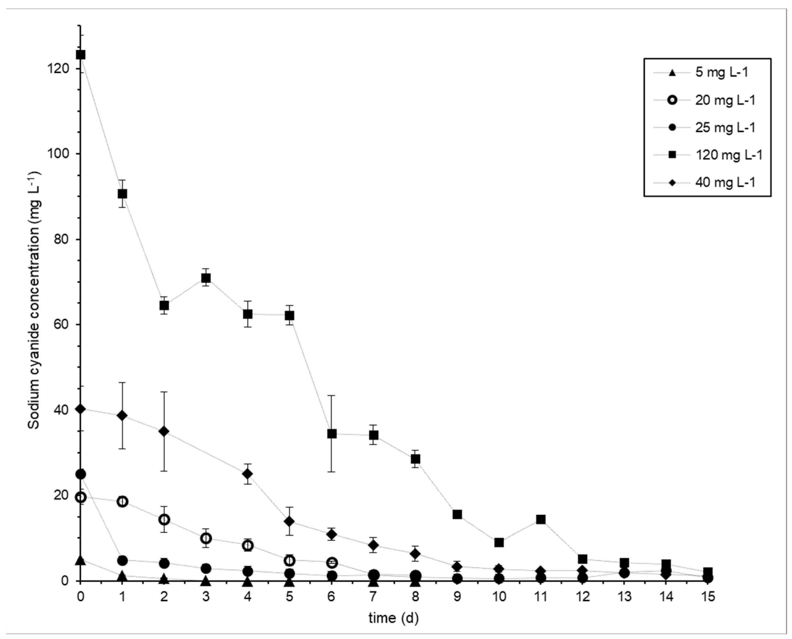
3.2. Cyanide Biodegradation

The native cyanotrophic consortium was shown to degrade F-CN by up to 120 mg L−1. The free cyanide biodegradation performance was the highest with 25 mg L−1 of F-CN initial cyanide concentration (0.19 mg L−1 OD540nm−1 d−1) and the lowest with 5 mg L−1 of F-CN initial cyanide concentration (0.05 mg L−1 OD540nm−1 d−1). The total F-CN efficiency was greater than 93% for every initial cyanide concentration evaluated, and the residual F-CN was lower than 0.05 mg L−1, except when the F-CN initial concentration was 20, 40 or 120 mg L−1. The performance coefficients and biodegradation efficiencies are listed in Table 2.

3.3. Kinetic Models

The F-CN biodegradation profiles are shown in Figure 1, in which it can be seen that as the initial cyanide concentration increased, the time required for full biodegradation also increased, but in each case, BE was above 93%. The F-CN biodegradation models presented first-order kinetics for every initial F-CN concentration tested and had the best fit according to a correlation coefficient R2 > 0.97. These models were not adjusted to the second kinetic order, since the R2 coefficients were smaller than those for the first kinetic order. The rate constants of each model, F-CN0, DO0 and correlation coefficients are presented in Table 2. The biomass growth profile could not be determined by OD measurement due to the formation of extracellular materials, which prevented us from taking a good measurement, and the samples were not sonicated. We followed the same procedures as those published previously [23,24,25]. The pH was maintained at 9.6 when biodegradation started and was monitored daily, being kept at over 9.2 and up to 10.5.

4. Discussion

Mine tailing cyanide biodegradation by native bacteria may be suitable for mine tailing biotreatment [5,26]; thus, the bacterial composition of tailing samples was assessed. During the isolation process, five groups of colonies were obtained (Table 1), which were identified as members belonging to the following species or genera: Microbacterium paraoxydans, Bacillus cereus, Brevundimonas vesicularis, Brevibacterium casei and Cellulosimicrobium sp. The results of the identification of the 16S rRNA genes, proteomic fingerprints and microbiological characteristics assayed in this study were consistent. For colony C16, the 16S rRNA identification showed a match indicating 99% identity with C. cellulans, C. funkei and C. aquatile. Given that we obtained more than one match, we decided to report it as Cellulosimicrobium sp. throughout the article. Microbacterium [27,28], Bacillus cereus [29], Brevibacterium [30] and Brevundimonas [27,31] are known to be cyanide degraders or found in native cyanotrophic consortia. However, to the best of our knowledge, this study is the first to report the presence of genera closely related to Cellulosimicrobium in a native cyanotrophic consortium. However, Cellulosimicrobium has previously been described as cyanogenic [32].
Additionally, the identified genera have previously been isolated from mining-related environments. Microbacterium was isolated from a coal mine [33], uranium mill tailing porewaters [34], mine tailing soil and native plant roots [35]. Bacillus cereus was isolated from mining soil, a lead–zinc factory and a nickel mine [29]. Brevundimonas sp. was found in the soil of a gold mine [36]. Brevibacterium casei was identified in the roots of Prosopis laegivata growing at the edge of a gold mine tailing [37].
It is noteworthy that the genera identified in previous studies describing cyanide-degrading consortia were absent from the composition of the consortium determined in this study. For instance, genera isolated from gold mine tailings such as Halomonas [38] and Pseudomonas [18] were not found in our consortium. This observation is in agreement with previously published literature that seems to indicate that the microbial compositions of native cyanotrophic consortia depend on the isolation source.
To determine the kinetics of cyanide biodegradation of the native cyanotrophic consortium, it was cultivated in a synthetic cyanide solution with cyanide concentrations ranging from 5 to 120 mg L−1 (Figure 1). The native cyanotrophic consortium degraded over 98% of a maximum initial F-CN of 120 mg L−1. No inhibition was observed at any of the initial F-CN concentrations tested. It is likely that the F-CN tolerance limit is higher than 120 mg L−1. There are reports of tolerance to F-CN in synthetic cyanide solution at concentrations up to 976 mg L−1 in the case of Bacillus sp. M01 isolated from gold mine soil [24]. However, the cyanide concentrations in gold and silver mine tailings are 116.13 mg L−1 on average [5,39], which is compatible with the maximum initial F-CN tested in this study. It is noteworthy that biomass evaluation through DO measurements was not possible due to the formation of extracellular material. It is likely that this is a strategy of protection and resistance to alkaline conditions [40]. Microbacterium, one of the bacterial genera identified in this study as part of the native cyanotrophic consortium, was previously reported as a biosurfactant and exopolysaccharide producer [41]. In future experiments, biomass growth will be studied through other techniques, such as total protein measurements. The highest cyanide biodegradation performance achieved by the native cyanotrophic consortium was 0.19 mg L−1 DO540−1 h−1, which is lower than the previously reported values [23,25]. This is probably due to the differences between previous studies, in which a single bacterial strain was used, and our study, in which a consortium was used.
The kinetic biodegradation profiles were adjusted to first- and second-order models (Table 2), resulting in a best fit with a first-order kinetic biodegradation model, with a high correlation (R2 > 0.97). The estimated kinetic rate constant k1 for F-CN biodegradation for the native cyanotrophic consortium was 0.12 ± 0.011 d−1. Cyanide kinetic biodegradation constants were reported for batch co-cultures of cyanotrophic bacteria, with the authors concluding that the phenol and cyanide biodegradation kinetics followed a three-half-order model at similar cyanide initial concentrations to those reported in this study [22]. This discrepancy can be explained by the fact that those authors simultaneously evaluated phenol and cyanide biodegradation. This is the first study to focus on kinetic biodegradation models of cyanide with a gold mine tailing native cyanotrophic consortium without the interference of other compounds, such as phenol.
For a consortium to be suitable for cyanide-contaminated mine tailing biotreatment, several conditions must be fulfilled, in addition to the ability of the consortium to resist and degrade cyanide. The ability to grow in highly alkaline conditions (pH > 9.2) is necessary to avoid the volatilization of HCN under less alkaline conditions. Additionally, the consortium must show resistance to multiple heavy metals that are often present in mine tailings [8,15].
Four of the species that composed the native cyanotrophic consortium were species that have previously been reported to resist alkaline conditions: Microbacterium paraoxydans [42], Bacillus cereus [43], Brevibacterium casei [37] and Cellulosimicrobium sp. [44]. The resistance of the latter to heavy metals such as zinc, nickel, copper, iron, lead and cadmium has been documented [45]. These compounds are usually found in mine tailings [5]. All five genera have previously been reported to be resistant to several heavy metals, such as chromium, nickel, zinc, copper, lead, cadmium, cobalt and mercury [36,37,46,47,48,49,50,51,52]. This characteristic renders them potentially suitable for the biotreatment of gold mine tailings. However, additional information is needed to assess their tolerance to all the conditions combined (alkaline medium, presence of heavy metals and cyanide).
To improve the sustainability and viability of the wastewater treatment bioprocess, it is desirable to couple cyanide biodegradation with valuable by-product formation. Several of the identified genera possess the capacity to produce potentially valuable molecules. Microbacterium was previously reported to produce biosurfactants [41] and metalloproteases [53] that can be used as alternative components of detergents and carotenoids [54] that can be used as food additives. Biosurfactant production by Microbacterium could explain our observation of the formation of extracellular materials that prevented us from conducting OD measurements. It is noteworthy that the biosurfactant secreted by Microbacterium is an exopolysaccharide. Brevundimonas vesicularis was previously reported to be a producer of polyhydroxyalcanoates using sawdust as a substrate [55]. Cellulosimicrobium sp. was previously reported to be a producer of lytic enzymes such as proteases [56], used as biocontrol agents. Further investigation is needed to determine whether the bacterial strains present in this consortium present with the same cyanide biodegradation efficiency under the combined conditions of high alkalinity with the presence of heavy metals and cyanide and, subsequently, under real mine tailing conditions. It is important to explore the effects of factors affecting biodegradation, such as aeration, alkalinity, heavy metal resistance, other carbon sources and interactions between strains. Additionally, kinetic models for growth that integrate biomass production should be determined. It would be useful to determine the nature of the extracellular material produced by the native cyanotrophic consortium and to identify its possible applications. Finally, the metabolic pathway implicated in the biodegradation of cyanide should also be identified.

5. Conclusions

A native cyanide-biodegrading consortium isolated from a gold mine tailing was identified at the species (Microbacterium paraoxydans, Bacillus cereus, Brevundimonas vesicularis, Brevibacterium casei) and genus (Cellulosimicrobium sp.) levels. The latter was identified as a member of a cyanide-biodegrading consortium for the first time. The consortium showed 98% biodegradation efficiency at 120 mg L−1 of F-CN with a first-order biodegrading kinetic model. The identified bacterial species had previously been reported as being resistant to heavy metals and alkaline conditions, rendering this consortium a suitable alternative for gold mine tailing treatment.

Author Contributions

All authors contributed to the study conception and design. Material preparation, data collection and analysis of cyanide degradation experiments were performed by M.J.A.-L., G.R.-O. and S.E.G.-H. Material preparation, data collection and analysis of the molecular biology experiments were performed by M.J.A.-L., E.G.E.-K., M.E.R.-G. and V.M.L.-A. The first draft of the manuscript was written by M.J.A.-L., and all authors commented on previous versions of the manuscript. All authors have read and agreed to the published version of the manuscript.

Funding

This study was funded by the Research Department of the Universidad Popular Autónoma del Estado de Puebla (UPAEP) and a scholarship grant awarded to Alvarado-López M.J. from the Consejo Nacional de Ciencia y Tecnología (CONACyT).

Data Availability Statement

The data presented in this study are available within this article.

Acknowledgments

The author would like to acknowledge the Don David Gold Mine for providing access to the mine tailings, Ozono Polaris for the technical support, and the Consejo Nacional de Ciencia y Tecnología (CONACyT) for the scholarship grant awarded to Alvarado-López. Many thanks to Ismael Ramírez de Jesús, Alejandra Torres Vivar, Giovana Sáez Ramos, Andrea Núñez Banuet Anduaga, and Diana Elena Enríquez Torres for their technical assistance.

Conflicts of Interest

The authors declare no conflict of interest.

References

  1. Sharma, M.; Akhter, Y.; Chatterjee, S. A review on remediation of cyanide containing industrial wastes using biological systems with special reference to enzymatic degradation. World J. Microbiol. Biotechnol. 2019, 35, 70. [Google Scholar] [CrossRef] [PubMed]
  2. Park, J.M.; Sewell, B.T.; Benedik, M.J. Cyanide bioremediation: The potential of engineered nitrilases. Appl. Microbiol. Biotechnol. 2017, 101, 3029–3042. [Google Scholar] [CrossRef] [PubMed]
  3. Kandasamy, S.; Dananjeyan, B.; Krishnamurthy, K.; Benckiser, G. Aerobic cyanide degradation by bacterial isolates from cassava factory wastewater. Braz. J. Microbiol. 2015, 46, 659–666. [Google Scholar] [CrossRef] [PubMed]
  4. Acheampong, M.A.; Paksirajan, K.; Lens, P.N.L. Assessment of the effluent quality from a gold mining industry in Ghana. Environ. Sci. Pollut. Res. 2012, 20, 3799–3811. [Google Scholar] [CrossRef] [PubMed]
  5. Alvillo-Rivera, A.; Garrido-Hoyos, S.; Buitrón, G.; Thangarasu-Sarasvathi, P.; Rosano-Ortega, G. Biological treatment for the degradation of cyanide: A review. J. Mater. Res. Technol. 2021, 12, 1418–1433. [Google Scholar] [CrossRef]
  6. Cyanide in Drinking-Water. Background Document for Development of WHO Guidelines for Drinking-Water Quality. World Health Organization. Available online: https://cdn.who.int/media/docs/default-source/wash-documents/wash-chemicals/cyanide-background.pdf?sfvrsn=29c5b9f4_4 (accessed on 29 January 2022).
  7. US-EPA. Cyanide Clarification of Free and Total Cyanide Analysis for Safe Drinking Water Act (SDWA) Compliance, Revision 1.0. Available online: https://www.epa.gov/sites/production/files/2016-08/documents/cyanide-clarification-free-and-total-cyanide-analysis-safe-drinking-water.pdf (accessed on 30 January 2023).
  8. Ganji, S.M.S.A.; Hayati, M. Selecting an appropriate method to remove cyanide from the wastewater of Moteh gold mine using a mathematical approach. Environ. Sci. Pollut. Res. 2018, 25, 23357–23369. [Google Scholar] [CrossRef]
  9. Kumar, R.; Saha, S.; Dhaka, S.; Kurade, M.B.; Kang, C.U.; Baek, S.H.; Jeon, B.-H. Remediation of cyanide-contaminated environments through microbes and plants: A review of current knowledge and future perspectives. Geosystem Eng. 2016, 20, 28–40. [Google Scholar] [CrossRef]
  10. Cabello, P.; Luque-Almagro, V.M.; Olaya-Abril, A.; Sáez, L.P.; Moreno-Vivián, C.; Roldán, M.D. Assimilation of cyanide and cyano-derivatives by Pseudomonas pseudoalcaligenes CECT5344: From omic approaches to biotechnological applications. FEMS Microbiol. Lett. 2018, 365, fny032. [Google Scholar] [CrossRef]
  11. Mekuto, L.; Ntwampe, S.; Akcil, A. An integrated biological approach for treatment of cyanidation wastewater. Sci. Total Environ. 2016, 571, 711–720. [Google Scholar] [CrossRef]
  12. Mahendran, R.; Bs, S.; Thandeeswaran, M.; Kg, K.; Vijayasarathy, M.; Angayarkanni, J.; Muthusamy, G. Microbial (Enzymatic) Degradation of Cyanide to Produce Pterins as Cofactors. Curr. Microbiol. 2019, 77, 578–587. [Google Scholar] [CrossRef]
  13. Ibrahim, K.K.; Syed, M.A.; Shukor, M.Y.; Ahmad, S.A. Biological Remediation of Cyanide: A Review. Biotropia 2015, 22, 151–163. [Google Scholar] [CrossRef]
  14. Lovasoa, C.R.; Hela, K.; Harinaivo, A.A.; Hamma, Y. Bioremediation of soil and water polluted by cyanide: A review. Afr. J. Environ. Sci. Technol. 2017, 11, 272–291. [Google Scholar] [CrossRef]
  15. Luque-Almagro, V.M.; Moreno-Vivián, C.; Roldán, M.D. Biodegradation of cyanide wastes from mining and jewellery industries. Curr. Opin. Biotechnol. 2016, 38, 9–13. [Google Scholar] [CrossRef]
  16. Gupta, N.; Balomajumder, C.; Agarwal, V.K. Enzymatic mechanism and biochemistry for cyanide degradation: A review. J. Hazard. Mater. 2010, 176, 1–13. [Google Scholar] [CrossRef] [PubMed]
  17. Luque-Almagro, V.M.; Cabello, P.; Sáez, L.P.; Olaya-Abril, A.; Moreno-Vivián, C.; Roldán, M.D. Exploring anaerobic environments for cyanide and cyano-derivatives microbial degradation. Appl. Microbiol. Biotechnol. 2017, 102, 1067–1074. [Google Scholar] [CrossRef]
  18. Moradkhani, M.; Yaghmaei, S.; Nejad, Z.G. Biodegradation of Cyanide under Alkaline Conditions by a Strain of Pseudomonas putida Isolated from Gold Mine Soil and Optimization of Process Variables through Response Surface Methodology (RSM). Period. Polytech. Chem. Eng. 2017, 62, 265–273. [Google Scholar] [CrossRef]
  19. Willey, J.; Sandman, K.; Wood, D.H. Prescott’s Microbiology, 12th ed.; McGraw Hill: New York, NY, USA, 2022. [Google Scholar]
  20. Frank, J.A.; Reich, C.I.; Sharma, S.; Weisbaum, J.S.; Wilson, B.A.; Olsen, G.J. Critical Evaluation of Two Primers Commonly Used for Amplification of Bacterial 16S rRNA Genes. Appl. Environ. Microbiol. 2008, 74, 2461–2470. [Google Scholar] [CrossRef]
  21. Madigan, M.T.; Bender, K.S.; Buckley, D.H.; Sattley, W.M.; Stahl, D. Brock Biology of Microorganisms, 15th ed.; Pearson: Upper Saddle River, NJ, USA, 2017. [Google Scholar]
  22. Singh, N.; Balomajumder, C. Batch growth kinetic studies for elimination of phenol and cyanide using mixed microbial culture. J. Water Process Eng. 2016, 11, 130–137. [Google Scholar] [CrossRef]
  23. Huertas, M.; Sáez, L.; Roldán, M.D.; Luque-Almagro, V.M.; Martínez-Luque, M.; Blasco, R.; Castillo, F.; Moreno-Vivián, C.; Garcia-Garcia, I. Alkaline cyanide degradation by Pseudomonas pseudoalcaligenes CECT5344 in a batch reactor. Influence of pH. J. Hazard. Mater. 2010, 179, 72–78. [Google Scholar] [CrossRef]
  24. Safa, Z.J.; Aminzadeh, S.; Zamani, M.; Motallebi, M. Significant increase in cyanide degradation by Bacillus sp. M01 PTCC 1908 with response surface methodology optimization. AMB Express 2017, 7, 200. [Google Scholar] [CrossRef]
  25. Razanamahandry, L.C.; Onwordi, C.T.; Saban, W.; Bashir, A.K.H.; Mekuto, L.; Malenga, E.; Manikandan, E.; Fosso-Kankeu, E.; Maaza, M.; Ntwampe, S.K.O. Performance of various cyanide degrading bacteria on the biodegradation of free cyanide in water. J. Hazard. Mater. 2019, 380, 120900. [Google Scholar] [CrossRef] [PubMed]
  26. Roldán, M.D.; Olaya-Abril, A.; Sáez, L.P.; Cabello, P.; Luque-Almagro, V.M.; Moreno-Vivián, C. Bioremediation of cyanide-containing wastes. EMBO Rep. 2021, 22, e53720. [Google Scholar] [CrossRef] [PubMed]
  27. Han, Y.; Jin, X.; Wang, F.; Liu, Y.; Chen, X. Successful startup of a full-scale acrylonitrile wastewater biological treatment plant (ACN-WWTP) by eliminating the inhibitory effects of toxic compounds on nitrification. Water Sci. Technol. 2013, 69, 553–559. [Google Scholar] [CrossRef]
  28. Sankaranarayanan, A.; Gowthami, M. Cyanide Degradation by Consortium of Bacterial Species Isolated from Sago Industry Effluenty. J. Environ. Treat. Tech. 2014, 3, 41–46. [Google Scholar]
  29. Dwivedi, N.; Balomajumder, C.; Mondal, P. Comparative evaluation of cyanide removal by adsorption, biodegradation, and simultaneous adsorption and biodegradation (SAB) process using Bacillus cereus and almond shell. J. Environ. Biol. 2016, 37, 551–556. [Google Scholar] [PubMed]
  30. He, Y.-C.; Pan, X.-H.; Xu, X.-F.; Wang, L.-Q. Biosynthesis of Benzoylformic Acid from Benzoyl Cyanide with a New Bacterial Isolate of Brevibacterium sp. CCZU12-1. Appl. Biochem. Biotechnol. 2014, 172, 3223–3233. [Google Scholar] [CrossRef]
  31. Zhao, J.; Tang, X.; Wang, D.; Gan, X.; Hua, R. Isolation and identification of malathion-degrading strain of Brevundimonas diminuta. In Proceedings of the 2011 International Conference on Electrical and Control Engineering, Yichang, China, 16–18 September 2011. [Google Scholar] [CrossRef]
  32. Yadav, A.N.; Sachan, S.G.; Verma, P.; Tyagi, S.P.; Kaushik, R.; Saxena, A.K. Culturable diversity and functional annotation of psychrotrophic bacteria from cold desert of Leh Ladakh (India). World J. Microbiol. Biotechnol. 2014, 31, 95–108. [Google Scholar] [CrossRef] [PubMed]
  33. Steinbock, B.; Bechtold, R.; Sevigny, J.L.; Thomas, D.; Thomas, W.K.; Ghosh, A. Draft Genome Sequences of 10 Bacterial Strains Isolated from an Abandoned Coal Mine in Southeast Kansas. Genome Announc. 2019, 8, e01001-19. [Google Scholar] [CrossRef]
  34. Martínez-Rodríguez, P.; Sánchez-Castro, I.; Descostes, M.; Merroun, M.L. Draft genome sequence data of Microbacterium sp. strain Be9 isolated from uranium-mill tailings porewaters. Data Brief 2020, 31, 105732. [Google Scholar] [CrossRef]
  35. Román-Ponce, B.; Ramos-Garza, J.; Arroyo-Herrera, I.; Maldonado-Hernández, J.; Bahena-Osorio, Y.; Vásquez-Murrieta, M.S.; Wang, E.T. Mechanism of arsenic resistance in endophytic bacteria isolated from endemic plant of mine tailings and their arsenophore production. Arch. Microbiol. 2018, 200, 883–895. [Google Scholar] [CrossRef]
  36. Irawati, W.; Patricia; Soraya, Y.; Baskoro, A.H. A Study on Mercury-Resistant Bacteria Isolated from a Gold Mine in Pongkor Village, Bogor, Indonesia. HAYATI J. Biosci. 2012, 19, 197–200. [Google Scholar] [CrossRef]
  37. Román-Ponce, B.; Li, Y.H.; Murrieta, M.S.V.; Sui, X.H.; Chen, W.F.; Santos, P.E.D.L.; Wang, E.T. Brevibacterium metallicus sp. nov., an endophytic bacterium isolated from roots of Prosopis laegivata grown at the edge of a mine tailing in Mexico. Arch. Microbiol. 2015, 197, 1151–1158. [Google Scholar] [CrossRef] [PubMed]
  38. Khamar, Z.; Makhdoumi-Kakhki, A.; Gharaie, M.M. Remediation of cyanide from the gold mine tailing pond by a novel bacterial co-culture. Int. Biodeterior. Biodegradation 2015, 99, 123–128. [Google Scholar] [CrossRef]
  39. Brüger, A.; Fafilek, G.; Restrepo, B.O.J.; Rojas-Mendoza, L. On the volatilisation and decomposition of cyanide contaminations from gold mining. Sci. Total Environ. 2018, 627, 1167–1173. [Google Scholar] [CrossRef]
  40. Yin, W.; Wang, Y.; Liu, L.; He, J. Biofilms: The Microbial “Protective Clothing” in Extreme Environments. Int. J. Mol. Sci. 2019, 20, 3423. [Google Scholar] [CrossRef]
  41. Ojha, S.; Mishra, S.; Kapoor, S.; Chand, S. Synthesis of hexyl α-glucoside and α-polyglucosides by a novel Microbacterium isolate. Appl. Microbiol. Biotechnol. 2013, 97, 5293–5301. [Google Scholar] [CrossRef]
  42. Buczolits, S.; Schumann, P.; Valens, M.; Rosselló-Mora, R.; Busse, H.-J. Identification of a bacterial strain isolated from the liver of a laboratory mouse as Microbacterium paraoxydans and emended description of the species Microbacterium paraoxydans Laffineur et al 2003. Indian J. Microbiol. 2008, 48, 243–251. [Google Scholar] [CrossRef]
  43. Bacillus Cereus and Other Bacillus Species, in Bad Bug Book, Foodborne Pathogenic Microorganisms and Natural Toxins. Food and Drug Administration. Available online: https://www.fda.gov/food/foodborne-pathogens/bad-bug-book-second-edition (accessed on 9 August 2022).
  44. Karthik, C.; Ramkumar, V.S.; Pugazhendhi, A.; Gopalakrishnan, K.; Arulselvi, P.I. Biosorption and biotransformation of Cr(VI) by novel Cellulosimicrobium funkei strain AR6. J. Taiwan Inst. Chem. Eng. 2017, 70, 282–290. [Google Scholar] [CrossRef]
  45. Bhati, T.; Gupta, R.; Yadav, N.; Singh, R.; Fuloria, A.; Waziri, A.; Chatterjee, S.; Purty, R. Assessment of Bioremediation Potential of Cellulosimicrobium sp. for Treatment of Multiple Heavy Metals. Microbiol. Biotechnol. Lett. 2019, 47, 269–277. [Google Scholar] [CrossRef]
  46. Flora, S.; Kaushik, P.; Mathur, M.; Bhatnagar, P.; Rawat, N.; Raghuvanshi, P.; Swarnkar, H. Arsenic hyper-tolerance in four Microbacterium species isolated from soil contaminated with textile effluent. Toxicol. Int. 2012, 19, 188–194. [Google Scholar] [CrossRef]
  47. Sathyavathi, S.; Manjula, A.; Rajendhran, J.; Gunasekaran, P. Extracellular synthesis and characterization of nickel oxide nanoparticles from Microbacterium sp. MRS-1 towards bioremediation of nickel electroplating industrial effluent. Bioresour. Technol. 2014, 165, 270–273. [Google Scholar] [CrossRef] [PubMed]
  48. Zhou, J.; Wang, H.; Yang, K.; Ji, B.; Chen, D.; Zhang, H.; Sun, Y.; Tian, J. Autotrophic denitrification by nitrate-dependent Fe(II) oxidation in a continuous up-flow biofilter. Bioprocess Biosyst. Eng. 2015, 39, 277–284. [Google Scholar] [CrossRef] [PubMed]
  49. Venugopal, S.; Irudayarajan, L.; Khambhaty, Y.; Palanivel, S. Batch experiments towards remediation of phenolic syntan using individual as well as co-culture of Bacillus cereus and Pseudomonas aeruginosa. World J. Microbiol. Biotechnol. 2019, 35, 137. [Google Scholar] [CrossRef]
  50. Shah, S.; Damare, S. Cellular response of Brevibacterium casei #NIOSBA88 to arsenic and chromium—A proteomic approach. Braz. J. Microbiol. 2020, 51, 1885–1895. [Google Scholar] [CrossRef] [PubMed]
  51. Rathi, M.; Yogalakshmi, K.N. Brevundimonas diminuta MYS6 associated Helianthus annuus L. for enhanced copper phytoremediation. Chemosphere 2020, 263, 128195. [Google Scholar] [CrossRef] [PubMed]
  52. Wu, X.; Huang, P.; Dong, C.; Deng, X. Nickel bioaccumulation by a marine bacterium Brevibacterium sp. (X6) isolated from Shenzhen Bay, China. Mar. Pollut. Bull. 2021, 170, 112656. [Google Scholar] [CrossRef] [PubMed]
  53. Saggu, S.K.; Jha, G.; Mishra, P.C. Enzymatic Degradation of Biofilm by Metalloprotease From Microbacterium sp. SKS10. Front. Bioeng. Biotechnol. 2019, 7, 192. [Google Scholar] [CrossRef]
  54. Ojha, S.; Kapoor, S.; Mishra, S. Carotenoid Production by a Novel Isolate of Microbacterium paraoxydans. Indian J. Microbiol. 2017, 58, 118–122. [Google Scholar] [CrossRef]
  55. Silva, J.A.; Tobella, L.M.; Becerra, J.; Godoy, F.; Martínez, M.A. Biosynthesis of poly-β-hydroxyalkanoate by Brevundimonas vesicularis LMG P-23615 and Sphingopyxis macrogoltabida LMG 17324 using acid-hydrolyzed sawdust as carbon source. J. Biosci. Bioeng. 2007, 103, 542–546. [Google Scholar] [CrossRef]
  56. Ferracini-Santos, L.; Sato, H.H. Production of alkaline protease from Cellulosimicrobium cellulans. Braz. J. Microbiol. 2009, 40, 54–60. [Google Scholar] [CrossRef]
Figure 1. Kinetics of cyanide biodegradation. The kinetic profile depends on the NaCN initial concentration tested: ▲ 5 mg L−1, 🞇 20 mg L−1, ● 25 mg L−1, 40 mg L−1, ■ 120 mg L−1. Each point represents the average of two independent experiments of cyanide biodegradation in synthetic water. The error bar represents the standard deviation.
Water 15 01595 g001
Table 1. Composition of the cyanotrophic native consortium.
ColoniesMicrobiological Characteristics16S rRNA IdentificationMALDI-TOF MS 4 Fingerprint
Appearance in Agar R2A 1Gram StainingSpore StainingSpecies/GenusIP 2 (%)Sim 3 (%)SpeciesMax. Factor 5
C1, C2, C3, C4, C5, C9, C11, C13, C14, C15Transparent yellowish, creamyPositivePositiveBacillus cereus100100Bacillus cereus2.441
C6, C7, C8, C18, C20, C21, C23Transparent yellow, smoothPositivePositiveMicrobacterium paraoxydans10099.61Microbacterium paraoxydans2.248
C10, C12, C17, C19Transparent pink to brownish, smoothNegativeNegativeBrevundimonas vesicularis99.87100Brevundimonas vesicularis2.063
C16Yellowish, smoothPositiveNegativeCellulosimicrobium sp.99.6199.61Cellulosimicrobium cellulans1.693
C22Whitish and stickyPositiveNegativeBrevibacterium casei100100Brevibacterium casei1.714
Notes: 1 R2A: Culture medium developed by Reasoner and Geldreich in 1985 for the bacterial count in treated water. 2 IP: Identity percentage found with NCBI Blast. 3 Sim: Similitude percentage found with the EZBioCloud 16S-based ID tool. 4 MALDI-TOF MS: Matrix-assisted laser desorption/ionization time-of-flight mass spectrometry. 5 Max. Factor: Maximum score using the BioTyper software version 2.2 of the strains assayed.
Table 2. F-CN biodegradation parameters.
F-CN0 1 (mg L−1)PF-CN 2 (mg L−1 OD540nm−1 d−1)BE 3 (%)Kinetic Model
OD540nm 4S05 (mg L−1)First-Order Kinetic ModelSecond-Order Kinetic Model
k1 6 (d−1)R2 7k2 8 (mg.mg−1 d−1)kS 9 (mg L−1)R2 7
50.0599.80.3015.920.11510.97730.2314307.7960.9058
20*93.20.24119.680.1330.97950.031931.8790.8987
250.19970.35525.130.1090.98980.137819.0020.8913
40*97.150.37940.350.11260.99240.054554.7920.8415
120*98.30.414123.370.13440.9880.0283137.1290.7826
Notes: 1 F-CN0: Free cyanide initial concentration. 2 PF-CN: Free cyanide biodegradation performance. 3 BE: Biodegradation efficiency. 4 OD540: Optic density at 540 nm. 5 S0: Free initial cyanide concentration (mg L−1). 6 k1: First-order rate constant (d−1). 8 k2: Second-order rate constant (mg.mg−1). 9 kS: Half-saturation coefficient (mg L−1). 7 R2: Linear regression correlation coefficient. * Residual concentration greater than 0.05 mg L−1 (not fully consumed) for the duration of the experiment.
Disclaimer/Publisher’s Note: The statements, opinions and data contained in all publications are solely those of the individual author(s) and contributor(s) and not of MDPI and/or the editor(s). MDPI and/or the editor(s) disclaim responsibility for any injury to people or property resulting from any ideas, methods, instructions or products referred to in the content.

