

Effects of Spontaneous Fluorescent Polystyrene on Nostocaceae and Daphnia Magna
by
Shaolin Qiu
1, 
Wenjiao Yuan
1,
Yujie Qin
1,
Xin Feng
1,
Meitong Li
1,2,3,4,* and
Yuhong Xie
1,2,3,4,* 1
School of Environmental Science and Safety Engineering, Tianjin University of Technology, Tianjin 300384, China
2
Easy Clear (Tianjin) Environmental Protection Science & Technology Co., Ltd., Tianjin 300384, China
3
Tianjin Key Laboratory of Hazardous Waste Safety Disposal and Recycling Technology, Tianjin 300384, China
4
Tianjin Engineering Research Center of Green Chemical Industry and Waste Recycling, Tianjin 300384, China
*
Authors to whom correspondence should be addressed.
Water 2023, 15(9), 1744; https://doi.org/10.3390/w15091744
Submission received: 30 March 2023
/
Revised: 22 April 2023
/
Accepted: 25 April 2023
/
Published: 30 April 2023
(This article belongs to the Special Issue Microplastic Pollutants in Aquatic Ecosystems: Present and Future Challenges)
Abstract
:Microplastic pollution is widespread around the world and inevitably comes into contact with organisms. With the accumulation of microplastics in the environment, the negative impact of microplastics on organisms has become the main focus in the field of microplastics. In this study, the different particle and concentration effects of fluorescent polystyrene microplastics (PS-MPs) on Nostocaceae and Daphnia Magna were researched. The results indicate that PS-MPs adhered to Nostocaceae through static electricity, which hindered the absorption of photons and CO2 by Nostocaceae, resulting in a decrease in chlorophyll, a low growth rate and high mortality for Nostocaceae. PS-MPs with very small particles may be integrated into the blood of Daphnia Magna, leading to an increasing trend of mortality and a decrease in spawning rate. The research provides basic data and a reference for the effect of PS-MPs on freshwater organisms and has implications for the further study of microplastics.
1. Introduction
Plastic has been widely produced and used since the 1950s, but the production of plastics has increased rapidly, outspending most other synthetic materials [1]. In 2021, the total global plastic production reached 390.7 million tons, and 90.2% of the world’s plastic production was fossil-based [2]. Due to the ease in manufacturing plastics and their good stability as well as low price, plastics have been widely used in various sectors [3]. In 2021, packaging and building and construction applications were the world’s two largest plastics markets, accounting for 44% and 18% of the annual plastic production, respectively [2].
Microplastics are defined as plastic fragments or debris smaller than 5 mm [4] and are widely distributed in the ocean and on land [5]. Microplastics from different sources vary in size, shape, density and composition [6]. Microplastics usually exist as particles, films or fibers, and their surfaces are mostly accompanied by pits and cracks due to mechanical erosion and chemical weathering [7,8]. Microplastics include primary and secondary microplastics, and primary microplastics are manufactured on a large scale and used in specific personal care products, such as exfoliants/abrasives, specific medical applications and others [6]. After being discarded, plastic particles are released directly into the environment. Secondary microplastics are a result of the fragmentation of larger plastic materials under the action of physical, chemical and biological processes [6].
Currently, microplastics pollution is widespread throughout the world [9]. At least 12.7 million tons of plastic without effective disposal flowed into the sea in 2010, and assuming no management measures are taken, the cumulative amount of plastic waste entering the sea from the land is expected to increase by an order of magnitude by 2025 [10].
As a common packaging material, polystyrene plays an important role in human production and life, with a global market value of more than USD 30 billion [11]. In a report on marine microplastics, polystyrene was one of the most common microplastics found in the ocean [12,13]. Polystyrene microplastics (PS-MPs) are considered carriers of hazardous contaminants due to their propensity for adsorbing xenobiotic chemicals [14]. PS-MPs can adsorb various hazardous contaminants, such as triazole fungicides (hexaconazole (HEX), myclobutanil (MYC) and triadimenol (TRI)), which are extensively utilized in agriculture [15]. Furthermore, smaller-sized PS-MPs have been demonstrated to possess a greater adsorption capacity, making them more likely to be ingested by organisms and accumulate in their intestines, potentially causing harm to aquatic life [16,17,18]. Furthermore, PS-MPs release numerous harmful substances, including dioctyl terephthalate (DEHT), diethyl hexyl phthalate (DEHP), diphenols (bisphenol A (BPA) and bisphenol S (BPS)) and polycyclic aromatic hydrocarbons (PAH) compounds, all of which are recognized carcinogens [19,20]. Simultaneously, aged PS-MPs release organic matter more rapidly than their non-aged counterparts, posing a heightened risk to aquatic environments [21].
At present, research in the field of polystyrene mainly focuses on the impact of polystyrene on organisms, including plankton, fish and land animals [22,23,24,25]. Microplastics in the environment can be ingested by organisms due to their small size, and there is compelling evidence that microplastics negatively impact the organisms [26]. Algae are the most important primary producers in the aquatic ecosystems [27]. Wang et al. proved that PS-MPs can inhibit the growth of Dunaliella salina and Phaeodactylum tricornutum and induce high reactive oxygen species (ROS) produced by algae cells to cause oxidative damage to them [28,29]. Similarly, the exposure of Scenedesmus obliquus to PS-MPs led to growth inhibition and reduced chlorophyll levels in the cells [30]. PS-MPs also promoted the production and release of volatile halocarbons (CHBrCl2, CHBr2Cl and CHBr3), indicating that PS-MPs in water may enhance volatile halogenated emissions [29]. As for aquatic animals, Martins et al. also found that exposure to microplastics causes a significant reduction in spawning rates of Daphnia and suggested that continued exposure to microplastics may cause population extinction [31]. More seriously, zebrafish exposed to microplastics face oxidative stress damage, and they induce gut microbiota dysbiosis, even leading to glomerulopathy [32,33]. Nanosized microplastics could breach the blood–brain barrier in crucian crap, resulting in a significant reduction in acetylcholine enzyme activity in crucian crap brain [25]. Jaehee et al. found that PS-MPs can also increase the toxicity of hexavalent chromium to aquatic organisms, resulting in critical inhibition of amphipod growth, severe necrosis in gill tissue and behavior changes of zebrafish [34]. More and more research has proved that microplastics pose great threats to animals, human health and ecosystems [22,35,36], especially after the discovery of quantifiable microplastics in human blood [37].
In this study, we prepared spontaneously fluorescent polystyrene, which is our team’s research and development. This fluorescent polystyrene does not release fluorescent dyes and can be stably tracked in organisms. We investigated the effects of polystyrene on autotrophs and heterotrophic organisms: algae represented by Nostocaceae and plankton represented by Daphnia Magna. We analyzed the effects of polystyrene on chlorophyll content, fresh weight, dry weight of Nostocaceae and on growth and spawning of Daphnia Magna. The research provides some data on the biological toxicology of polystyrene microplastics.
2. Materials and Methods
2.1. Materials
2.1.1. Nostocaceae
Nostocaceae (FACHB-106), Latin name Nostoc commune Vauch, were purchased from the Freshwater Algae Culture Collection at the Institute of Hydrobiology (FACHB) and transferred into a three-necked flask with BG-11 (blue-green medium) under sterile conditions. The Nostocaceae were cultivated for 20–30 d at 25 ± 1 °C and 1000–2000 lux illumination with a 12 h/12 h (light/dark) cycle. After 20–30 d of cultivation, we continued to expand the cultivation for standby.
2.1.2. Daphnia Magna
Daphnia Magna, Latin name Daphnia Magna, was cultivated. Daphnia Magna was provided by the Microbiology Laboratory of the School of Environmental Science and Safety Engineering, Tianjin University of Technology. We controlled the water temperature at (22 ± 1) °C and put the box containing Daphnia Magna in a bright place with good ventilation.
2.1.3. Microplastics
Aluminium chloride (AlCl3, AR) and acetyl chloride (AC, AR) were purchased from the Aladdin reagent website; carbon disulfide (CS2, AR) was purchased from Fuchen (Tianjin) Chemical Reagent Co., Ltd.; PS was purchased from Chemart (Tianjin) Chemical Technology Co., Ltd. and separated into different sizes of microplastics with sieves for standby. The particle size range is shown in Table 1.
2.2. Methods
2.2.1. Microplastic Treatment
We acetylated the screened PS-MPs using the method of Yang so that the PS-MPs could spontaneously fluoresce. First, dry PS-MPs were placed in a three-necked flask, and CS2 was added to soak the PS-MPs for 12 h. Then, 5.805 g of AlCl3, 175 mL of CS2 and 5.855 g of AC were added. After 10 min of stirring, the cyclic cooling reaction was carried out in a water bath at 50 °C for 5 h. The solvent was removed by vacuum filtration with a microporous membrane. Subsequently, the product was washed with 3% dilute hydrochloric acid, deionised water and absolute ethanol. Finally, the product was dried in a vacuum drying chamber at 40 °C for 5 h to obtain acetylated polystyrene [38]. We stored different-sized spontaneous fluorescent PS-MPs separately for standby.
2.2.2. Nostocaceae
We transferred Nostocaceae into the three-necked flask with BG-11 (blue-green medium) under sterile conditions. Spontaneously fluorescent PS-MPs were individually added to the algae cultures to reach concentrations of 0.02, 0.04, 0.06, 0.08, 0.10, 0.12, 0.16 and 0.20 g/25 mL, respectively. The Nostocaceae medium with 0 g/L was used as the blank, and all operations were carried out under sterile conditions to avoid contamination from bacteria. The algae were cultivated at 25 ± 1 °C and 1000–2000 lux illumination with a 12 h/12 h (light/dark) cycle.
- (1)
- Microscopic observation
After a 24 h exposure experiment, we applied some Nostocaceae solution under the fluorescent inverted microscope to observe the adsorption of Nostocaceae and PS-MPs.
- (2)
- Chlorophyll content
An aliquot of 2 mL of Nostocaceae solution was added into a centrifuge tube and centrifuged for 5 min at 12,000 rpm. We put the Nostocaceae in the centrifuge tube with acetone solution for resuspension and wrapped the centrifuge tube with tin foil; the tube was then heated in a 55 °C water bath for 30 min and centrifugated again at 12,000 rpm for 5 min. Following this, we placed the centrifuged supernatant in a test tube and acetone (80%) was added to a volume of 5 mL. The ultraviolet spectrophotometer was used to measure the absorbance value of the Nostocaceae sample at 663 nm. The measurement result was used to calculate the content of chlorophyll A according to Equation (1). Since Cyanobacteria only have chlorophyll A, the measured content of chlorophyll A was the chlorophyll content of Nostocaceae
Note: CA is the chlorophyll A content in Equation (1); OD663 is the absorbance value; 82 is the specific absorption coefficient.
- (3)
- Nostocaceae density
Algal density was measured via blood count plate. One drop of shaken Nostocaceae solution was pipetted onto the blood count plate and covered with a coverslip and the blood count plate was placed under a microscope to pass through a 40× mirror and counted. Each sample was counted in three replicates and the average value was used for the calculation of Nostocaceae solution density, using the Equation (2)
Note: The biomass in Equation (2) is the number of Nostocaceae in one large square.
- (4)
- Fresh and dry weight
The 5 mL centrifuge tube was weighed, and the weight was signed as M0. Two mL of Nostocaceae solution was added into the centrifuge tube. The tube in which there was 2 mL of Nostocaceae was centrifugated at 12,000 rpm for 5 min and then weighed. The weight was signed as M1. The weight of Nostocaceae was M1–M0. After weighing, we put the centrifuge tube containing Nostocaceae in a drying oven for 2 h and then put it in a drying dish to cool for 30 min. We then weighed the centrifuge tube containing Nostocaceae signed M2. The Nostocaceae in the centrifuge tube was cleaned and the tube was placed in a drying oven for 2 h and cooled for 30 min. We then weighed the centrifuge tube signed M3. The dry weight of Nostocaceae was M2–M3.
2.2.3. Daphnia Magna
Ten young fleas aged 6–24 h were selected and transferred into 50 mL cultivated medium. Spontaneously fluorescent PS-MPs were individually added to the cultivated medium to reach concentrations of 0.2, 0.4, 0.6, 0.8 and 1.0 mg/L, respectively. The cultivated medium with 0 mg/L was used as blank, and all operations were carried out under sterile conditions to avoid contamination from bacteria. We sealed the beaker with cling film and opened 6 holes in the cling film to ensure that there was enough oxygen for the growth of Daphnia Magna. The beaker with the solution mixture was cultivated in the shaking incubator for 9 days at a temperature of (22 ± 1) °C, pH 7.84 ± 0.46 with natural light. Fluorescence was observed and we recorded the growth and spawning conditions of Daphnia Magna every day.
- (1)
- Fluorescence observation
During the exposure experiment, we took the Daphnia Magna on the slide with droppers every day, and the uptake of PS-MPs by Daphnia Magna was observed under the inverted fluorescent microscope through a 10-times eyepiece and 40-times objective lens.
- (2)
- Growth and spawning conditions
The growth and spawning of Daphnia Magna within 9 days were counted by visual inspection (excluding the second and third generation) to characterize the toxicity of fluorescent polystyrene. The criterion for judging the death of Daphnia Magna was a loss of activity of Daphnia Magna after repeatedly shaking the beaker within 15 s. Then, we observed the Daphnia Magna under the microscope, and the heart stopped beating, which was regarded as death.
3. Results and Discussion
3.1. Effects on Nostocaceae
3.1.1. Observation of Fluorescent PS-MPs and Nostocaceae
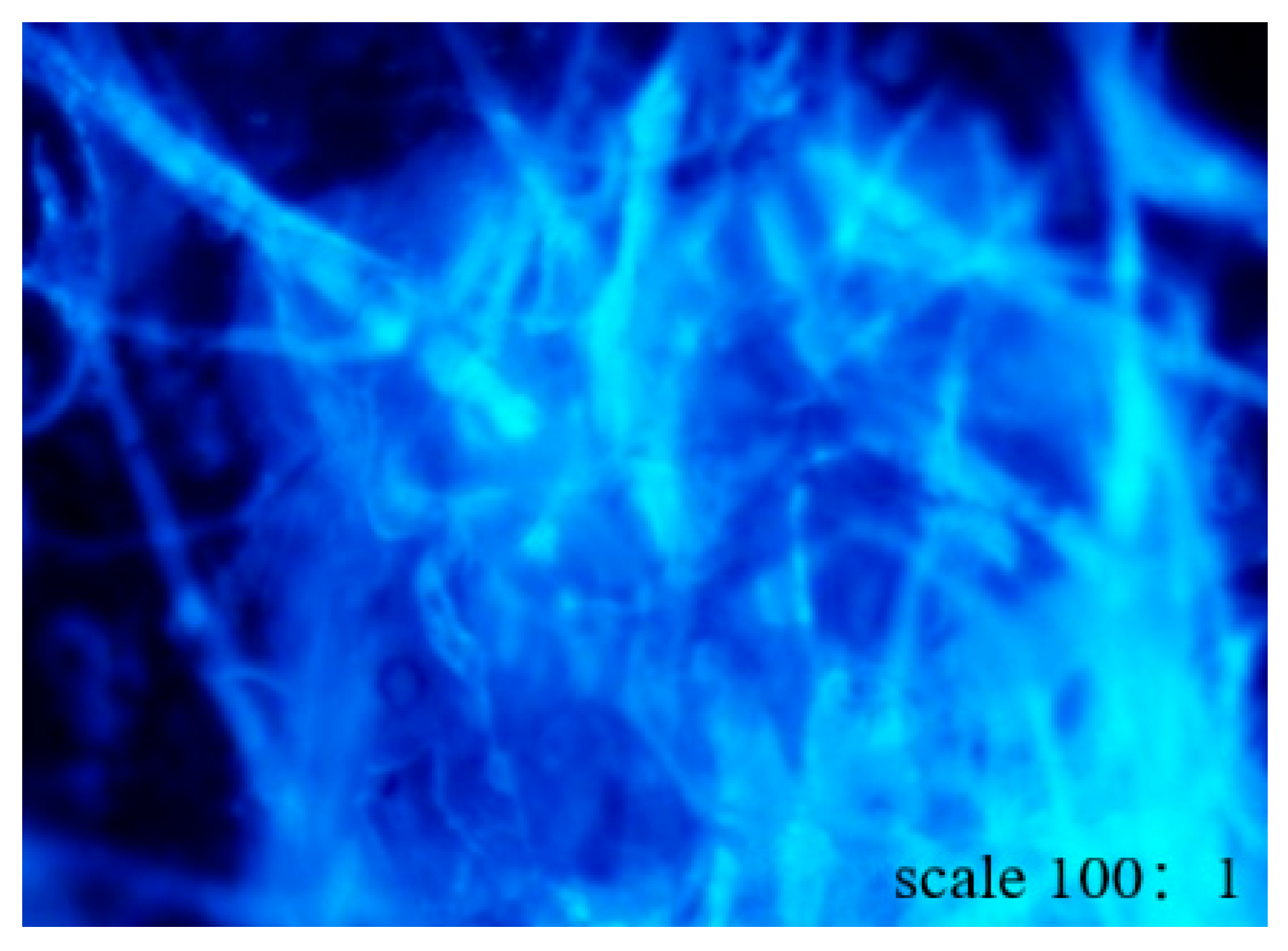
By acetylating polystyrene, we obtained stable fluorescent polystyrene, and significant fluorescence of acetylated polystyrene (see Figure 1) was observed through an inverted fluorescence microscope.
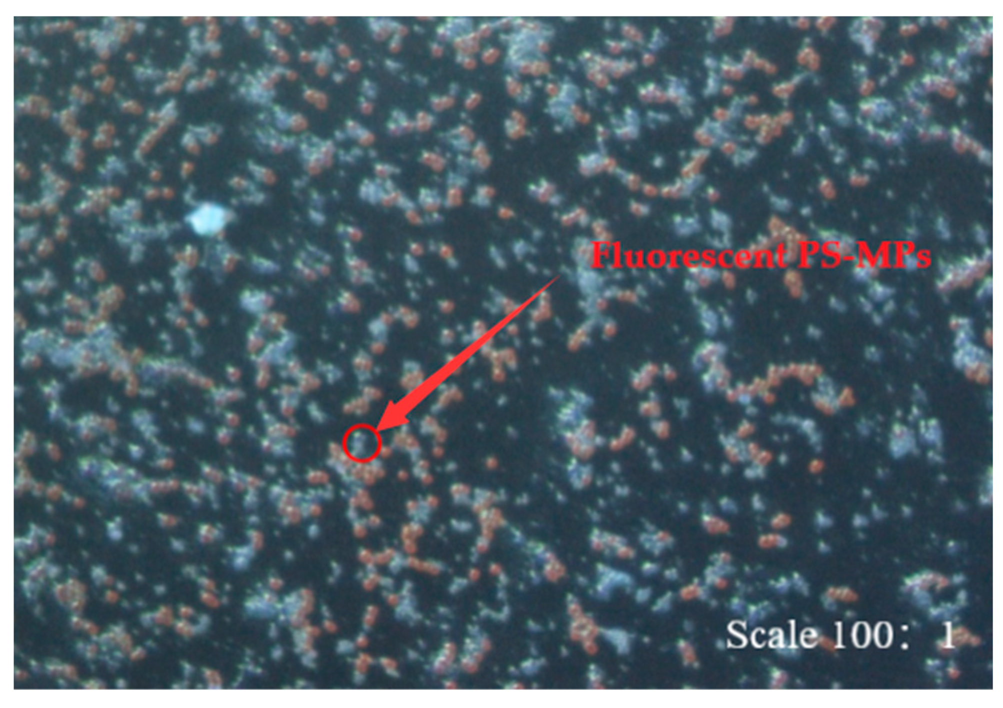
The adsorption of PS-MPs by Nostocaceae was observed through an inverted fluorescence microscope after exposure to PS-MPs for 24 h (see Figure 2). The blue particles were PS-MPs, and the yellow particles were Nostocaceae.
3.1.2. Changes in Chlorophyll Content
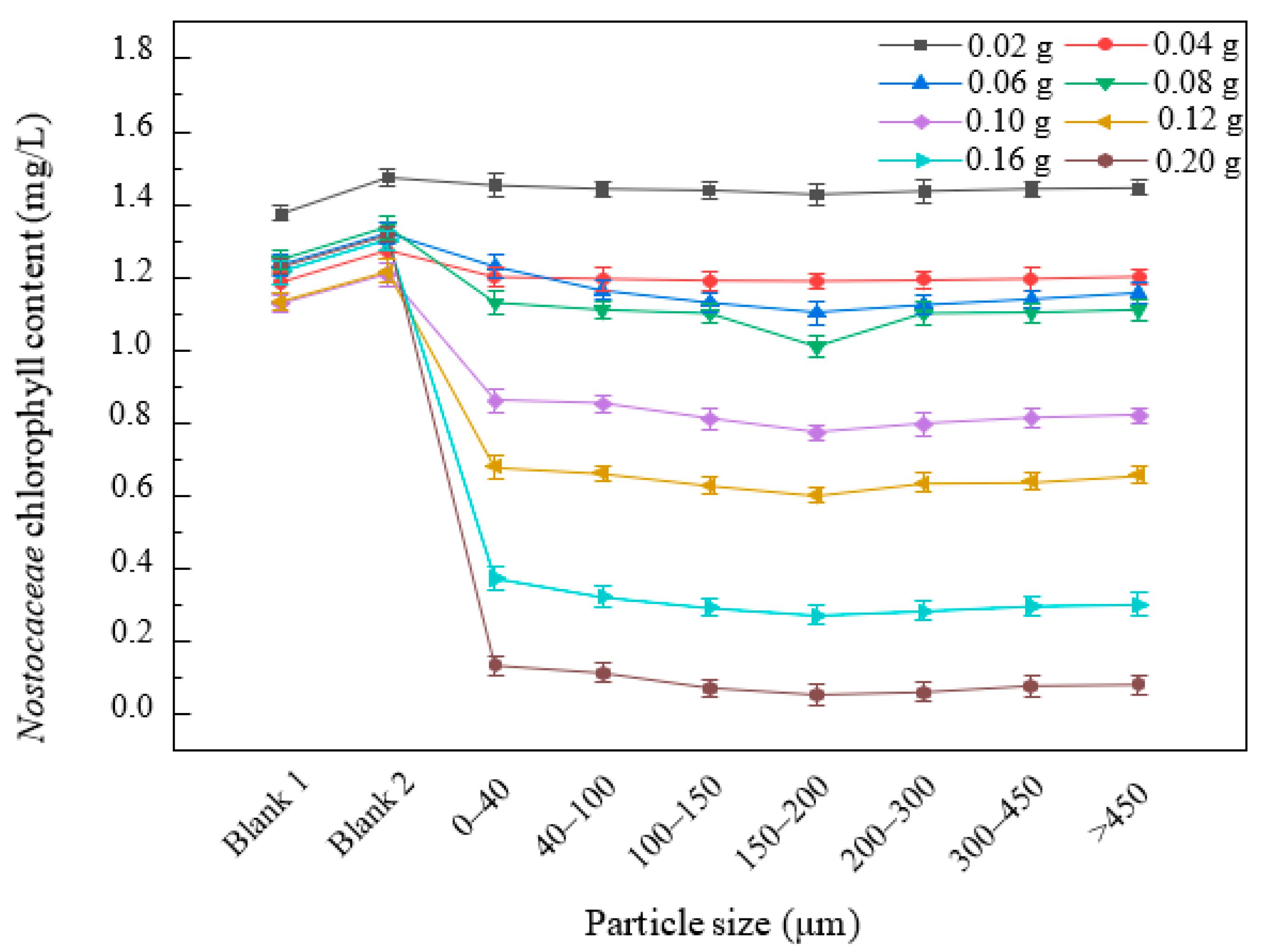
In the Nostocaceae experiment, the medium of Nostocaceae with the same concentration and different particle-sized microplastics demonstrated that the chlorophyll decreased with a rise in particle size. The chlorophyll reached the minimum value at 150–200 μm when the Nostocaceae were exposed to the same concentration of PS-MPs with 0–200 μm particle size (see Figure 3). The chlorophyll increased gradually when the particle size of PS-MPs was larger than 200 μm, which proved that 150–200 μm PS-MPs had the greatest effect on the chlorophyll of Nostocaceae.
The change in chlorophyll content was not obvious when the mass of PS-MPs was 0.02 g and the chlorophyll was lower than blank group 2. However, the chlorophyll was above blank group 1, which proved that the number of Nostocaceae and chlorophyll still increased when the PS-MP weight was less than 0.02 g. The increase in Nostocaceae number and chlorophyll in the low-concentration group may be due to the fact that the algae are resistant to microplastic stress through self-regulation [39].
3.1.3. Changes in Density, Fresh and Dry Weight
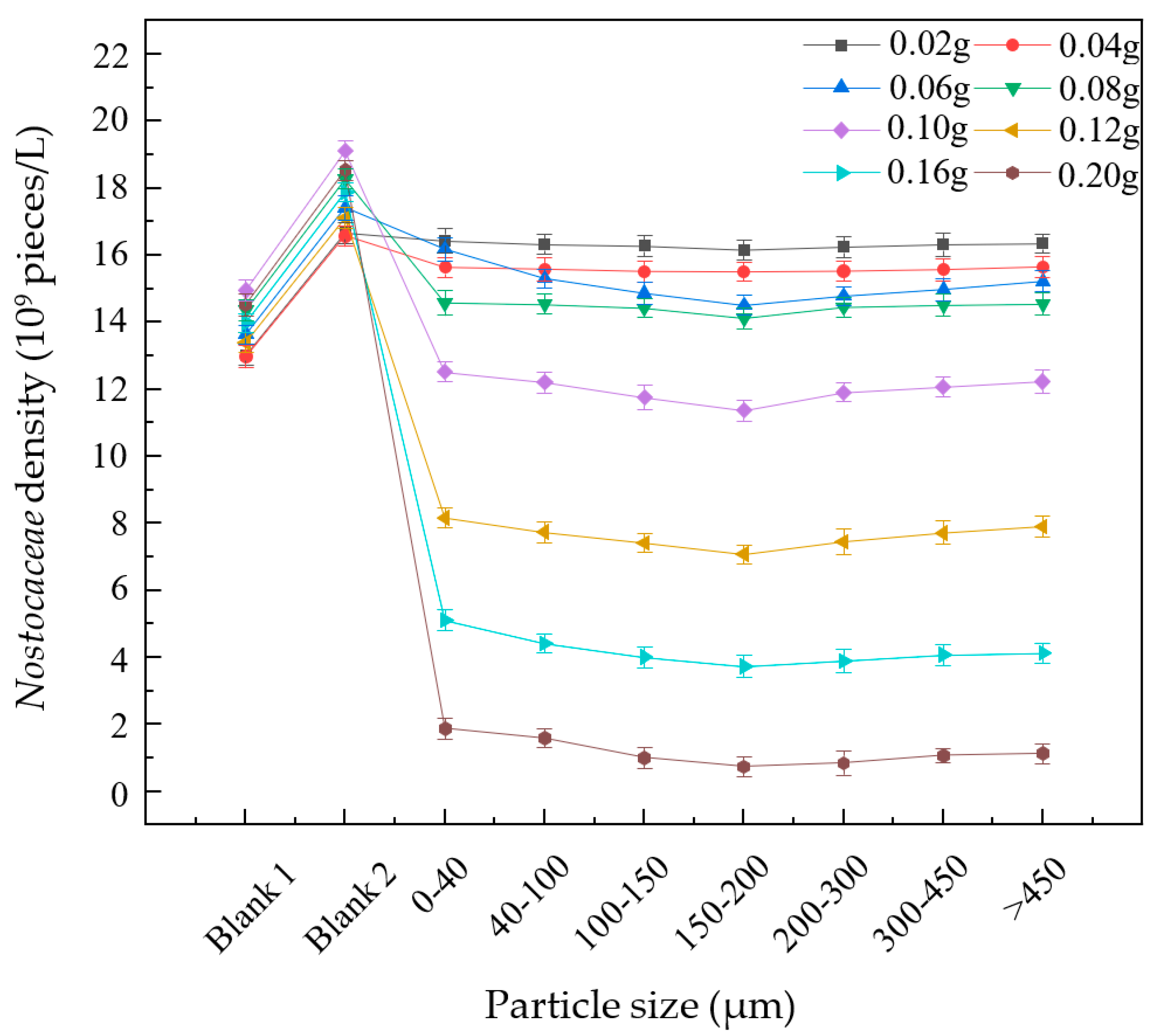
The chlorophyll decreased rapidly, and a large number of Nostocaceae died when the additional mass of PS-MPs was more than 0.08 g. If the PS-MP mass reached 0.12 g, the chlorophyll decreased by about 50% and half of the Nostocaceae died (see Figure 4). When the PS-MP mass reached 0.20 g, the content of chlorophyll tended towards 0, and almost all the Nostocaceae died. The result indicated that high concentrations of PS-MPs had a lethal effect on Nostocaceae. The reason may be that polystyrene particles adhering to algae can exacerbate light attenuation and reduce nutrient and gas swapping in algal cells, which may adversely affect Nostocaceae respiration and photosynthesis [40,41].
Keeping the concentration constant, the density of Nostocaceae decreased with the increasing particle size and reached a minimum at 150–200 μm. If the particle size was greater than 200 μm, the density of Nostocaceae slowly rose by 1–2% compared to the particle size of 150–200 μm, which proved that the PS-MPs of 150–200 μm had a significant effect on the density of Nostocaceae. The density of Nostocaceae did not change significantly, which was 3%, 6% and 15% lower than that of blank 2 after 0.02 g/0.04 g/0.06 g of PS-MPs with different particle sizes.
After exposure to 0.02–0.08 g of PS-MPs for 24 h, the density of Nostocaceae was significantly lower than blank 2 but higher than blank 1, which proved that the growth of Nostocaceae was inhibited when the dosage of PS-MPs was less than 0.08 g. If the mass of PS-MPs was more than 0.10 g, a large number of Nostocaceae died, indicating that PS-MPs had a lethal effect on Nostocaceae. The mortality rate of Nostocaceae was about 50% compared to that before the exposure experiment after 0.12 g of 150–200 μm PS-MPs was added. If 0.20 g of 150–200 μm PS-MPs was added, the Nostocaceae density tended towards 0.
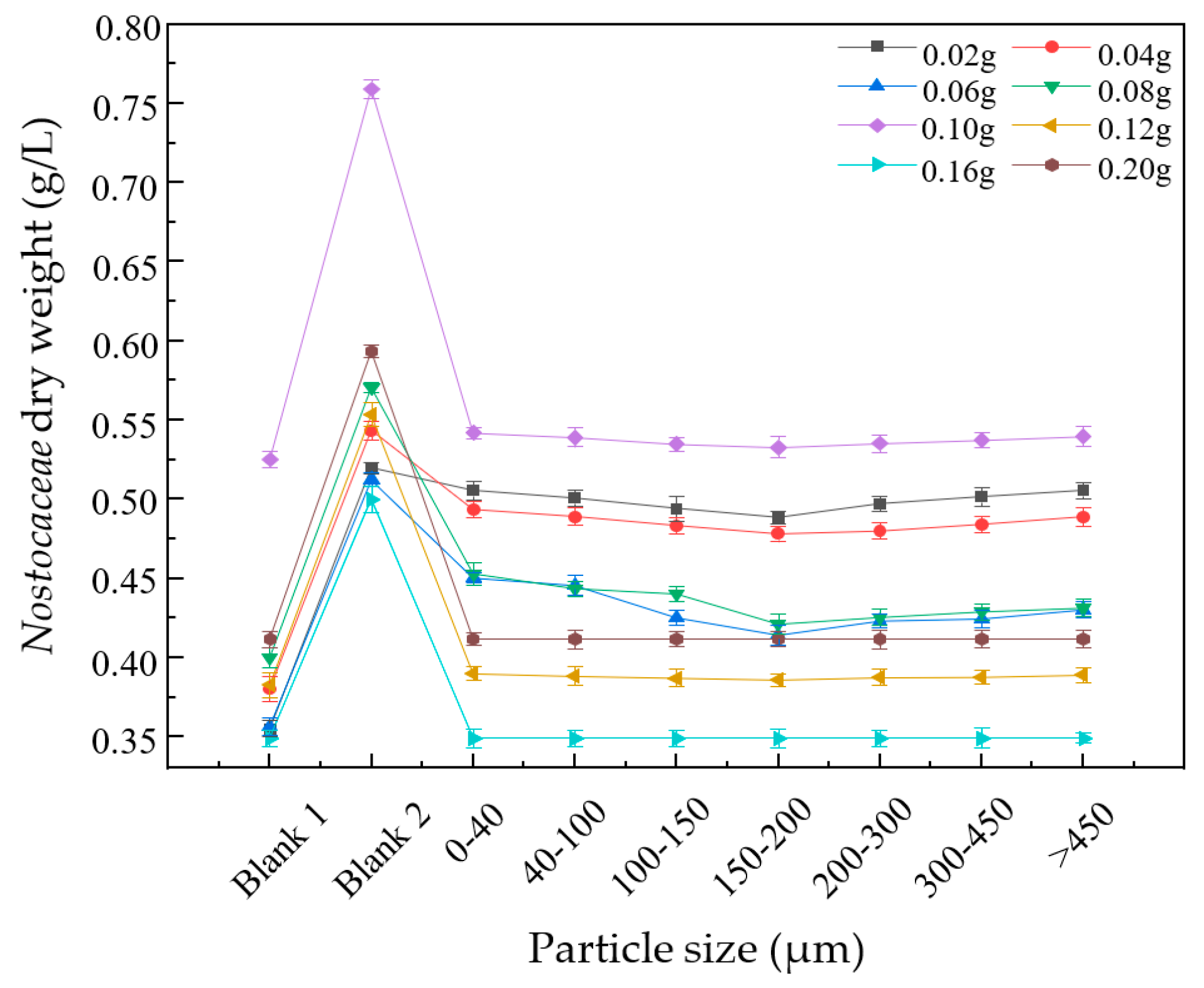
Studies have reported that PS-MPs can inhibit the respiration and photosynthesis of algae, interfere with the amino acid metabolism pathway of algae, indirectly inhibit cell division and hinder algae growth [24,28]. When the mass of PS-MPs was more than 0.10 g, the chlorophyll content of Nostocaceae decreased significantly, regardless of the particle size (see Figure 3), and a large number of Nostocaceae ceased to grow and thus died, resulting in the fresh and dry weight of Nostocaceae being close to that before the exposure experiment (see Figure 5 and Figure 6). In addition, polystyrene microplastics and algae extracellular polymers were able to form heterogeneous aggregates, leading to physical damage, such as cell wall damage and membrane structure changes [9], which may also contribute to the death of large numbers of Nostocaceae.
3.2. Effects on Daphnia Magna
3.2.1. Observation of the Intestine of Daphnia Magna
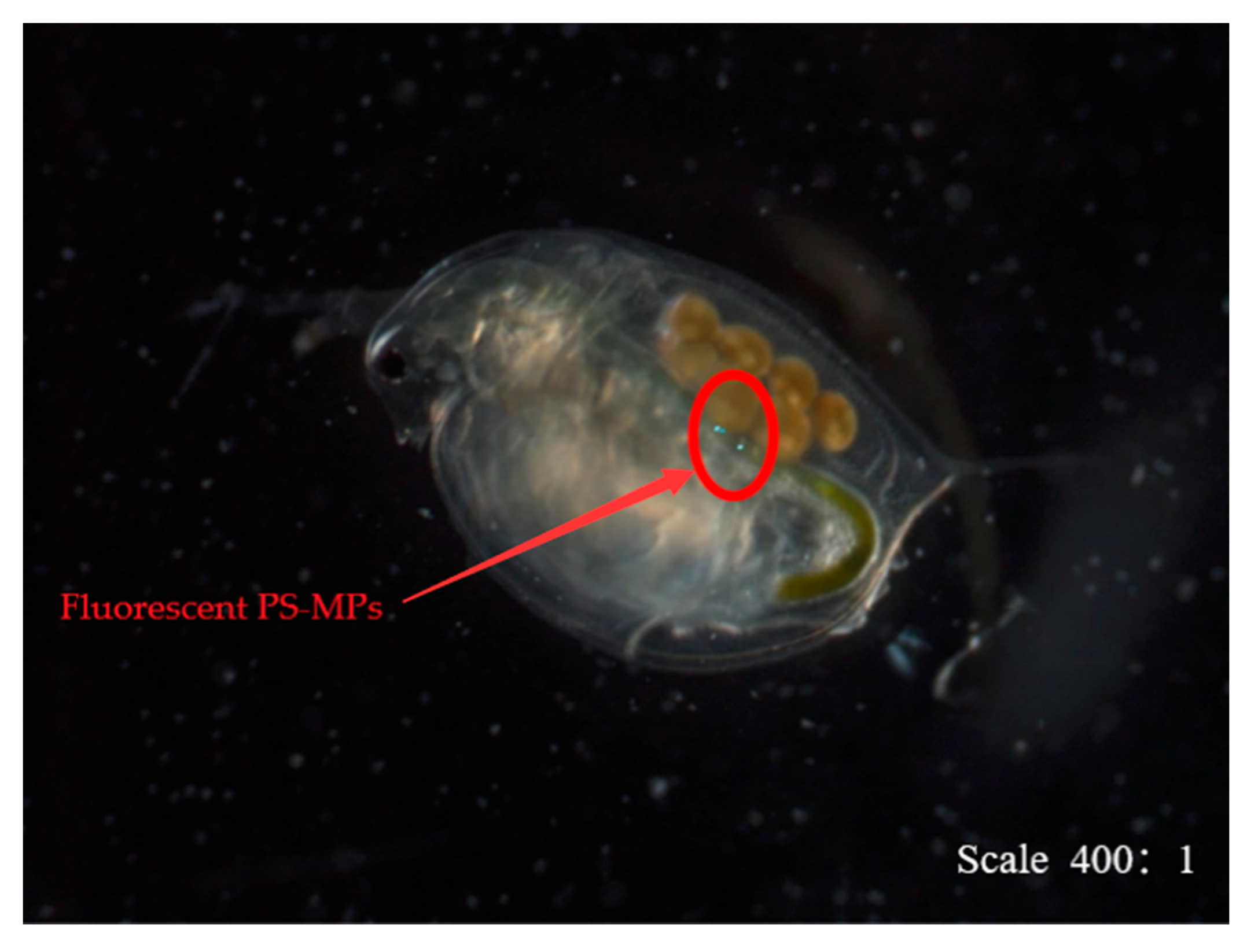
Fluorescent PS-MPs were observed in the intestines of Daphnia Magna with the 0–40 μm group by using a fluorescence inverted microscope (see Figure 7) There was no fluorescence in the intestines of Daphnia Magna when the particle size was more than 40 μm, indicating that MPs were not easily ingested by Daphnia Magna when the particle size was too large.
3.2.2. Changes in the Number of Survivors
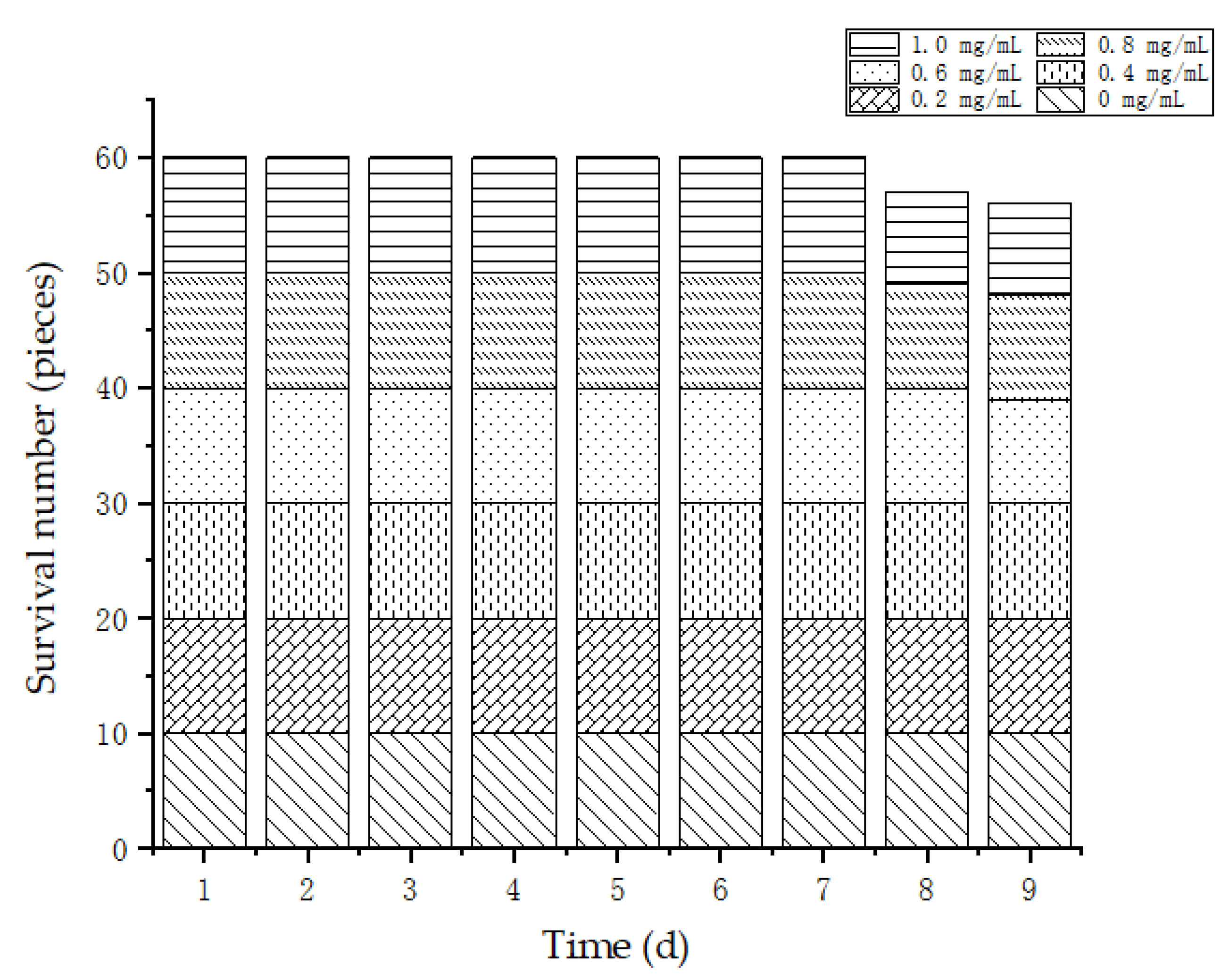
According to Figure 8, one or two Daphnia Magna died when the particle size of PS-MPs was over 0–40 μm and the concentration was higher than 0.6 mg/mL. Fluorescent PS-MPs were observed in the dead Daphnia Magna, which proved that PS-MPs could be ingested by Daphnia Magna and gathered in the intestine, causing intestinal obstruction [42]. Larger PS-MP aggregates would cause greater damage to the intestines of organisms [43]. Under high concentrations of PS-MPs, an accumulation of PS-MPs in the intestine and the death of Daphnia Magna were observed, which may be because the accumulation of PS-MPs and other substances caused intestinal obstruction of Daphnia Magna and the destruction of intestinal microvilli and epithelial cells, leading to the death of Daphnia Magna [42].
3.2.3. Changes in the Egg-Laying Amount
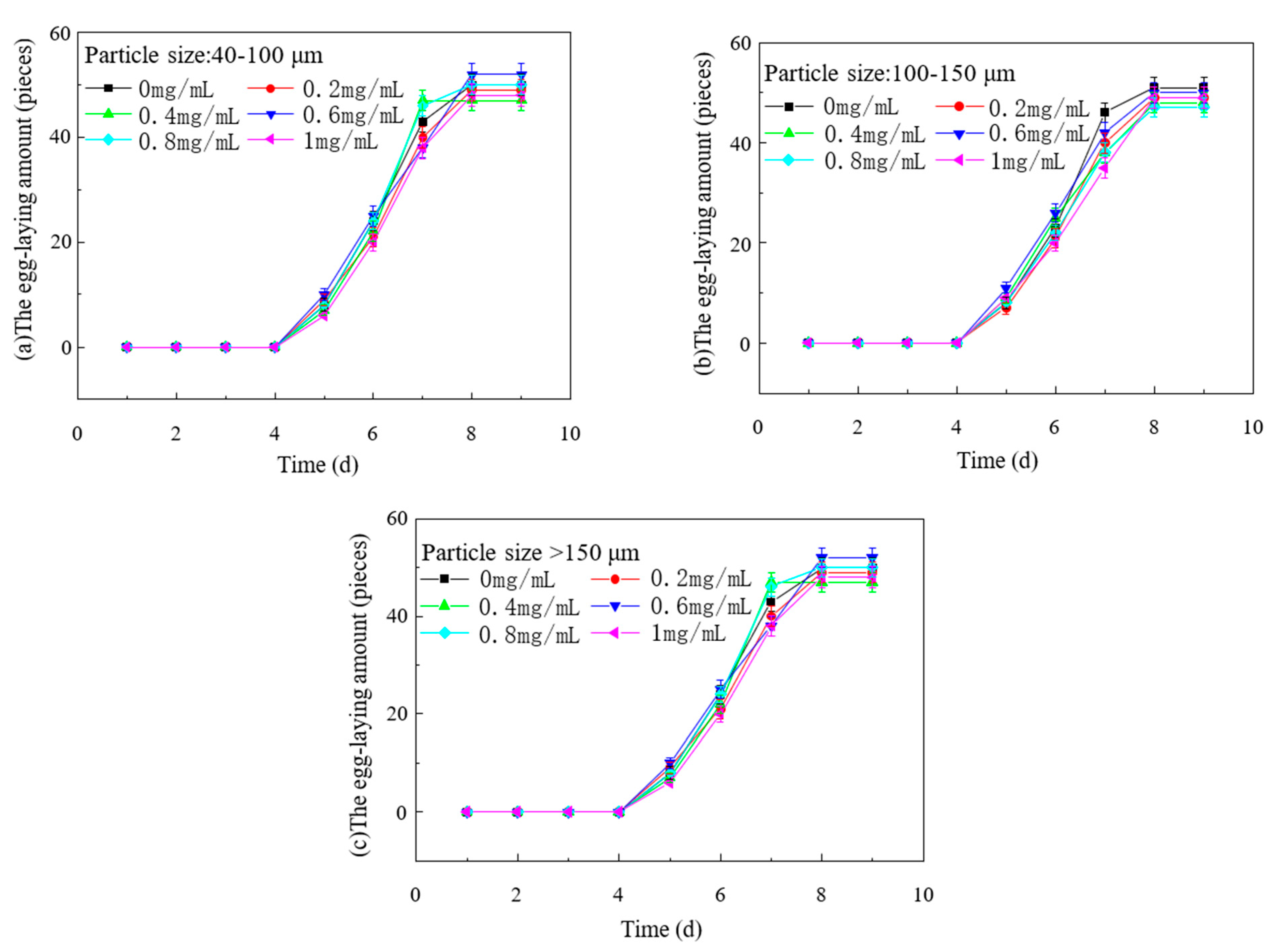
The effect of PS-MPs on the spawning rate of Daphnia Magna was researched. We maintained the same concentration; the spawning rate of the 0–40 μm group was lower than other groups (see Figure 9), and the spawning time was delayed, meaning that the oviposition of Daphnia Magna was inhibited. However, in groups larger than 40 μm, the spawning rate was not affected (see Figure 10). At the same particle size, the higher the concentration, the stronger the inhibitory effect on spawning.
The reasons for the decreased spawning rate of Daphnia Magna were diverse, which may include the effects of PS-MPs on the nervous system and behavior of Daphnia Magna, leading to a decrease in spawning rate, and continuous exposure to MPs over generations could cause population extinction [31]. PS-MPs may influence the Daphnia Magna in the content of acetyl cholinesterase (Acetylcholinesterase, AchE) to produce neurotoxicity. AchE is one of the most important enzymes in invertebrates [44,45]. Exposure of Daphnia Magna to a PS environment can inhibit the activity of AchE, causing oxidative stress and nervous system disorder [44].
In addition, changes in the behavior of Daphnia Magna may also be responsible for the decrease in the spawning rate. PS-NPs can break the blood–brain barrier of aquatic organisms [46] and cause neuritis [47]. After crucian carp was exposed to 100 mg/L PS-NPs for 64 days, it was found that the crucian carp brain mass decreased, behavior changed and cerebral gyri enlarged, which proved that PS-MPs produced significant neurotoxicity in the crucian carp [25]. Although MPs did not block the absorption of nutrients from fish, they can break down the innate immune system, damage cells, weaken the barrier function of the gut, lead to more harmful substances and pathogens entering the body [33] and cause intestinal microbial flora imbalance and inflammatory reaction [48,49]. NPs could cause oxidative stress, destroy the antioxidant system and inhibit AchE activity after entering the zebrafish brain [50]. Brain histology showed inflammatory cell infiltration, neuronal degeneration and necrosis and cytoplasmic vacuoles. Abnormal zebrafish behavior, such as seizures, was also observed in the large MP group [51,52]. The effects of PS-MPs on the spawning rate of Daphnia Magna suggested that the high concentration and long-term effect of MPs on the population of Daphnia Magna cannot be ignored.
4. Conclusions
In conclusion, the toxicity of PS-MPs on autotrophs and heterotrophic organisms in fresh water was illustrated from the individual level to the population level in this research. In the test group, the chlorophyll content of Nostocaceae, was significantly reduced, and the density was gradually decreased or even dead when PS-MPs were more than 0.08 g in weight. The dry weight and fresh weight of Nostocaceae were close to the levels before the exposure experiment, and the growth of Nostocaceae was greatly inhibited at the same time. Fluorescent PS-MPs were observed in the intestine of Daphnia Magna, and with an increase in the PS-MP concentration, some of the Daphnia Magna died. The test group Daphnia Magna had adverse reactions, such as a decreased oviposition rate and prolonged spawning time, under the high PS-MP concentration. In a word, considering these changes, it is necessary to further study the molecular mechanisms by which polystyrene microplastics affect the algal chlorophyll content and the resulting neurotoxicity toward Daphnia Magna, to provide a theoretical reference for further exploring the threat of microplastics to other animals.
Author Contributions
Conceptualization, M.L. and Y.X.; Data curation, S.Q. and Y.Q.; Formal analysis, Y.Q.; Funding acquisition, M.L.; Investigation, Y.Q.; Methodology, Y.X.; Project administration, W.Y.; Resources, S.Q., W.Y. and Y.X.; Supervision, W.Y.; Validation, Y.Q., X.F. and Y.X.; Visualization, S.Q.; Writing—original draft, S.Q. and Y.Q.; Writing—review and editing, X.F. and Y.X. All authors have read and agreed to the published version of the manuscript.
Funding
This work was supported by the Tianjin Science and Technology Planning Project (22YDTPJC00760); Tianjin Technical Innovation Guidance Special Project (20YDTPJC01790); Tianjin Key R&D Program—Social Development and Agriculture Project (21YFSNSN00190); National Key R&D Program ‘Technology Boosts Economy 2020′ key Special Project (SQ2020YFF0401503); Tianjin Science and Technology Planning Project (18ZXSZSF00090); Tianjin Technical Innovation Guidance Special Project (20YDTPJC00150).
Data Availability Statement
The data used to support the findings of this study were included within the article.
Conflicts of Interest
The authors declare that there were no conflict of interest regarding the publication of this paper.
References
- Geyer, R.; Jambeck, J.K.L. Law, Production, use, and fate of all plastics ever made. Sci. Adv. 2017, 3, e1700782. [Google Scholar] [CrossRef] [PubMed]
- PlasticsEurope. Plastics—The Facts 2022, PlasticsEurope. 2022. Available online: https://plasticseurope.org/knowledge-hub/plastics-the-facts-2022/ (accessed on 3 March 2023).
- Wang, L.; Wu, W.-M.; Bolan, N.S.; Tsang, D.C.; Li, Y.; Qin, M.; Hou, D. Environmental fate, toxicity and risk management strategies of nanoplastics in the environment: Current status and future perspectives. J. Hazard. Mater. 2021, 401, 123415. [Google Scholar] [CrossRef] [PubMed]
- Thompson, R.C.; Olsen, Y.; Mitchell, R.P.; Davis, A.; Rowland, S.J.; John, A.W.G.; McGonigle, D.; Russell, A.E. Lost at Sea: Where Is All the Plastic? Science 2004, 304, 838. [Google Scholar] [CrossRef] [PubMed]
- Kögel, T.; Bjorøy, O.; Toto, B.; Bienfait, A.M.; Sanden, M. Micro- and nanoplastic toxicity on aquatic life: Determining factors. Sci. Total. Environ. 2020, 709, 136050. [Google Scholar] [CrossRef]
- Duis, K.; Coors, A. Microplastics in the aquatic and terrestrial environment: Sources (with a specific focus on personal care products), fate and effects. Environ. Sci. Eur. 2016, 28, 1–25. [Google Scholar] [CrossRef]
- Wang, J.; Peng, J.; Tan, Z.; Gao, Y.; Zhan, Z.; Chen, Q.; Cai, L. Microplastics in the surface sediments from the Beijiang River littoral zone: Composition, abundance, surface textures and interaction with heavy metals. Chemosphere 2017, 171, 248–258. [Google Scholar] [CrossRef]
- Ballent, A.; Corcoran, P.L.; Madden, O.; Helm, P.A.; Longstaffe, F.J. Sources and sinks of microplastics in Canadian Lake Ontario nearshore, tributary and beach sediments. Mar. Pollut. Bull. 2016, 110, 383–395. [Google Scholar] [CrossRef]
- Mao, Y.; Ai, H.; Chen, Y.; Zhang, Z.; Zeng, P.; Kang, L.; Li, W.; Gu, W.; He, Q.; Li, H. Phytoplankton response to polystyrene microplastics: Perspective from an entire growth period. Chemosphere 2018, 208, 59–68. [Google Scholar] [CrossRef]
- Jambeck, J.R.; Geyer, R.; Wilcox, C.; Siegler, T.R.; Perryman, M.; Andrady, A.; Narayan, R.; Law, K.L. Plastic waste inputs from land into the ocean. Science 2015, 347, 768–771. [Google Scholar] [CrossRef]
- Raju, P.; Santhanam, P.; Pandian, S.S.; Divya, M.; Arunkrishnan, A.; Devi, K.N.; Ananth, S.; Roopavathy, J.; Perumal, P. Impact of polystyrene microplastics on major marine primary (phytoplankton) and secondary producers (copepod). Arch. Microbiol. 2021, 204, 84. [Google Scholar] [CrossRef]
- Kelpsiene, E.; Ekvall, M.T.; Lundqvist, M.; Torstensson, O.; Hua, J.; Cedervall, T. Review of ecotoxicological studies of widely used polystyrene nanoparticles. Environ. Sci. Process. Impacts 2022, 24, 8–16. [Google Scholar] [CrossRef]
- De-La-Torre, G.E.; Dioses-Salinas, D.C.; Castro, J.M.; Antay, R.; Fernández, N.Y.; Espinoza-Morriberón, D.; Saldaña-Serrano, M. Abundance and distribution of microplastics on sandy beaches of Lima, Peru. Mar. Pollut. Bull. 2020, 151, 110877. [Google Scholar] [CrossRef]
- Lim, C.; Kim, N.; Lee, J.; Yoon, Y. Potential of Adsorption of Diverse Environmental Contaminants onto Microplastics. Water 2022, 14, 4086. [Google Scholar] [CrossRef]
- Fang, S.; Yu, W.; Li, C.; Liu, Y.; Qiu, J.; Kong, F. Adsorption behavior of three triazole fungicides on polystyrene microplastics. Sci. Total. Environ. 2019, 691, 1119–1126. [Google Scholar] [CrossRef]
- Llorca, M.; Schirinzi, G.; Martínez, M.; Barceló, D.; Farré, M. Adsorption of perfluoroalkyl substances on microplastics under environmental conditions. Environ. Pollut. 2018, 235, 680–691. [Google Scholar] [CrossRef] [PubMed]
- Chen, Z.; Yang, J.; Huang, D.; Wang, S.; Jiang, K.; Sun, W.; Chen, Z.; Cao, Z.; Ren, Y.; Wang, Q.; et al. Adsorption behavior of aniline pollutant on polystyrene microplastics. Chemosphere 2023, 323, 138187. [Google Scholar] [CrossRef] [PubMed]
- Kik, K.; Bukowska, B.; Sicińska, P. Polystyrene nanoparticles: Sources, occurrence in the environment, distribution in tissues, accumulation and toxicity to various organisms. Environ. Pollut. 2020, 262, 114297. [Google Scholar] [CrossRef] [PubMed]
- Chaukura, N.; Gwenzi, W.; Bunhu, T.; Ruziwa, D.T.; Pumure, I. Potential uses and value-added products derived from waste polystyrene in developing countries: A review. Resour. Conserv. Recycl. 2016, 107, 157–165. [Google Scholar] [CrossRef]
- Gulizia, A.M.; Patel, K.; Philippa, B.; Motti, C.A.; van Herwerden, L.; Vamvounis, G. Understanding plasticiser leaching from polystyrene microplastics. Sci. Total. Environ. 2023, 857, 159099. [Google Scholar] [CrossRef]
- Bandow, N.; Will, V.; Wachtendorf, V.; Simon, F.-G. Contaminant release from aged microplastic. Environ. Chem. 2017, 14, 394–405. [Google Scholar] [CrossRef]
- Zhang, Y.; Yin, K.; Wang, D.; Wang, Y.; Lu, H.; Zhao, H.; Xing, M. Polystyrene microplastics-induced cardiotoxicity in chickens via the ROS-driven NF-kappaB-NLRP3-GSDMD and AMPK-PGC-1alpha axes. Sci. Total Environ. 2022, 840, 156727. [Google Scholar] [CrossRef]
- Nogueira, D.J.; Silva, A.C.d.O.d.; da Silva, M.L.N.; Vicentini, D.S.; Matias, W.G. Individual and combined multigenerational effects induced by polystyrene nanoplastic and glyphosate in Daphnia magna (Strauss, 1820). Sci. Total. Environ. 2022, 811, 151360. [Google Scholar] [CrossRef]
- Guschina, I.A.; Hayes, A.J.; Ormerod, S.J. Polystyrene microplastics decrease accumulation of essential fatty acids in common freshwater algae. Environ. Pollut. 2020, 263, 114425. [Google Scholar] [CrossRef]
- Mattsson, K.; Johnson, E.V.; Malmendal, A.; Linse, S.; Hansson, L.-A.; Cedervall, T. Brain damage and behavioural disorders in fish induced by plastic nanoparticles delivered through the food chain. Sci. Rep. 2017, 7, 11452. [Google Scholar] [CrossRef]
- Rillig, M.C. Microplastic in Terrestrial Ecosystems and the Soil? Environ. Sci. Technol. 2012, 46, 6453–6454. [Google Scholar] [CrossRef] [PubMed]
- Chang, Y.-H.; Ku, C.-R.; Lu, H.-L. Effects of aquatic ecological indicators of sustainable green energy landscape facilities. Ecol. Eng. 2014, 71, 144–153. [Google Scholar] [CrossRef]
- Wang, S.-C.; Liu, G.-Z.; Liu, F.-F. Physiological and metabolic toxicity of polystyrene microplastics to Dunaliella salina. Environ. Pollut. 2023, 316, 120544. [Google Scholar] [CrossRef] [PubMed]
- Lang, X.; Ni, J.; He, Z. Effects of polystyrene microplastic on the growth and volatile halocarbons release of microalgae Phaeodactylum tricornutum. Mar. Pollut. Bull. 2022, 174, 113197. [Google Scholar] [CrossRef]
- Besseling, E.; Wang, B.; Lurling, M.; Koelmans, A. Nanoplastic affects growth of S. obliquus and reproduction of D. magna. Environ. Sci. Technol. 2014, 48, 12336–12343. [Google Scholar] [CrossRef]
- Martins, A.; Guilhermino, L. Transgenerational effects and recovery of microplastics exposure in model populations of the freshwater cladoceran Daphnia magna Straus. Sci. Total. Environ. 2018, 631-632, 421–428. [Google Scholar] [CrossRef]
- Pei, X.; Heng, X.; Chu, W. Polystyrene nano/microplastics induce microbiota dysbiosis, oxidative damage, and innate immune disruption in zebrafish. Microb. Pathog. 2022, 163, 105387. [Google Scholar] [CrossRef]
- Zhu, M.; Chernick, M.; Rittschof, D.; Hinton, D.E. Chronic dietary exposure to polystyrene microplastics in maturing Japanese medaka (Oryzias latipes). Aquat. Toxicol. 2020, 220, 105396. [Google Scholar] [CrossRef]
- Kim, J.; Haque, N.; Lee, S.; Lee, D.-H.; Rhee, J.-S. Exposure to Environmentally Relevant Concentrations of Polystyrene Microplastics Increases Hexavalent Chromium Toxicity in Aquatic Animals. Toxics 2022, 10, 563. [Google Scholar] [CrossRef]
- Nor, N.M.; Kooi, M.; Diepens, N.; Koelmans, A.A. Lifetime Accumulation of Microplastic in Children and Adults. Environ. Sci. Technol. 2021, 55, 5084–5096. [Google Scholar]
- Zhu, X.; Wang, C.; Duan, X.; Liang, B.; Xu, E.G.; Huang, Z. Micro- and nanoplastics: A new cardiovascular risk factor? Environ. Int. 2023, 171, 107662. [Google Scholar] [CrossRef]
- Leslie, H.A.; van Velzen, M.J.; Brandsma, S.H.; Vethaak, A.D.; Garcia-Vallejo, J.J.; Lamoree, M.H. Discovery and quantification of plastic particle pollution in human blood. Environ. Int. 2022, 163, 107199. [Google Scholar] [CrossRef]
- Feng, X.; Yang, X.; Li, M.; Qin, Y.; Li, H.; Xie, Y. Production and method optimization of fluorescent polystyrene. J. Mol. Struct. 2021, 1243. [Google Scholar] [CrossRef]
- Wu, Y.; Guo, P.; Zhang, X.; Zhang, Y.; Xie, S.; Deng, J. Effect of microplastics exposure on the photosynthesis system of freshwater algae. J. Hazard. Mater. 2019, 374, 219–227. [Google Scholar] [CrossRef]
- Chen, P.; Powell, B.A.; Mortimer, M.; Ke, P.C. Adaptive Interactions between Zinc Oxide Nanoparticles and Chlorella sp. Environ. Sci. Technol. 2012, 46, 12178–12185. [Google Scholar] [CrossRef]
- Bhattacharya, P.; Lin, S.; Turner, J.P.; Ke, P.C. Physical Adsorption of Charged Plastic Nanoparticles Affects Algal Photosynthesis. J. Phys. Chem. C 2010, 114, 16556–16561. [Google Scholar] [CrossRef]
- Chen, C.C.; Shi, Y.; Zhu, Y.; Zeng, J.; Qian, W.; Zhou, S.; Ma, J.; Pan, K.; Jiang, Y.; Tao, Y.; et al. Combined toxicity of polystyrene microplastics and ammonium perfluorooctanoate to Daphnia magna: Mediation of intestinal blockage. Water Res. 2022, 219, 118536. [Google Scholar] [CrossRef] [PubMed]
- Wright, S.L.; Thompson, R.C.; Galloway, T.S. The physical impacts of microplastics on marine organisms: A review. Environ. Pollut. 2013, 178, 483–492. [Google Scholar] [CrossRef] [PubMed]
- Liu, J.; Yang, H.; Meng, Q.; Feng, Q.; Yan, Z.; Liu, J.; Liu, Z.; Zhou, Z. Intergenerational and biological effects of roxithromycin and polystyrene microplastics to Daphnia magna. Aquat. Toxicol. 2022, 248, 106192. [Google Scholar] [CrossRef] [PubMed]
- Richter, N.; Beckers, N.; Onur, O.; Dietlein, M.; Tittgemeyer, M.; Kracht, L.; Neumaier, B.; Fink, G.R.; Kukolja, J. Effect of cholinergic treatment depends on cholinergic integrity in early Alzheimer’s disease. Brain 2018, 141, 903–915. [Google Scholar] [CrossRef] [PubMed]
- Kashiwada, S. Distribution of Nanoparticles in the See-through Medaka (Oryzias latipes). Environ. Health Perspect. 2006, 114, 1697–1702. [Google Scholar] [CrossRef]
- Prüst, M.; Meijer, J.; Westerink, R.H.S. The plastic brain: Neurotoxicity of micro- and nanoplastics. Part. Fibre Toxicol. 2020, 17, 24. [Google Scholar] [CrossRef]
- Xie, S.; Zhou, A.; Wei, T.; Li, S.; Yang, B.; Xu, G.; Zou, J. Nanoplastics Induce More Serious Microbiota Dysbiosis and Inflammation in the Gut of Adult Zebrafish than Microplastics. Bull. Environ. Contam. Toxicol. 2021, 107, 640–650. [Google Scholar] [CrossRef]
- Jin, Y.; Xia, J.; Pan, Z.; Yang, J.; Wang, W.; Fu, Z. Polystyrene microplastics induce microbiota dysbiosis and inflammation in the gut of adult zebrafish. Environ. Pollut. 2018, 235, 322–329. [Google Scholar] [CrossRef]
- Yu, H.; Chen, Q.; Qiu, W.; Ma, C.; Gao, Z.; Chu, W.; Shi, H. Concurrent water- and foodborne exposure to microplastics leads to differential microplastic ingestion and neurotoxic effects in zebrafish. Water Res. 2022, 219, 118582. [Google Scholar] [CrossRef]
- Umamaheswari, S.; Priyadarshinee, S.; Bhattacharjee, M.; Kadirvelu, K.; Ramesh, M. Exposure to polystyrene microplastics induced gene modulated biological responses in zebrafish (Danio rerio). Chemosphere 2021, 281, 128592. [Google Scholar] [CrossRef]
- Chen, Q.; Lackmann, C.; Wang, W.; Seiler, T.-B.; Hollert, H.; Shi, H. Microplastics Lead to Hyperactive Swimming Behaviour in Adult Zebrafish. Aquat. Toxicol. 2020, 224, 105521. [Google Scholar] [CrossRef]
Figure 1.
Fluorescent microplastics under fluorescence microscope.

Figure 2.
Adsorption of PS-MPs by Nostocaceae after exposure.

Figure 3.
Changes in chlorophyll content before and after 24 h exposure of Nostocaceae. Blank 1 was Nostocaceae chlorophyll content in 0 h without adding polystyrene microplastic. Blank 2 was Nostocaceae chlorophyll content in 24 h without adding polystyrene microplastic. The error bar represents the standard deviation of measurement for chlorophyll content in three separate sample runs.

Figure 4.
Changes in algal density before and after 24 h exposure of Nostocaceae. Blank 1 was non-dosed polystyrene microplastic 0 h Nostocaceae density; blank 2 was non-dosed polystyrene microplastics. The error bar represents the standard deviation of measurement for Nostocaceae density in three separate sample runs.

Figure 5.
Changes in fresh weight before and after 24 h exposure of Nostocaceae. Blank 1 was non-dosed polystyrene microplastic 0 h Nostocaceae fresh weight. Blank 2 was non-dosed polystyrene microplastic 24 h Nostocaceae fresh weight. The error bar represents the standard deviation of measurement for fresh weight in three separate sample runs.

Figure 6.
Changes in dry weight before and after 24 h exposure of Nostocaceae. Blank 1 was non-dosed polystyrene microplastic 0 h dry weight of Nostocaceae. Blank 2 was non-dosed polystyrene microplastic 24 h dry weight of Nostocaceae. The error bar represents the standard deviation of measurement for dry weight in three separate sample runs.

Figure 7.
Uptake of polystyrene particles by Daphnia Magna.

Figure 8.
The survival number of Daphnia Magna after the addition of different concentrations of polystyrene with a particle size of 0–40 μm.
Figure 8.
The survival number of Daphnia Magna after the addition of different concentrations of polystyrene with a particle size of 0–40 μm.

Figure 9.
The egg-laying status of Daphnia Magna with different concentrations of fluorescent polystyrene particles at a particle size of 0–40 μm. The number of eggs laid by Daphnia Magna was counted visually. The error bar represents the standard deviation of measurement for egg-laying amount in three separate sample runs.

Figure 10.
The egg-laying status of Daphnia Magna with different concentrations of fluorescent polystyrene particles at a particle size of >40 μm. The number of eggs laid by Daphnia Magna was counted visually. The error bar represents the standard deviation of measurement for egg-laying amount in three separate sample runs.


Table 1.
The sizes of PS microplastics.
| Distribution of Size | D1 | D2 | D3 | D4 | D5 | D6 | D7 |
|---|---|---|---|---|---|---|---|
| Size (μm) | 0–40 | 4–100 | 100–150 | 150–200 | 200–300 | 300–450 | 450+ |
Disclaimer/Publisher’s Note: The statements, opinions and data contained in all publications are solely those of the individual author(s) and contributor(s) and not of MDPI and/or the editor(s). MDPI and/or the editor(s) disclaim responsibility for any injury to people or property resulting from any ideas, methods, instructions or products referred to in the content. |
© 2023 by the authors. Licensee MDPI, Basel, Switzerland. This article is an open access article distributed under the terms and conditions of the Creative Commons Attribution (CC BY) license (https://creativecommons.org/licenses/by/4.0/).
Share and Cite
MDPI and ACS Style
Qiu, S.; Yuan, W.; Qin, Y.; Feng, X.; Li, M.; Xie, Y. Effects of Spontaneous Fluorescent Polystyrene on Nostocaceae and Daphnia Magna. Water 2023, 15, 1744. https://doi.org/10.3390/w15091744
AMA Style
Qiu S, Yuan W, Qin Y, Feng X, Li M, Xie Y. Effects of Spontaneous Fluorescent Polystyrene on Nostocaceae and Daphnia Magna. Water. 2023; 15(9):1744. https://doi.org/10.3390/w15091744
Chicago/Turabian StyleQiu, Shaolin, Wenjiao Yuan, Yujie Qin, Xin Feng, Meitong Li, and Yuhong Xie. 2023. "Effects of Spontaneous Fluorescent Polystyrene on Nostocaceae and Daphnia Magna" Water 15, no. 9: 1744. https://doi.org/10.3390/w15091744
Note that from the first issue of 2016, this journal uses article numbers instead of page numbers. See further details here.